Microscopes and Endodontics
Sunny Dental & Implant Centre, Kathua (J & K) Dr Neeraj Narang (Private Practice), s Microdentistry & Implant Clinic, Meham, Rohtak (Haryana)
Abstract
Use of Microscope in the field of medical science is not new. Introduction of Dental Operating Microscope (DOM) in Endodontics has changed the field completely and started a new era of Endodontics. This has brought revolution in the field of endodontics with an improved success rate for clinicians. Clinicians using microscope are more confident about their work quality. Endodontic treatment before microscopes was never so reliable & predictable and less invasive during surgical procedures. The future in microscopic endodontics will involve the use of even finer microscopic instruments and the development of even more sophisticated techniques & will make it mandatory in every modern endodontic practice.
INTRODUCTION
Root canal treatment was thought to be a blind procedure and was taught as a blind procedure as it was not possible to see beyond the canal orifices. Even sometimes it was not possible to explore all the canals, leading to failure of root canal treatment.
Success of root canal treatment depends on the locating all the canals, cleaning & shaping the canals and filling the canals with a biocompatible material in 3 dimensions.
Introduction of Dental Operating Microscope (DOM) in Endodontics has changed the field completely and started a new era of Endodontics. DOM has its own use from diagnosis to completion of treatment viz, diagnosis of cracks, locating extra/ missed canals, removal of separated instruments, Re-treatment of failed root canal treatments, proper cleaning of root canal system & 3-D obturation. DOM has changed both non-surgical and surgical endodontics. In nonsurgical endodontics, every challenge existing in the straight portion of the root canal system, even if located in the most apical part, can be easily seen and managed competently under the microscope. In surgical endodontics, it is possible to carefully examine the apical segment of the root-end and perform an apical resection of the root without an exaggerated bevel, thereby making Class I cavity preparations along the longitudinal axis of the root easy to perform. DOM with Greater Taper instruments and Ultrasonics has changed the complete concept of Endodontics. Microsurgery involves less traumatic procedures with better success rate.
HISTORY
Use of Microscope in the field of medical science is not new. It was being used in the field Ophthalmology, Otolaryngology & Neurosurgery since 1920s. According to the Zeiss Company, the microscope was first introduced to otolaryngology around 1950, then to neurosurgery in the 1960s and to endodontics in the early 1990s. It was capable of only one magnification (8x), was positioned on a floor stand and poorly balanced, had only straight binoculars, and had a fixed focal length of 250 mm. This microscope used angled illumination instead of co focal illumination. It did not gain wide acceptance and the manufacturer ceased manufacturing them shortly thereafter their introduction.. Dentistry, therefore, is about 40 years behind medicine in this respect. In 1991 Gary Carr introduced an operating microscope with Galilean optics and ergonomically configured for dentistry with several advantages that allowed for easy use of the scope for nearly all endodontic and restorative procedures. This microscope had a magnification changer that allowed for five discrete magnifications(3.5-30x), had a stable mounting on either the wall or ceiling, hadangled binoculars allowing for sit-down dentistry, Then it becamepopular by 1995 amongst Endodontists. However surgical loupes were being used in dentistry long back.
DENTAL OPERATING MICROSCOPE
:
Why A Microscope! :
The Limits Of Human Vision: Resolution is defined as the ability of an optical system to make clear and distinguishable two separate entities. The resolving power of the unaided human eye is only 0.2 mm. In other words, most people who view two points closer than 0.2 mm will see only one point. A microscope can improve resolution by many orders of magnitude. For example, a common operating microscope can raise the resolving limit from 0.2 mm to 0.006 mm (6 microns), it’s a dramatic improvement.
Either a general practitioner or an endodontist. All of us routinely perform procedures requiring resolution well beyond 0.2 mm, the limit of human sight. Crown margins, scaling procedures, incisions, root canal location, caries removal, furcation and perforation repair, post placement or removal, and bone-and soft-tissue grafting procedures are only a few of the procedures that demand tolerances well beyond the 0.2 mm limit.
WAYS FOR MOUNTING THE MICROSCOPE :
It can be:
- Floor Microscope
- Ceiling Mounted
- Wall Mounted
- Attached with Dental Chair Unit (Available with few brands of chair and microscopes).
Microscope should be firmly fixed to a rigid surface to minimize the small movements of the head of the microscope during working.


PARTS OF DOM:
Operating Microscope has two main components
- Light Source/ Illumination
- Head of The Microscope
LIGHT SOURCE/ ILLUMINATION:
It’s a box having illumination system for the Microscope. It is cooled by an integrated fan in it. A fiber optic cable connects it to the head of the microscope.
The light source is one of the most important features of an operating microscope. Besides optics, the light source is responsible for operating in operative fields that are small and deep like the root canal.
Light source in it can be:
- Halogen: It is used very less now a days. It is yellow in colour.
- Metal Halide: White in colour but short life, about 6,000 to 15,000 Hrs.
- LED: Most commonly used. White in colour. It has long life, about 50,000 Hrs.
- Xenon: It is much more powerful and provides a brighter light at about 5,000° Kelvin approximating day light.
Intensity of light is controlled by a rheostat. Light from the light source after being transferred to head of the microscope by fiber optic cable is reflected through the condensing lens to a series of prisms and then to the objective lens to the surgical field. Light from the surgical field is reflected back through the objective lens through the magnification changer lenses and through the binoculars and then exits to the eye as two separate beams of light.
HEAD OF THE MICROSCOPE (THE LENS SYSTEM):
It consist of
- Eyepiece Lenses/ Binoculars
- Objective Lens
- Body Tube Optics
- Magnification Changers
- Beam Splitter (Optional)

EYEPIECE LENSES/ BINOCULARS:
Function of binoculars is to hold eyepiece lenses. It is the part of microscope through which operator see. Operator can adjust interpupillary distance (IPD) according to him.
Eyepiece lenses are available in various powers ranging 10X, 12.5X, 15X. Powers of these lenses contribute to the final magnification value with power of magnification changer and focal length of objective lens. As we increase the magnification, focal length will increase and will lead to narrower field of view.
Binoculars can be
- Rigid/ Fixed
- Inclinable
Rigid/ Fixed Binoculars:
These binoculars are so oriented that the tubes are offset at 45 degree angle with the head of the microscope. They can be tiring for long procedures but these are economical as easy to engineer.
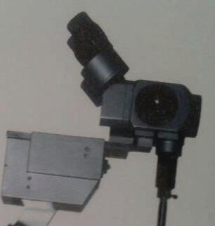
Inclinable Binoculars:
These binoculars are inclinable to an angle of 0 – 210 degrees depending on the brand. It provides better ergonomics and additional


postural comfort during long procedures. However these are difficult to engineer and quite expensive.
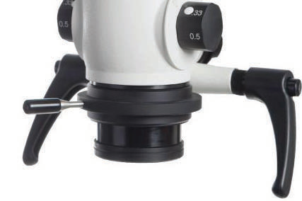
OBJECTIVE LENS:
This lens is towards the patient. These are available in variety of foal lengths ranging from 100mm to 400mm. However 200mm or 250mm is most commonly used.
The fine focus can be done manually using the device integrated in the objective lens, or rotating a fine focus knob, which raises the entire body of the microscope, or by an electric foot control.
MAGNIFICATION CHANGERS:
According to the levels/ steps of magnification, it can be 3 or 5 step magnification changers. It consists of different lenses that are mounted on turret.
3 Step changers has one set of lenses and one blank space on the turret which are factored in three fixed powers of magnification, two from each lens pair combination and one from the blank space. 5 step magnification changer has a second set of lenses mounted on the turret and produces five fixed powers of magnification.
Power Zoom Changer:
Few of microscopes also offers zoom ranging from 5X to 25X. it uses the mechanism of optical zoom moving the lenses back and forth on a focusing ring to give a wide range of magnification factors. Power zoom changer provide progressive up and down in magnification and avoid the momentary visual disruption or jump that occurs as clinician rotates the turret.
The total magnification (TM) of a microscope depends on the combination of the four variables: 1) focal length of binocular (FLB); 2) focal length of objective lens (FLOL); 3) eyepiece power (EP); magnification factor of the changer (MF). The total magnification can be represented by the following formula:
TM = (FLB/FLOL) x EP x MF
BODY TUBE OPTICS:
Body tube has a no of lenses and prisms to convey the light from the fiber optic cable to the patient via objective lens and to the operator from the patient with the desired level of magnification.

A beam splitter is an optical device that splits a beam of light in two. It is integrated between binoculars and objective lens. It splits the light beam which is being transferred from objective lens to binoculars and supplies the light to an accessory such as camera or an auxiliary observation tube. Most commonly used beam splitter is 20:80.
Its main role is in documentation. It can be single sided or double sided. In double sided beam splitter one can be used for camera for still photography and other side for video camera for video documentation and live display or an be used for attachment of
other eyepiece for assistant. LCD screens can also be attached with the video camera and screen mounted on the ceiling so the patient can also see ongoing treatment. When viewing the LCD screen, the assistant sees without having to take his eyes away from the operating field.
ADVANTAGES OF MICROSCOPE:
Postural:
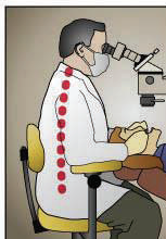
The microscope forces us to work at the same distance from the object all the times, avoiding tiring eyes. Microscope provides us better ergonomics so less back strain.
Procedural:
It considerably improves manual abilities as the operating field is magnified.
Lighting is magnificent, as it is always in the field of view andwithout shadows. With recording operations we can the techniques followed & detect procedural errors or problems & so clinical outcome.
Psychological:
Decreasing occupational, physical and postural stress.
Increase in personal & professional satisfaction.
Patient feels more comfortable.
Educational:
Microscope makes it easy for us to gather clinical images withmagnification having minute details which can be used for makingpresentations and other educational purposes.
DISADVANTAGES:
There are no such disadvantages of using DOM. However there is a learning curve to master its use, once adapted it is fun to use the scope and exploring the tooth under magnification. The more the microscope is used, the shorter the learning curve will be. Intermittent use, on the other hand, frequently leads to only frustration.
Also competency in microscope use can be acquired very quickly with a disciplined and professional approach to training. Rarelydoes one struggle for more than a month or two if a conscientious effort is made.
The Laws of Ergonomics:
An understanding of efficient work flow using a microscope entails the knowledge of the basics of ergonomic motion. Ergonomic motion is divided up into five classes of motion:
1. Class I Motion: Moving only the fingers
2. Class II Motion: Moving only the fingers and wrists
3. Class III Motion: Movement originating from the elbow
4. Class IV Motion: Movement originating from the shoulder
5. Class V Motion: Movement that involve twisting or bending at the
waist
To develop the highest efficiency, most endodontic procedures should be performed utilizing only Class I and Class II motions.Therefore the operator should never lose the distance relationshipbetween his or her hands and the mouth of the patient and every instrument should be positioned by the dental assistant not just in his or her hands, but in his or her fingers. This is very easy to accomplish if the doctor restricts the motions to only Class I and Class II motions. When Class III motions creep in, inefficiencies increase exponentially.
THE USE OF THE OPERATING MICROSCOPE IN ENDODONTICS:
- Diagnosis
- Locating calcified/ extra canals
- Retreatment
- Repairing of perforations
- Removing fractured instruments
- Surgical Endodontics
Diagnosis:
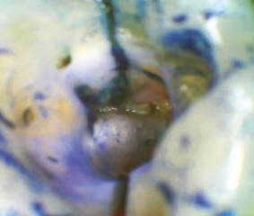
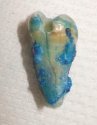

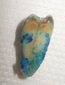
The operating microscope enables the endodontist to assess themarginal integrity of restorations and to detect cracks or fractures.The use of a dye like methylene blue or a caries detector can be very helpful to better visualize the crack and to follow its length to its termination.
Locating Calcified/ Extra Canals:
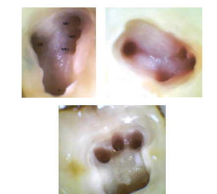
Accessing the pulp chamber and locating the canal orificesconstitute important visual phases of endodontic therapy. Mostof the endodontic treatments failures are due to inability of the operator to find and clean the canals. With the aid of operating microscope it becomes easy to find and explore a canal. Using the scope one can easily distinguish calcified pulp and dentine due to colour difference, calcified pulp being whiter and yellower dentine. Commonly “missed” canals frequently seen with the use ofmicroscope are the MB2/MB3 and the DB1/DB2 of maxillary molars, and the MM and deep splitting of D canal of mandibular molars. According to recent studies, the second canal of mesiobuccal root of maxillary first molars is present and can be clinically negotiated in almost 100% of cases. If we compare this result with those published 10 or 15 years before, we can conclude that the higher percentage of these findings today is due to nothing else than the use of the operating microscope
Retreatment:
The biggest advantage in using the microscope is during retreatment. To perform a retreatment can be as simple as the removal of guttapercha from a poorly obturated canal to more complex, delicate and time consuming procedures, like removing screw posts, separated instruments, silver points, carbon fiber posts, or repairing a perforation or obturating an immature open apex. These all procedures can be performed more confidently using a microscope. With microscope any challenge in the straight part of the canal can be easily seen and resolved with the help of magnification and coaxial illumination.
Surgical Endodontics:
While the origins of apical surgery can be traced to pre-Colombian times. Contemporary surgical endodontics began its journey in the early 1960s, along with the recognition of endodontics as a specialty in the United States in 1964. It is said that “necessity is the mother of invention”. Previously surgical instruments designed were bigger in size leading to more trauma to the tissue but now with the advancements of DOM, smaller instruments has been designed and so is the result, lesser trauma to the tissue and faster healing.
Apical microsurgery can be divided into 20 stages or sections. These are flap design, flap reflection, flap retraction, osteotomy, periapical curettage, biopsy, hemostasis, apical resection, resected apex evaluation, apical preparation, apical preparation evaluation, drying the apical preparation, selecting retrofilling materials, mixing retrofilling materials, placing retrofilling materials, compacting retrofilling materials, carving retrofilling materials, finishing retrofilling materials, documenting the completed retrofill, and tissue flap closure.
Surgical endodontics demands an entirely different set of skills than conventional endodontics. The practitioner must have a comprehensive knowledge and understanding of the multiple parameters involved in the management of both, hard and soft tissue, as well as an appreciation for the many factors involved in surgical wound healing. The operating microscope enhances surgical skill in both soft and hard tissue management. Light and visibility are critical for any surgical procedure; the operating microscope provides levels of illumination and magnification that are appropriate for surgical endodontics. When the flap is handled under the microscope, the physical trauma is lessened and gentle manipulation is assured. Microsurgical suturing techniques take advantage of smaller gauge tapered-point needles and smaller suture sizes. Suture sizes of 5-0 and 6-0 are handled with ease; sutures are placed much more accurately than with the naked eye. If one follows an atraumatic microsurgical flap management technique, sutures can be removed in 24 to 48 hours, with startling healing rates evident.
CONCLUSION:
“You can only treat what you can see!”
- Prof. Kim
Today, a small but growing number of dentists are providing endodontic treatment using DOM. There is a huge shift on magnification from conventional endodontics to vision based endodontics. This has brought revolution in the field of endodontics with an improved success rate for clinicians. Clinicians using microscope are more confident about their work quality. Endodontic treatment before microscopes was never so reliable & predictable and less invasive during surgical procedures. There are only advantages of using microscope in endodontics however initial cost can be a barrier in the path which can be recovered very easily getting referrals. All endodontic postgraduate programs are now teaching its use as part of their curriculum.
The next stage in microscopic endodontics will involve the use of even finer microscopic instruments and the development of even more sophisticated techniques. Eventually, endodontists will be able to re-vascularize the pulp and grow dentin. These procedures will most certainly be microscopic in nature and will be quickly embraced by a specialty already well trained in microscopic procedures.
Disclaimer from Guident
The views and opinions expressed in articles published in Guident are solely those of the authors and do not necessarily reflect the views of the editors, the editorial board or the publisher, Ivory India. Guident does not endorse any product, technique or treatment described. Clinical content is intended for qualified dental and medical professionals and is not a substitute for individual clinical judgement. Guident, its editors and the publisher accept no liability for any consequence arising from the use of information published in the journal.
How to cite this article
Dr Abhishek Mahajan, Dr Narang. Microscopes and Endodontics. Guident. 2014 Jan;7(2). Available from: https://guident.in/articles/endodontics/microscopes-and-endodontics.html