Peripheral Ossifying Fibroma
Department of Oral and Maxillofacial Surgery K.D. Dental College and Hospital, Mathura, UP
Peripheral ossifying fibroma is defined as a non- neoplastic enlargement of gingiva that is classified as a reactive hyperplastic inflammatory lesion 1. The synonyms of POF are peripheral cementifying fibroma, calcifying or ossifying fibroid epulis, and peripheral fibroma with calcification. These lesions may arise as a result of irritants such as trauma, micro- organisms, plaque, calculus, faulty restorations, and dental appliances 2,3. It is typically seen on the interdental papilla and is believed to comprise about 9% of all gingival growths4. The pathogenesis of this lesion is uncertain and is thought to arise from the periosteal or the periodontal membrane5. It has been reported that it represents a maturation of a pre- existing pyogenic granuloma or a peripheral giant cell granuloma 4. Peripheral ossifying fibroma mainly affects women in the second decade of life ( 50% of all patients between 5- 25 years of age ). The lesions are most often found in the gingiva, located anterior to the molars and in the maxilla 6,7,8,9,10. Clinically the POF are sessile or it may be pedunculated and are usually less than 2 cm but occasionally can reach the size of 6 cm.
CASE REPORT
A female patient age about 26 years reported with the chief complaint of painless growth on the gingiva in the upper left front region of mouth since 6 months . It had gradually progressed & attained the present size. The growth was associated with occasional bleeding. The patient did not give any history of trauma. Intraoral examination revealed a solitary, sessile growth present i.r.t 21 and 24 , extending mesiodistally from the distal aspect of 21 up to mesial aspect of 24 region. Superioinferiorly, extending superiorly from marginal gingiva of 21,22,22 & 24 and inferiorly from 2 mm above the incisor line. Anteroposteriorly, extending from inner aspect of upper lip to the labial surface of 21 to 24 . The color of the growth is same as that of the adjacent gingiva. The growth was oval in shape and approximately 2.5*1.5 cm in size in greatest dimensions. The growth was firm & non tender on palpation. It was nonfluctuant, nonreducible & noncompressible.
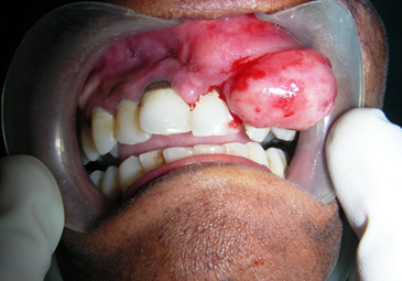
Fig.1 Preoperative photograph
Clinically, differential diagnoses for the growth were peripheral giant cell granuloma, pyogenic granuloma, peripheral odontogenic fibroma, peripheral ossifying fibroma, traumatic or irritational fibroma.
Radiological investigations done include maxillary occlusal view and intraoral periapical radiograph

Fig. 2 Occlusal view
The growth was excised conservatively followed by extraction of 11,21,22,23,24 .
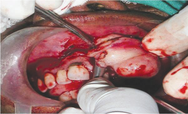
Fig. 3 Intraoperative photograph showing excision of growth
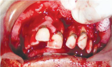
Fig. 4 Intraoperative photograph after excision of growth
The excised tissue was sent for histopathological examination and the area was sutured with 3-0 vicryl suture.

Fig. 5 Excised tissue specimen
Histopathologic examination showed parakeratinized stratified squamous epithelium with proliferation at few places. The underlying connective tissue shows loosely arranged wavy collagen fibres, dense chronic inflammatory cell infiltrate also exhibits thin endothelial blood vessels & deep in the connective tissue were seen numerous ossifications in the form of bony trabaculae.
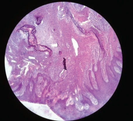
Fig. 6 Histopathological slide
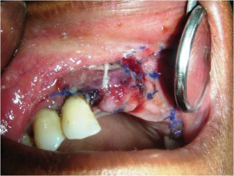
Fig. 7 Postoperative photograph after 24 hours
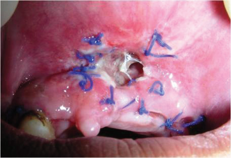
Fig. 8 Postoperative photograph after one week

Fig. 9 Postoperative photograph after one month
DISCUSSION-
Menzel first described the lesion ossifying fibroma in 1872, but its terminology was given by Montgomery in 192711. Ossifying fibroma occurs mostly in craniofacial bones and is generally categorized in to two types : central & peripheral. The central type of ossifying fibroma arises from the endosteum or the periodontal ligament adjacent to the root apex and expands from the medullary cavity of the bone. On the other hand, the peripheral type shows a contiguous relationship with the PDL, occurring solely on the soft tissues overlying the alveolar process. The reasons for considering a PDL origin for POF include : exclusive occurrence of POF in the gingiva ( interdental papilla ); the proximity of the gingival lesion to the periodontal ligament; the presence of oxytalan fibers within the mineralized matrix of some lesions; age distribution, which is inversely related to the number of lost permanent teeth; and the fibrocellular response in POF, which is similar to the other reactive gingival lesions of PDL origin12,13,14. The POF may occur at any age range, but exhibits a peak incidence between the second & third decades. The average age is around 28 years, with females being affected more than males. Most of lesions are usually more than 1.5 cm. Radiologically and depending on the size of the ossification foci, radiopaque stains can be seen on the periapical or panoramic X-ray15,16. Clinical differential diagnosis for gingival growths includes peripheral giant cell granuloma, pyogenic granuloma, peripheral odontogenic fibroma, peripheral ossifying fibroma, traumatic or irritational fibroma. The definitive diagnosis of POF is made by histologic evaluation12, 17. Histologically, the key feature of this lesion is exceedingly cellular mass of connective tissue comprising large numbers of plump, proliferating fibroblasts intermingled throughout with delicate fibrillar stroma17. Treatment consists of conservative surgical excision & scaling of adjacent teeth. The rate of recurrence is high, therefore, regular follow- up is required4,3,18.
REFERENCES
- Buduneli E, Buduneli N, Unal T. Long-term follow-up of peripheral ossifying fibroma: report of three cases. Periodontol Clin Investig. 2001;23(1):11-14.
- L. R. Eversole and S. Rovin, “Reactive lesions of the gingiva,” Journal of oral pathology, vol. 1, no. 1, pp. 30–38, 1972.
- D. G. Gardner, “The peripheral odontogenic fibroma: an attempt at clarification,” Oral Surgery Oral Medicine and Oral Pathology, vol. 54, no. 1, pp. 40–48, 1982.
- Bhaskar SN, Jacoway JR. Peripheral fibroma and peripheral fibroma with calcification: report of 376 cases. J Am Dent Assoc.1966;73(6):1312-1320.
- Kendrick F, Waggoner WF. Managing a peripheral ossifying fibroma. J Dent Child 1996; 63: 135- 138.
- Batsakis JG (1999). Non- odontogenic tumors: Clinical evaluation and pathology in comprehensive management of head and neck tumors, 2nd ed, Thawley SE, Panje WR, Batsakis JG, Lindberg RD eds, W.B. Saunders, Philadelphia, 1641-1642.
- Marx RE, Stern D (2003). Oral and Maxillofacial pathology: A rationale for diagnosis and treatment. Quintessence, Chicago, 23- 25.
- Passos M, Azevedo R, Janini ME, Maia LG (2007). Peripheral cement- ossifying fibroma: A case report. J Clin Pediatr Dent 32, 57- 59.
- Kumar SK, Ram S, Jorgensen MG, Shuler CF, Sedghizadeh PP (2006). Multicentic peripheral ossifying fibroma. J Oral Sci 48, 239- 243.
- Zhang W, Chen Y, An Z, Geng N, Bo D (2007). Reactive gingival lesions: a retrospective study of 2,439 cases. Quintessence Int 38, 103- 110.
- G. Sujatha, G. Sivakumar, J. Muruganandhan, J. Selvakumar, and M. Ramasamy, “Peripheral ossifying fibroma-report of a case,” Indian Journal of Multidisciplinary Dentistry, vol. 2, no. 1, pp. 415–418, 2012.
- Saito I, Ide F, Inoue M, et al. Periosteal ossifying fibroma of the palate. J Periodontol.1984;55(12):704-707.
- Killi V Prabhakar Rao, Penmatsa Tanuja, M Narendra Reddy. Peripheral ossifying fibroma: A case report. IJCRI 2012: 3 (7) : 11- 14.
- Miller CS, Henry RG, Damm DD. Proliferative mass found in the gingiva. J Am Dent Assoc. 1990;121(4):559-560.
- Leena Bari, Namita Rathod, Sonam C. Kapse, Akram Khan, Bindiya Narang, Vaibhav Bari, Ajit V. Koshy. Peripheral ossifying firoma: maxilla. A case report. Int J Dent case report s 2013: 3(1) :50-54.
- Kumar SK, Ram S, Jorgensen MG, et al. Multicentric peripheral ossifying fibroma. J Oral Sci. 2006;48(4):239-243.
- Rajendran R. Benign and malignant tumors of oral cavity. In: Shafer WG, Hine MK, Levy BM. Shafer’s Textbook of Oral Pathology. 5th ed. Philadelphia, PA: W.B. Saunders Co; 2006:113-308.
- J. N. Kenney, G. E. Kaugars, and L. M. Abbey, “Comparison between the peripheral ossifying fibroma and peripheral odontogenic fibroma,” Journal of Oral and Maxillofacial Surgery, vol. 47, no. 4, pp. 378–382, 1989
Disclaimer from Guident
The views and opinions expressed in articles published in Guident are solely those of the authors and do not necessarily reflect the views of the editors, the editorial board or the publisher, Ivory India. Guident does not endorse any product, technique or treatment described. Clinical content is intended for qualified dental and medical professionals and is not a substitute for individual clinical judgement. Guident, its editors and the publisher accept no liability for any consequence arising from the use of information published in the journal.
How to cite this article
Dr. Sonu Punia, Dr. Bhupender Harjani, Dr. Shishirmohan Garg. Peripheral Ossifying Fibroma. Guident. 2014 Mar;7(4). Available from: https://guident.in/articles/endodontics/peripheral-ossifying-fibroma.html