Electronic Apex Locators - an Insight
Department of Conservative Dentistry & Endodontics D.J College of Dental Sciences and Research, Modinagar, U.P, India
Abstract
Electronic apex locators are considered an accurate tool for determining the working length. Studies have reported that its use resulted in a higher quality of obturation length control and an overall reduction in the number of radiographs taken. There are several variables which affect their accuracy (example immature roots). Therefore, combined use of apex locators and radiographs results in greater accuracy.
INTRODUCTION
Locating the appropriate apical position always has been a challenge in clinical endodontics. Sjogren in 1990 stated that the outcome of treatment of roots with necrotic pulps and periapical lesions is influenced significantly by the apical level of the root filling.Traditionally, the point of termination or endodontic instrumentation and obturation has been determined by taking radiographs. Endodontic radiography often leads to inaccuracies because they provide a two-dimensional representation of a three-dimensional object.1,2
The development of the electronic apex locator (EAL) has helped make the assessment of working length more accurate and predictable, particularly useful when the apical portion of the canal system is obscured by certain anatomic structures:Impacted teeth, tori ,zygomatic arch, excessive bone density, overlapping roots and shallow palatal vault. The objective of working length determination is to establish the length (distance from the apex) at which canal preparation and subsequent obturation are to be terminated. Methods for determining working length are radiographs , electronic apex locators, tactile sense, mathematics method, apical periodontal sensitivity, paper points, microscopic magnification and average tooth length.3
Grove (1930) stated that ‘the proper point to which root canals should be filled is the junction of the dentin and the cementum and that the pulp should be severed at the point of its union with the periodontal membrane’.The cemento-dentinal junction (CDJ) is the point in the canal where cementum meets dentin; it is the point where pulp tissue ends and periodontal tissues begin. The location of the CDJ in the root canal varies considerably.
MECHANISM OF ACTION OF ELECTRONIC APEX LOCATORS
Root canals are surrounded by dentine and cementum that are insulators to electric current. At the apical foramen there is a small hole in which conductive materials within the canal are electrically connected to the periodontal ligament that is a conductor of electric current. The resistive material of the canal (dentine, tissue, fluid) with a particular resistivity
forms a resistor, the value of which depends on the length, cross-sectional area and the resistivity of the materials .
If an endodontic file penetrates inside the canal, and approaches the canal terminus, the resistance between the end of the instrument and the apical portion of the canal decreases, because the effective length of the resistive material inside the canal decreases (figure 1).
An apex locator has 4 parts (1) the lip clip, (2) the file clip, (3) the instrument (4) a cord connecting the three parts (figure 2).
Apex locators use the human body to complete an electrical circuit.One side of the circuitry is connected to an endo instrument & the other end to the patients body-- patients lip or by an electrode held in the patients hand.The impedance between the lip and the periodontal ligament (PDL) is a known value, so as the file tip is advanced toward the PDL, the EAL
detects the changing impedance values and indicates the approach to the apex on its screen.
TYPES OF ELECTRONIC APEX LOCATOR
There are six generations of the EALs.
The first generation
Resistance between the periodontium and the oral mucous membrane in humans was constant at 6.5 K Ohm, regardless of the age of the patients or
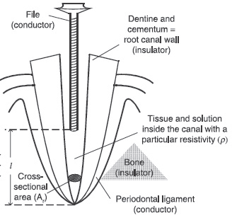
Figure 1
the shape and type of teeth.4 Contents of the canal (vital pulp vs. necrotic pulp) also had no effect upon the resistance. First-generation apex location devices measure the opposition to the flow of direct current or resistance. The resistance was measured between the two electrodes to determine location within a canal. Pain was often felt with this type of apex locator. In addition, these devices gave unreliable measurements in case of wet canals.5 Examples are Dentometer, Endo Radar.
The second generation
Second-generation apex locatorsmeasure the opposition to the flow of alternating current or impedance.This generation contains 2 types of apex locator: low frequency and high frequency apex locator. Low frequency AL is based on the assumption that the impedance between the oral mucous membrane and the depth of the gingival sulcus closely resembles theimpedance between the canal terminus and the oral mucous membrane.6While high frequency AL used an insulator to cover most of the surface of the file(Hasegawa, 1979).Examples are Endocator, Apex finder, Endo analyzer, Digipex and Exact-A-Pex.
The third generation
The 3rd generation apex locator has been called “frequency dependent” apex locators. This type was supplied by 2 frequencies to measure the impedance in the canal. There are 2 types of the 3rd generation ALs: impedance difference type and impedance ratio type. Impedance difference AL measures the impedance value at two different frequencies and calculates the difference between the two values (Yamashita, 1990) while impedance ratio type measured the position of the file from the ratio between these two impedances.7 Examples are Root ZX,Endex, Neosono ultimo Ez (figure 3) and Apex finder A.F.A (All Fluid Allowed).
The fourth generation
The 4th generation apex locator measures the impedance characteristics using more than two frequencies (Welk et al. 2003). A significant disadvantage of the fourth generation devices is that they need to perform in relatively dry or in partially dried canals (Vera and Gutierrez, 2004). Examples are Elements Diagnostic Unit and the Bingo 1020/Ray-Pex 4.
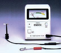
Figure 2

Figure 3
The fifth generation
5th generation apex locator was developed in 2003. It measures the capacitance and resistance of the circuit separately. It is supplied by diagnostic table that includes the statistics of the values at different positions to diagnose the position of the file (Gordon and Chandler, 2004). Devices employing this method experience considerable difficulties while operating in dry canals (Slavcho and Dimitur, 2009).
The sixth generation
Currently, fourth and fifth generation devices are mostly employed. A disadvantage of the fourth generation devices is that they need to perform in relatively dry or in partially dried canals. In some cases, this necessitates additional drying, and with heavy exudate or blood the method becomes inapplicable. 5th generation perform very well in the presence of blood and exudate but they experience considerable difficulties while operating in dry canals. Therefore, additional insertion of liquids in the canal is exerted almost always. Hence the sixth generation was developed which has a measuring method that combines the advantages of the methods for measuring in dry canals and in moist canals.8 Example Adaptive Apex Locator (fig. 4).
ACCURACYOF THEEAL
Most studies have reported that pulpal vitality does not affect EAL accuracy.Mayeda et al (1993) conducted a study to determine whether the pulp status makes a difference in the determination but no statistical difference in the measurements was found between the vital & necrotic canals.
Meares and Steiman(2002) used 2.125% and 5.25% sodium hypochlorite and no significant difference was found. Ngyyenetal (1996), Felippe MC. (1994), Lee et al (2002) evaluated the possible influence of the size of the instrument on the measurementsreported that there was no difference was found between the smaller and larger apical foramen.
Goldberg et al (2002) evaluated the accuracy of an apex locator in determining the working length in teeth with apical root resorption, the measurements were accurate in 62.7% of cases hence the authors suggested that the accuracy of EAL may depend more on the operators experience.
In the primary teeth the apical aperture as exposed to continuous and sometimes irregular resorption. Therefore it is even more imperative to minimize the periapical damage to protect the succedaneous tooth bud.
COMMON PROBLEM SOLVING WITH EAL
Unstable electronic signal with rapid wandering signs
This occurs if a file touches the metallic restorations or if there is a cervical leak through subgingival caries.
Sharp drop of the signal at the apical foramen
This happens if there is a very dry canal and hence no electric contact occurs. As soon as the file meets the apical tissue there is a sudden circuitbreak out which brings the signal to APEX mark therefore to avoid this gentlerrigation should be done for the normal operation of the unit.
Apex sign from the beginning
This occurs when there is a severely bleeding or exudating canal. The canal is then irrigated with sodium hypochlorite or saline until the drainage is
controlled reasonably.
Premature reading
Premature reading is there when there is an open apex it occurs due to the sharp drop in the gradient of impedance ratio at the thin dentin walls
CONCLUSION
Apex locators have a definite use in endodontics,butEAL give short reading compared with x-ray in case of presence of normal or inflamed pulp tissue in the canal. While it gives over reading compared with x-ray in case of presence of periapical radiolucency.9 EAL Cannot determine the canal width,canal curvature,or number of canals.Apex locators are not meant to replace radiographs, but to add to the information obtained by radiographs( Fouad et al).
REFERENCES
1. Tidmarsh BG. Radiographic interpretation of endodontic lesions--a shadow of reality. Int Dent J. 1987 Mar;37(1):10-5.
2. Goldman M, Pearson AH, Darzenta N. Endodontic success--who’s reading the radiograph?. Oral Surg Oral Med Oral Pathol. 1972 Mar;33(3):432-7.
3. Ricucci D. Apical limit of root canal instrumentation and obturation, part 1. Literature review. IntEndod J 1998;31:384-93.
4. Sunada, I. New method for measuring the length of the root canal. J Dent Res 1962; 41:375.
5. Suchde R.V. and Talim S.D.(1977). Electronic ohmmeter: an electronic device for the determination of the root canal length. Oral Surg., OralMed., and OralPathol. 43, 141–149
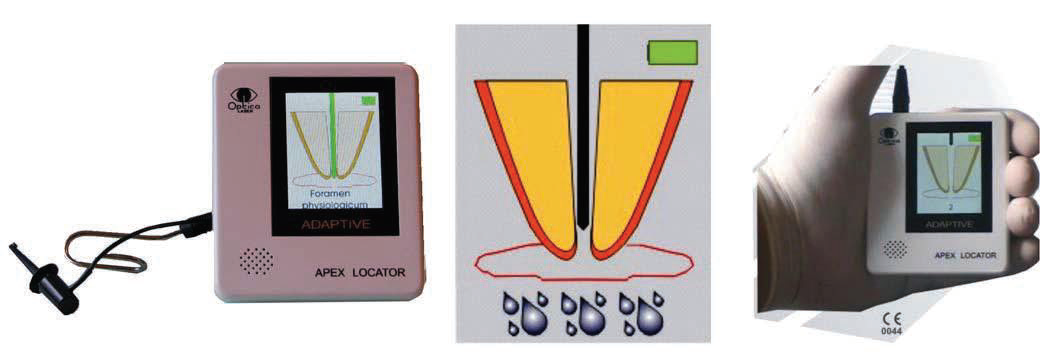
Figure 4
Disclaimer from Guident
The views and opinions expressed in articles published in Guident are solely those of the authors and do not necessarily reflect the views of the editors, the editorial board or the publisher, Ivory India. Guident does not endorse any product, technique or treatment described. Clinical content is intended for qualified dental and medical professionals and is not a substitute for individual clinical judgement. Guident, its editors and the publisher accept no liability for any consequence arising from the use of information published in the journal.
How to cite this article
Dr. Anil Dhingra Dr. Nayasha Manchanda. Electronic Apex Locators - an Insight. Guident. 2014 Mar;7(4). Available from: https://guident.in/articles/endodontics/electronic-apex-locators-an-insight.html