Management of Labially Placed Type -3 Talons Cusp on Permanent Maxillary Central Incisor: A Case Study with a Clinical Evaluation
Dept. of Pediatric and Preventive Dentistry, PDM Dental College, Bahadurgarh, Haryana, India, PGIMS, Rohtak, Haryana India, College of Pharmacy, Pt B.D. Sharma University of Health Sciences, Rohtak, Haryana India
ABSTRACT
Aim and background: The talon cusp on labial aspect is an uncommon, properly delineated odontogenic anomaly that is mostly located on the permanent maxillary incisors. Its presence leads to problems in esthetics, the prevention of caries, an occlusal hindrance for the affected individual, and troubles in the assessment and clinical management of the dentist.
Case description: An 8-year-old boy complains of the pain, swelling, and his bad looking maxillary central incisor of right side. Talons cusp is not indicated for any treatment, but as it was creating a problem for the child and hindering complete eruption, treatment was planned for the patient. The mucoperiosteal flap was reflected and raised, and with a low-speed dental hand drill and a carbide bur used to cut the talons cusp-like attached structure attached to the tooth, the mucoperiosteal flap was reflected and the talons cusp completely removed.
Conclusion: It's possible or unlikely that talon cusps are related to the symptoms described in this case study. Early diagnosis and treatment can lessen symptoms in symptomatic cases, stop tooth loss before it happens, enhance aesthetics, and generally improve the patient's health.
Clinical significance: To avoid occlusal interference, impaired esthetics, and the development of carious developing grooves, talon cusps should be diagnosed and treated early.
INTRODUCTION
The talon cusp is an unusual odontogenic aberration that is most frequently present at the palatal surfaces of the maxillary incisors and consists of an additional cusp-like form. It looks like an eagle's talon and projects like a cusp from the cingulum of the maxillary or mandibular anterior teeth. It normally extends lingually or incisally in the primary and secondary dentition. Mitchell1 originally identified this unusual dental aberration in 1892, which showed an additional structure resembling a cusp protruding from the cingulum to the cutting edge. Due to its likeness to an eagle's talon, Mellor and Ripa2 later gave it the moniker Talon Cusp. because this odontogenic anomaly has been described in many ways, including as having an exceptional accent cusp-like shape, an enlarged cusp-like hyperplasia and cingula, an accessory cusp, and a supernumerary cusp. Due to the possibility of a large range in the size and shape of this aberration due to this change, Hattab et al. have divided it into three types in an effort to have diagnostic standards.3
Type 1: Talon is the name given to an extra cusp with a distinctive morphology that prominently protrudes from the palatal (or face) floor of an anterior primary or permanent enamel and takes up at least half of the space between the CEJ and the incisal region.
Type 2: Semi-talon describes an extra cusp that extends at least one millimeter past the CEJ but not quite halfway to the incisal side. It could stand apart from the rest of the crown or merge in with the palatal floor.
Type 3: Trance Talon, which can be conical, bifid, or tubercle-like, is an enlarged or distinctive cingula.
The research claims that TCs on the face, labial region, and mandibular anterior teeth are uncommon.
CASE REPORT
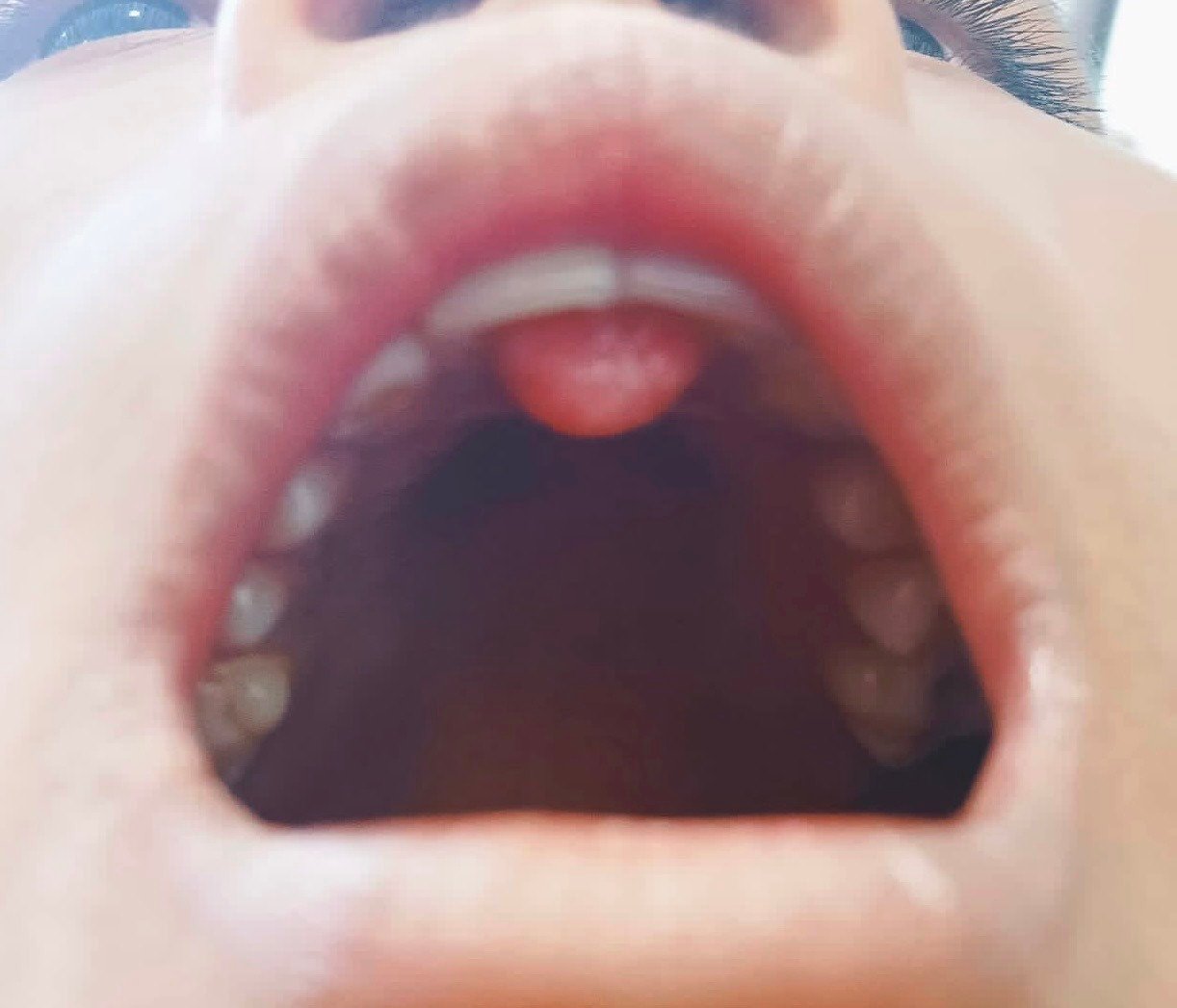
An old boy in good health visited our pediatric dentistry department with his mother. She complained of the pain, swelling, and unesthetic upper right central incisor. According to the history, there was minor pain and discomfort on the upper lip near the projection of the maxillary right central incisor. During the clinical examination, a noticeable labial projection that was partially lodged in the periodontium was found.
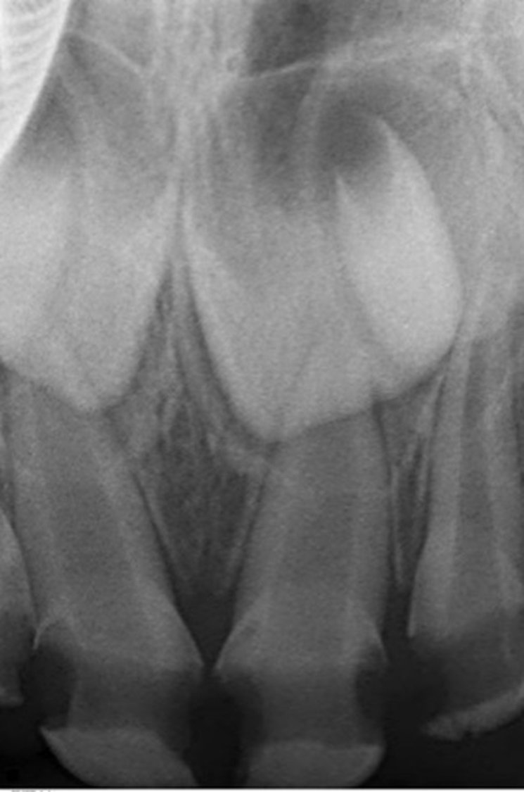
The projection extended from the cervical area of the central incisor. However, it was set apart from the remainder of the buccal surface by deep grooves (Fig. 1). It was also discovered that the upper lip had an indentation that matched the site of the talons cusp and that it was palpably sensitive. Periodontal probing depths around the tooth ranged from 2 to 3 mm. A labial TC was identified through clinical examination. On radiographs, the central region of the tooth's crown was covered by a clearly defined cusp-like structure. The dentine was prominently covered by an enamel layer, and the projection's contour was smooth. The tooth's root development was almost finished, and it matched the growth of the opposing central incisor (Fig. 2).

Talon's cusp is not a condition that needs to be treated, but because it was bothering the patient and preventing a full eruption, treatment was planned. Parents were made aware of the potential pulp exposure, and any necessary therapy for pulp can take place. Only a tiny reflection of the gingival on that day helped the eruption of the teeth, and analgesic medication was provided.
After a week had passed and the parent had completed the informed consent form, anesthesia was administered using ( lidocaine 2% with epinephrine 1:100,000). A raised and mirrored mucoperiosteal flap was observed. (Fig. 3) as opposed to using a carbide bur and a moderate-speed dental hand drill to trim the talon's cusp. (Fig. 4) The area was then rinsed with physiological saline, the gingiva was moved back into place, and 3.0 silk sutures were used to close the wound. (Fig.5), and perio pak was placed for fast healing of the gingival area. (fig6)

The exposed enamel was etched for 20 seconds using Scotchbond Universal Etchant, 3M ESPE, St. Paul, USA, which contains 34% phosphoric acid. As a bonding agent, light-cured (Adaper Scotchbond 1XT adhesive from 3M ESPE, St. Paul, USA ), was employed.
The restoration was subsequently polished and finished after one last light curing. The direct resin veneer was polished and finished using an ISO 806 314 249 504 012 diamond finishing bur with a stripe (yellow in color) from Dentsply Sirona in Ballaigues, Switzerland, and the Enhance Polishing System from Dentsply Sirona in Milford, USA. The patient's and the patient's parents early post-operative results were quite positive.followup was done after 1 month.
DISCUSSION
The talon cusp malformation, which is unusual and somewhat uncommon, most usually affects the maxillary permanent central incisor. The same situation as dens evaginatus, but on the anterior tooth, is referred to as the talon cusp. It is made up of pulp tissue, dentine, and variable amounts of tooth.6 This patient had no systemic conditions that were related to them, and there are no records of incidents with any family members that might be used to support the case. Treatment is not necessary for a minor, asymptomatic talon cusp. Large talon cusps can also result in carious lesions in the developmental grooves that define the cusp, pulpal necrosis, periapical pathosis, attrition of the opposing tooth, and periodontal problems as a result of excessive occlusal forces. These medical problems include occlusal interference, displacement of the affected teeth, irritation of the tongue during speech and chewing, irritation of the tongue during speech, and periodontal disease.7 If there are deep, developing grooves, fissure sealing and composite resin restoration can be done. Reducing the bulk of the cusp gradually and frequently, applying topical fluoride, or overall reducing the cusp and performing a calcium hydroxide pulpotomy are essential steps, particularly in cases of occlusal interference.8 Every case requires a different approach to treatment. When there is no occlusal interference and the talon's cusp is asymptomatic, there is no need for treatment, and regular checkups and good dental hygiene are appropriate.9
CONCLUSION
Talon's cusps either won't exhibit any symptoms or may be connected to the symptoms listed in this circumstance report. Early identification and prompt treatment can lessen symptoms in cases where they are present, stop premature tooth loss, enhance esthetics, and generally improve the patient's health.
Clinical significance: It's critical to diagnose and treat talon cusps early in order to avoid complications including poor esthetics, carious developing grooves, and occlusal interference.
REFERENCES
- Subramanian S,Manoharan GM, An atypical presentation of talon cusp.Double talon cusps.Indian Journal of Case Reports.2023;9(1):5-8.
- Dashatwar P.,Gadodoa.P,Patil N,Murgod V Journal of Interdisciplinary Dental Science 2019;8(3):09-12.
- Jingarwar M. M., Bajwa N. K., Pathak A. Minimal intervention dentistry—a new frontier in clinical dentistry. Journal of Clinical and Diagnostic Research. 2014;8(7):4–8. doi: 10.7860/jcdr/2014/9128.4583.
- Hattab FN, Yassin OM, al-Nimri KS. Talon cusp in permanent dentition associated with other dental anomalies: Review of literature and reports of seven cases. ASDC J Dent Child 1996;63:368-76.
- Segura-Egea J. J., Jiménez-Rubio A., Velasce-Ortega E., Ríos-Santos J. V. Talon cusp causing occlusal trauma and acute apical periodontitis: report of a case. Dental Traumatology. 2003;19(1):55–59. doi: 10.1034/j.1600-9657.2003.00110.x.
- Davis PJ, Brook AH. The presentation of talon cusp: Diagnosis, clinical features, associations and possible aetiology. Br Dent J 1986;160:84-8.
- Wakhloo T. Facial Cellulitis Due to Type I Talon Cusp in a Pediatric Patient: A Case Report. Cureus. 2023 Jan 20;15(1):e34011. doi: 10.7759/cureus.34011. PMID: 36814740; PMCID: PMC9939955.
- Chen RJ, Chen HS. Talon cusp in primary dentition. Oral Surg Oral Med Oral Pathol 1986;62:67-72.
- Jowharji N, Noonan RG, Tylka JA. An unusual case of dental anomaly: A facial talon cusp. ASDC J Dent Child 1992;59:156-8.
- Dankner E, Harari D, Rotstein I. Dens evaginatus of anterior teeth. Literature review and radiographic survey of 15,000 teeth. Oral Surg Oral Med Oral Pathol Oral Radiol Endod 1996;81:472-5.
Disclaimer from Guident
The views and opinions expressed in articles published in Guident are solely those of the authors and do not necessarily reflect the views of the editors, the editorial board or the publisher, Ivory India. Guident does not endorse any product, technique or treatment described. Clinical content is intended for qualified dental and medical professionals and is not a substitute for individual clinical judgement. Guident, its editors and the publisher accept no liability for any consequence arising from the use of information published in the journal.
How to cite this article
Dr. Preetika Yadav, Dr. Geetanjali Yadav, Mrs. Veena Yadav. Management of Labially Placed Type -3 Talons Cusp on Permanent Maxillary Central Incisor: A Case Study with a Clinical Evaluation. Guident. 2024 Apr;17(5). Available from: https://guident.in/articles/pedodontics/management-of-labially-placed-type-3-talons-cusp-on-permanent-maxillary-central-incisor-a-case-study-with-a.html