Fibroma- a Rare Case Report in Pediatric Patient
Department of Pediatric & Preventive Dentistry, Department of Oral Pathology and Microbiology, Dental College, RIMS, Imphal India
ABSTRACT
Fibromas, often regarded as ‘Irritation fibroma’, are one of the most common tumor-like growth in the oral cavity. It causes esthetic and functional problems when present at specific locations. It’s size varies from few millimeters to centimeter. It is a slow growing, painless, with surface comparatively pale than the unaffected adjacent areas and smooth surface. Majority of the case reports as a reaction to local irritation or trauma. The clinical appearances and pathogenesis of the fibroma, however, suggests more of focal hyperplasia accurately, which is less used. Treatment involves complete excision of the growth and meticulous removal of adjacent tissues, to prevent recurrence, which is rare. We present a case of a 13 year old male patient with fibroma on the maxillary incisor region in the palatal aspect.
Keywords: Fibroma, benign neoplasm, fibrous hyperplasia.
INTRODUCTION
Considered one of the most common benign soft tissue growth is the Fibromas. It originates from the gingival connective tissue or from the periodontal ligament. They exhibit a slow-growing and spherical tumor that tends to be firm and nodular but that may be soft and nodular. The terminology rather indicating a benign neoplasm, however, most of the growth results from the responds to local irritation or trauma.1
It shows a slightly female predilection, mostly affecting age group beyond 30 yrs. The size of the growth varies from few centimetres in diameter, may be pedunculated or sessile, and mostly occurs on the gingival or buccal mucosa. Daley2 suggested the term ‘focal fibrous hyperplasia, which implies a reactive tissue response, is preferable to term, ‘Fibroma’, which implies incorrectly, a benign neoplastic proliferative fibrous connective tissue.
The lesion presents as painless, sessile, round or ovoid with broad base and lighter in colour relative to surrounding mucosa, due to reduced vascularity.
CASE REPORT
A 13 year old male patient reported to the department of Pediatric dentistry, Dental college RIMS, Manipur, with a chief complaint of swelling on his upper front tooth region since 2 months. (fig. a)
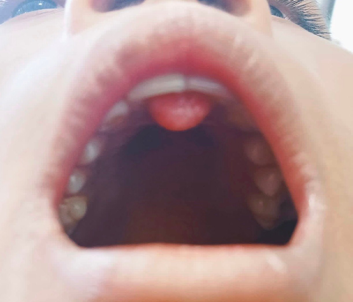
Fig-a
There was no history suggestive of any systemic diseases and no family history of gingival epulis. The patient was apparently alright 2 months back and then he noticed a swelling on his upper front tooth region, which remained constant. There was no suggestive history of any trauma or dental treatment in the past.
On inspection a localised growth of dome-shaped, approximately meauring 10mm•10mm is present on the maxillary palatal surface irt 11, 21 above the incisive foramen region.
On palpation the growth was soft and nodular however there was no local rise in temperature. Bleeding only occurs when probed using a Sharper’s hook explorer.
On learning the blood investigations were normal and considering radiographs, the treatment plan was executed under local anaesthesia (lignocaine 2% with Adrenaline)
Surgical procedure An excisional biopsy was performed using a no. 15 surgical blade, and thorough removal of the remnant tissues were done, to prevent recurrence. The surgical region prepared was then properly irrigated with a N/S and betadine solution (fig b). And he was kept under antibiotic therapy for 5 days (i,e Amoxycillin+ clavulanic acid) with a painkiller for 3 days. The patient was also booked for a follow up. (fig. d)
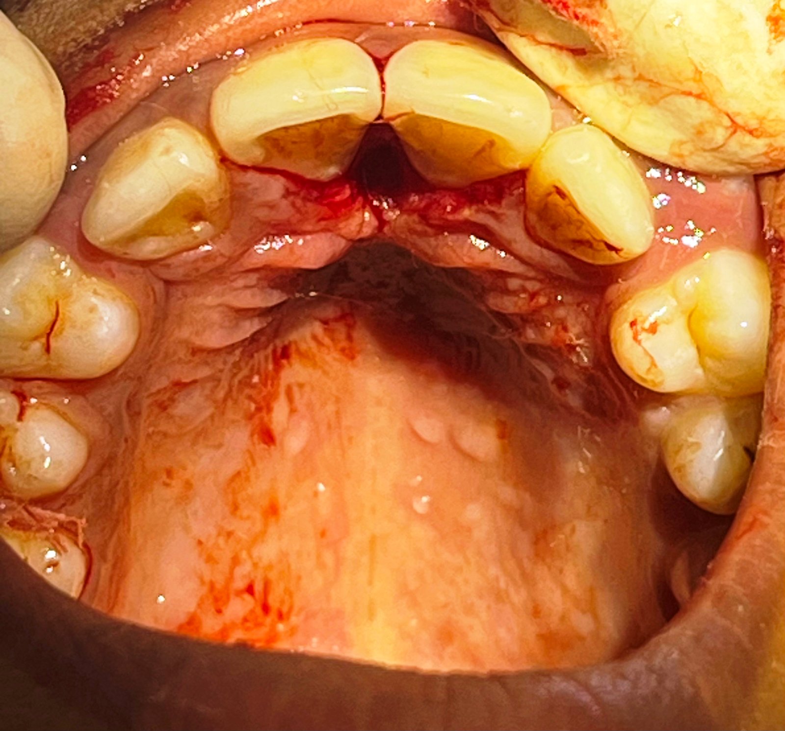
Fig-b
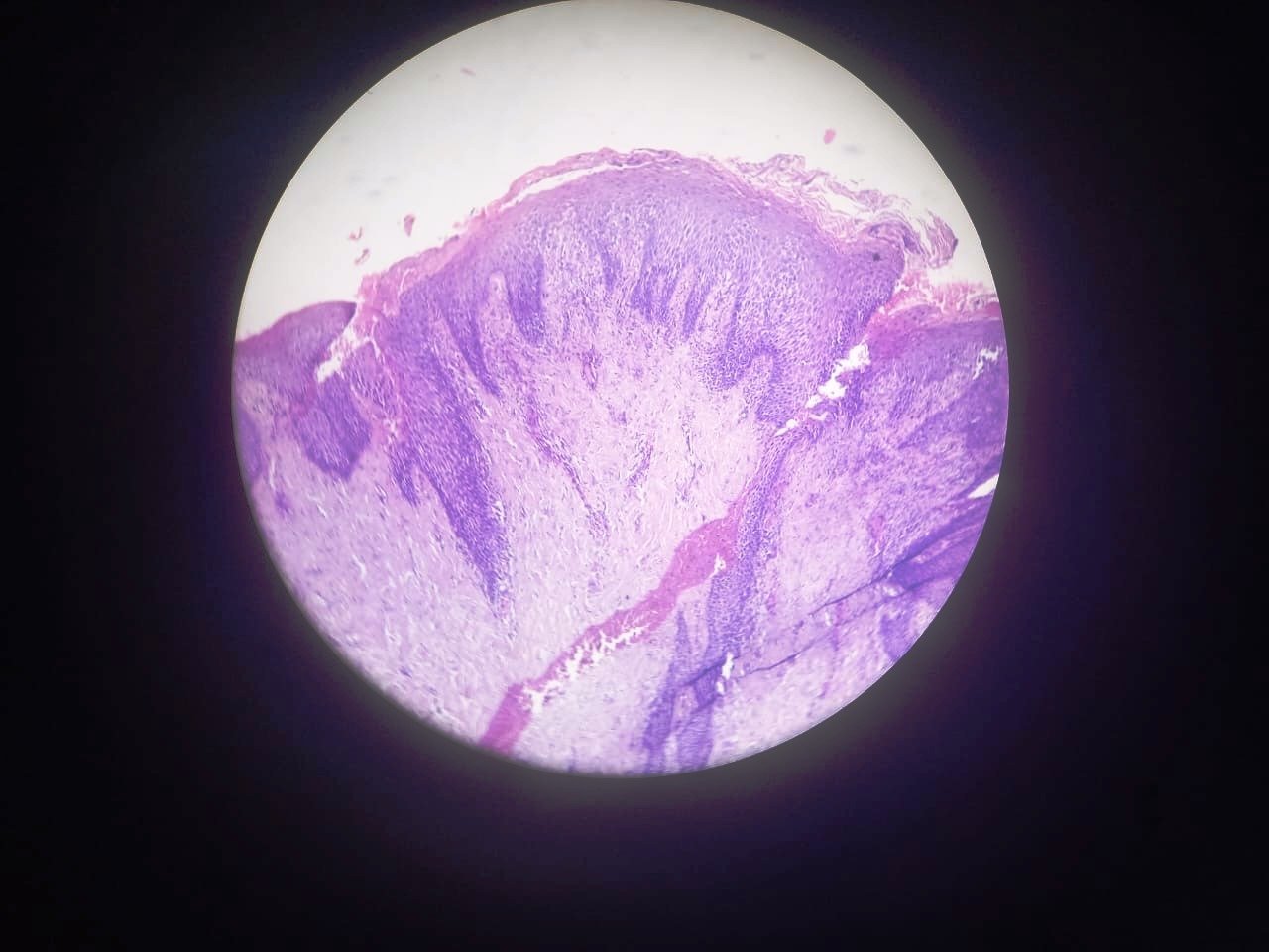
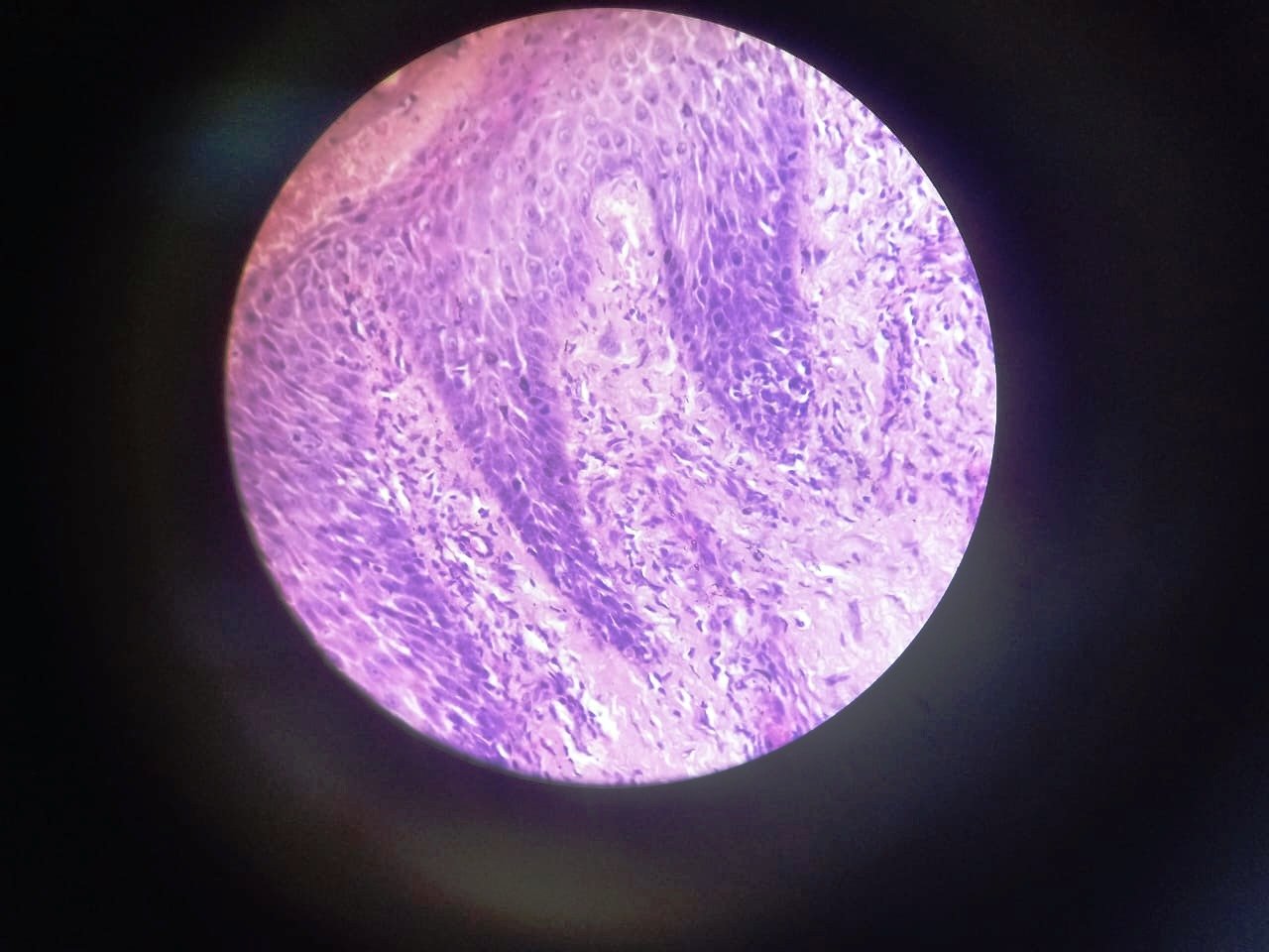
Fig-c
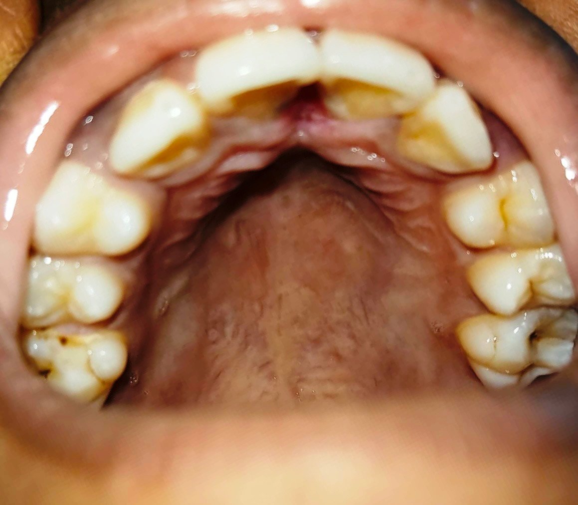
Fig-d
DIFFERENTIAL DIAGNOSIS
- Irritation fibroma
- Peripheral giant cell granuloma
- Peripheral ossifying fibroma
- Pyogenic granuloma
- Papilloma
- Gingival cyst
HISTOPATHOLOGICALLY
Presence of parakeratinised stratified squamous epithelium supported by fibrocellular connective tissue stroma. Epithelium shows focal areas of hyperplasia with long reteridges and bacterial colonies on surface in focal area. In the connective tissue there is presence of chronic inflammatory cells. Dense collagen fibres with few endothelial lined blood vessels and areas of haemorrhage are also evident. (fig. c)
DISCUSSION
It is a concern by areas of pathological expertise, that fibroma exists either in both hyperplastic as well as neoplastic form. However, fails to differentiate if a benign neoplasm actually occurs. Barker1 proved a very low incident rate of true fibromas (benign fibromas). A case study conducted by Christopoulos, Sklavounou & Patrikiou3 among 171 Fibroma specimens reported only 2 cases of true fibroma occurence. Reactive or Irritational Fibromas has as etiology, and they exhibit certain pattern. 2 patterns are identified- Radiating and Circular pattern. The presence of capsule around the fibroma is a proof that benign neoplasm can exist, and is differentiated from the normal tissue.
REFERENCES
- Barker D.S. & Lucas R. B. (1967) ‘’ Localised Fibrous overgrowths of the oral mucosa, ‘’British Journal of Oral surgery, vol. 5, no. 2, p. 8692
- Daley TD, Wysocki GP, Wysocki PD, Wysocki DM. The major epulides; Clinico pathological corelations. J Can Dent Assoc 1990; 56: 627-30
- Christopoulos P., Sklavounou A., Patrikiou A. (1994). ‘’True Fibroma of the Oral mucosa: a case report, ‘’International Journal of Oral and Maxillofacial Surgery, vol. 23, no. 2, p.98-9
- Newman and Carranza’s Clinical Periodontology 3rd South Asia Edition
- Riya verghese, Amitha Ramesh, Rahul Bhandary, Biju Thomas & Nishita L. Philip: A case report of a 13 year old lesion.
- Khaled Mohamed, Ahmed Ismail, Rayan Okla, ‘Fibroma of buccal mucosa: case report and literature review’, March 2017.
- SHAFER’s Textbook of Oral Pathology 8th edition.
Disclaimer from Guident
The views and opinions expressed in articles published in Guident are solely those of the authors and do not necessarily reflect the views of the editors, the editorial board or the publisher, Ivory India. Guident does not endorse any product, technique or treatment described. Clinical content is intended for qualified dental and medical professionals and is not a substitute for individual clinical judgement. Guident, its editors and the publisher accept no liability for any consequence arising from the use of information published in the journal.
How to cite this article
Dr. Wairokpam Bhoomika Devi, Dr. Veto Vadeo, Dr. Sumita Banerjee. Fibroma- a Rare Case Report in Pediatric Patient. Guident. 2023 Dec;17(1). Available from: https://guident.in/articles/pedodontics/fibroma-a-rare-case-report-in-pediatric-patient.html