Immediate Temporization in the Aesthetic Zone: A Case Report
Jaipur, India
ABSTRACT
This case report describes removal of anterior bridge, followed by immediate placement of a dental implant in the prepared socket and temporization by a bonded restoration. The socket was prepared to the required depth and two NobleBiocare Implants were inserted followed by temporization by a bonded restoration. An impression was made 3 months after implant insertion, and a definitive restoration was placed. The atraumatic operating technique and the immediate insertion of the Implant resulted in the preservation of the hard and soft tissues at the extraction site. The dental implant and provisional restoration provided the patient with immediate aesthetics, function, comfort, and most importantly preservation of tissues.
Keywords: Implant, Osseointegration, Temporization.
INTRODUCTION
Implant dentistry nowadays is considered to be an integral part of dentistry. The use of dental implants in the aesthetic zone has overcome many disadvantages of conventional restorative techniques. That is why implants are considered as an alternative treatment option for replacement of tooth as it restores both the function and aesthetics.1 Conventionally, after the placement of implant, it is recommended to have 3-6 months of load free healing period for successful osseointegration to occur. But numerous practitioners now advocate immediate loading of implants i.e. the implants were restored following the surgical placement which allow the maintenance of soft tissue contours along with the interdental alveolar contours.2,3
The purpose of this case report is to demonstrate key points of consideration when replacing a maxillary anterior tooth with a dental implant. Implant timing, selection, method of placement, and management of hard and soft tissues are paramount to achieving success. Additionally, the method and timing of temporization, incorporation of technologies, and restoration are equally crucial to achieving a long-term, aesthetic outcome.
The CAD-CAM technology is a computerized technique that allows to obtain a three-dimensional object from a vector drawing performed on the computer. Both CAD and CAM are acronyms, respectively, for Computer Aided Design and Computer Aided Manufacturing. Born in the 60s of last century, this technology is now used in industry for the production of an infinite number of objects also in the dental field. In the 80s the first systems in the dental industry, CEREC have been commercialized.4
The evolution of techniques and of implant components is making the implant-prosthetic rehabilitation increasingly complete and reliable. The increased demands of rehabilitation can be processed more serenely by operators in the industry. In this regard a valuable aid for the prosthodontist comes from a rapidly expanding sector which is that of the CAD-CAM applications in dentistry.5
CASE REPORT
A 20-year-old girl reported to clinic with a chief complaint of loss of upper front teeth due to road accident 2 years back. On detailed intra oral examination, patient had bridge on left and right incisors. The patient general periodontal condition was healthy. Patient had previously undergone RCT w.r.t 12,21Patient was wearing a bridge w.r.t 11,12,21,22 for 2 years and wants durable and fixed alternative to replace missing upper front tooth for aesthetics reasons. Clinical and Radiographic evaluation revealed adequate alveolar bone in the future implant site. (Figure 1)
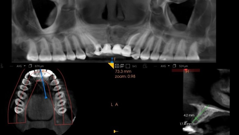
Figure 1: CBCT
So, it was planned to place implants measuring 3.5 x 15 mm,3.5 X 15 mm in dimension along with immediate temporisation to maintain the emergence profile and to preserve the alveolar bone. Following local Anaesthesia was administered in the area of missing teeth. Mid crystal incision was given with releasing incision and full thickness flap was raised and underlying bone was inspected for adequate perfusion. A punch osteotomy was made using a round punch drill in the Centre of edentulous span. A 2 mm pilot drill was used to make the initial osteotomy till a depth of 11.5 mm. Subsequent drilling was done till diameter of 2.8mm and a counter sink was given with 3.0 mm drill. (Figure 2 a, b)
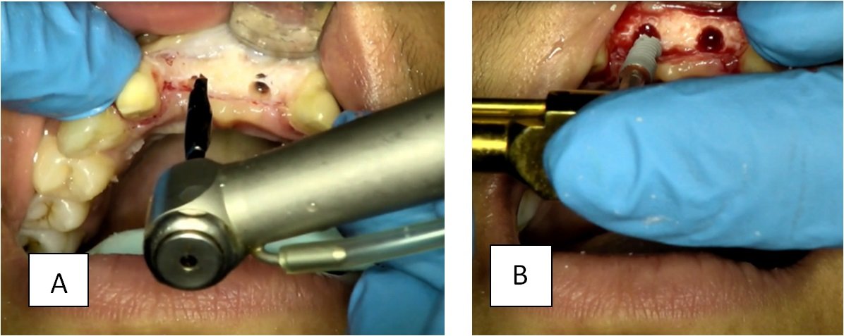
Figure 2 a,b:Implant placement
Implant insertion in the osteotomy site was done using torque ratchet till all threads were submerged into bone and insertion torque of 45 Ncm was achieved. ISQ was also in desired rang. (Figure 3)
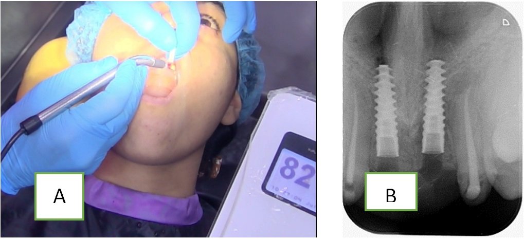
Figure 3:(a)ISQ was taken,(b)X-ray after implant placement
Immediate loading with 15 degree of angulated abutment was done and flap was closed using 3-0 silk suture (Figure 4)
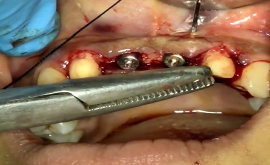
Figure 4: Suture placement
Impression was made to make check cast and temporary was fabricated using PMMA. It was verified in patient mouth and occlusion adjustments were done and was cemented with temporary cement. (Figure 5)
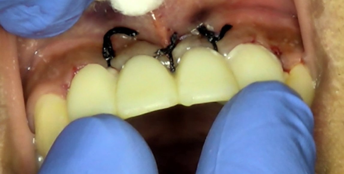
Figure 5: Temporization
Antibiotic (Amoxicillin & Clavulanic Acid 625 mg, twice daily for 5 days) and Anti-inflammatory (Zerodol SP, thrice daily for 5 days) were prescribed and post-operative instructions were given. The patient was seen post surgically after 1 week for suture removal and no signs of superinfection were noted.
Patient was recalled after 3 months for prosthetic procedures and adequate amount of interdental papilla and the buccal contours were observed similar to the adjacent tooth, the abutment was removed and an impression coping placed, followed by a digital impression. The impression coping was removed and abutment was replaced.
Digital impression was taken as an alternative to a conventional, rubber-based technique, a scan body was attached to the implant after removal of the provisional crown (Figure 6).
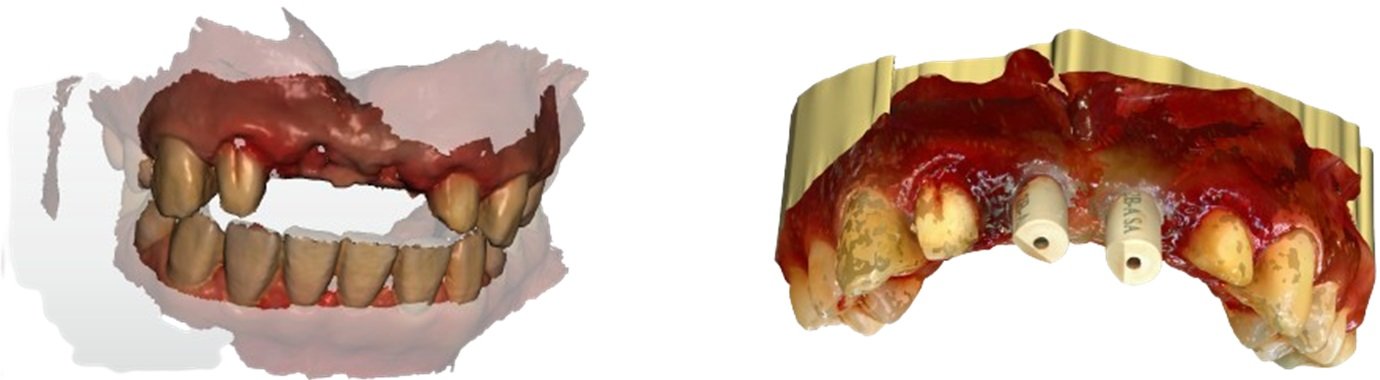
Figure 6: Digital impression
With a digital scanner capturing the position of the scanbody, a digital ?le was created, facilitating CAD/CAM fabrication of a custom zirconia abutment. The ?nal crown, porcelain pressed to an all-zirconia core was seated on the ?nal abutment. Then the ?nal crown was cemented onto the custom abutment. Excess cement was removed and the occlusion was again verified. (Figure 7)
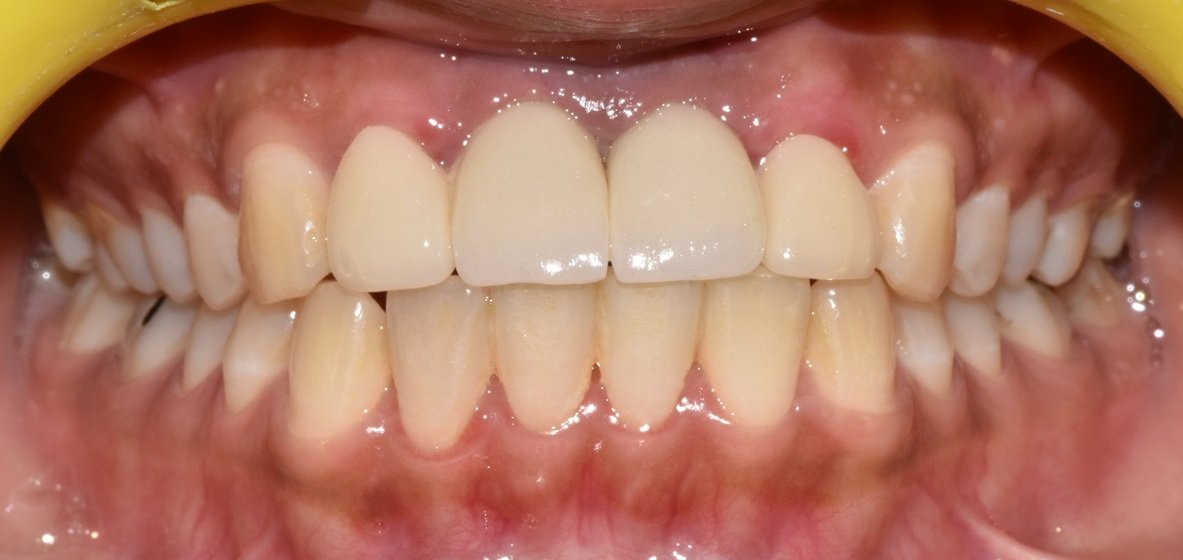
Figure 7: Occlusion was check
After 7 days, patient was recalled for follow up. The patient was very happy with the final aesthetic and functional outcome. (Figure 8)
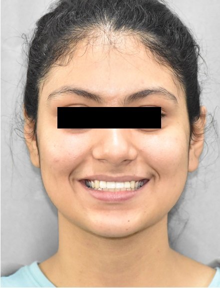
Figure 8: Final photo
DISCUSSION
The Nobel Active implant design offers a useful system for immediate placement and temporization at the time of extraction. The hexagonal anti-rotational connection with the conical seal and built in bone platform shift helps to prevent crestal bone loss. The large pitch variable thread design provides excellent initial stability. High torque test values can be achieved in minimal bone. The torque value of up to 70 Ncm aids to assist the restorative dentist to immediately temporize the implant eliminating second stage surgery. It has been shown that adjacent papilla and peri-implant tissues will be better supported using an immediate temporization.6 The Nobel Active™ implant system appears to have some very promising design features to aid in immediate implant placement and temporization.
An additional aspect to this treatment, which is relatively current, is the digital impression technique, rather than traditional, rubber-based impressions. The accuracy of digital scanners when measuring soft-tissue parameters was recently demonstrated by Schneider etal.7 Ramsey and Ritter8 demonstrated the digital fabrication of a 3-dimensional model, facilitating fabrication of a virtual abutment with the production of a CAD/CAM-designed custom abutment and all-ceramic crown, delivered to the clinician, seated on a stereolithographic model.
The obvious advantages to the digital impression technique include patient and operator comfort. The unpleasantness of rubber-based impressions is eliminated with this technology. The impression procedure is also of shorter duration, resulting in a more pleasant patient experience. The distortion of conventional impression materials is not a factor with digital impressions, contributing to their accuracy. Also, distortion related to contraction of gypsum-based stone models is obviated. The virtual reproduction of a working cast, which facilitates virtual abutment design, negates the need to pour traditional models, shortening laboratory working time, and avoids the possible dimensional changes associated with these materials.9
As with any procedure, case selection is critical to achieving optimal results. The patient featured in this case report was one with a thick periodontal biotype, lowering the risks of signi?cant postoperative recession.10 The extraction socket was intact after tooth removal, and his occlusal scheme permitted the placement of a provisional restoration out of contact with the mandibular antagonist teeth. Had any of these criteria not been met, a deviation from the current procedure may have been necessary. This may have included flap reflection and concurrent guided bone regeneration, delayed placement with socket augmentation, or the need for a transitional removable partial denture.11
CONCLUSION
Placing dental implant in the maxillary anterior region requires precise planning, surgery, and prosthetic treatment. This case report has illustrated the steps needed to create ideal aesthetics in the maxillary anterior region. The prosthetic restoration of a dental implant must be ideal to achieve the desired aesthetic result. This case report has discussed the importance of a comprehensive and interdisciplinary approach to treatment planning, surgery, and restoration of dental implants in the maxillary anterior region of the mouth.
REFERENCES
- El Askary AS. Aesthetic considerations in anterior single tooth replacement. Implant Dent 1999,8:61-67.
- Spielman HP. Influence of the implant position on the aesthetics of the restoration. Pract Periodontics Aesthet Dent. 1996; 8:897-904.
- S. Interdisciplinary treatment planning for single-tooth J Esthet Restor Dent. 2007;19(2):79-88.
- Murata H, Kawamura M, Hamada T, Saleh S, Kresnoadi U, Toki K. Dimensional stability and weight changes of tissue conditioners. J Oral Rehabil. 2001;28(10):918–23. [PubMed] [Google Scholar]
- Guzzo F, DE Luca G, Barnaba P, Severino D. Cad-cam procedure and implant-prosthetic rehaBilitation. Case report. Oral Implantol (Rome). 2016 Nov 13;9(1):27-32. doi: 10.11138/orl/2016.9.1.027. PMID: 28042428; PMCID: PMC5159934
- Wang H, Ormianer Z, Patti A. et a Consensus conference on immediate loading: The single tooth and partial edentulous areas. Implant Dent 2006; 15:324-333.
- Brunski, J. B., In vivo bone response to biomechanical loading at the bone/dental-implant interface. Adv Dent Res, 1999. 13: p. 99-119.
- Szmuckler-Moncler S, Salama, H. Reingewirtz Y., Dubruille JH. Timing of loadingand effects on micromotion on bone-dental implant interface: review of experimental literature. J Biomed Mater Res 1998; 43:192-203
- Kan JY, Roe P, Rungcharassaeng K, et al. Classification of sagittal root position in relation to the anterior maxillary osseous housing for immediate implant placement: a cone beam computed tomography study. Int J Oral Maxillofac Implants. 2011;26(4):873-876.
- Kan JY, Rungcharassaeng K, Lozada JL, Zimmerman G. Facial gingival tissue stability following immediate placement and provisionalization of maxillary anterior single implants: a 2- to 8-year follow-up. Int J Oral Maxillofac Implants. 2011;26(1):179-187.
- Gimenez B, Ozcan M, Martinez-Rus F, Pradies G. Accuracy of a digital impression system based on parallel confocal laser technology for implants with consideration of operator experience and implant angulation and depth. Int J Oral Maxillofac Implants. 2014;29(4):853-862.
Disclaimer from Guident
The views and opinions expressed in articles published in Guident are solely those of the authors and do not necessarily reflect the views of the editors, the editorial board or the publisher, Ivory India. Guident does not endorse any product, technique or treatment described. Clinical content is intended for qualified dental and medical professionals and is not a substitute for individual clinical judgement. Guident, its editors and the publisher accept no liability for any consequence arising from the use of information published in the journal.
How to cite this article
Dr. Gaurav Gupta, Dr. D.K. Gupta, Dr. Neelja GuptaWisdom Dental Clinics ®. Immediate Temporization in the Aesthetic Zone: A Case Report. Guident. 2023 Dec;17(1). Available from: https://guident.in/articles/implantology/immediate-temporization-in-the-aesthetic-zone-a-case-report.html