A Giant Compound Odontome in Posterior Mandible
Department of Oral & Maxillofacial Surgery Govt. Dental College & Hospital, Ahmedabad, India
Abstract
Odontomas, the most common odontogenic tumors, occur more frequently in the maxilla and are rarely larger than a tooth. They are usually found incidentally on routine dental radiographs during the second decade of life. The treatment of choice is surgical removal, and large tumors are challenging as damage may occur to surrounding anatomic structures. In this article we describe a "giant" compound odontoma occupying the entire body of the mandible and ramus in a 14-year-old boy. Because of its radiographic mixed radiolucent and radiopaque appearance and its expansive growth, it is imperative to make the differential diagnosis of giant complex odontoma for other more aggressive mixed odontogenic tumors, such as ameloblastic fibro-odontoma, odotoameloblastoma and cystic calcified odontogenic tumor.A conservative intraoral enucleation was used to avoid facial scarring and preserve mandibular continuity and function. The huge bony defect healed completely without a bone graft or further intervention. This case report emphasizes the importance of the early diagnosis of these lesions and a conservative surgical approach as well as the extraordinary healing potential of young patients.
INTRODUCTION
Odontomas are non-aggressive, hamartomatous developmental malformations or lesions of odontogenic origin, which consists of enamel, dentin, cementum and pulpal tissue. These lesions are composed of more than one type of tissue and for this reason, have been called as composite odontomas.1 The World Health Organization has subdivided odontomas into a compound and complex type based on histologic criteria. Compound odontomas consist of multiple miniature or rudimentary teeth, whereas complex odontomas appear as an amorphous and disorderly pattern of calcified dental tissues. The etiology is not clear, although local trauma, infection and genetic factors have been suggested as possible causes 2. Odontomas show no gender predilection. Odontomas are usually detected in school-age children, and the mean age at the time of diagnosis is 14 years 3,4. The majority of odontomas are asymptomatic and seldom causes swelling, pain, suppuration, bony expansion, and displacement of teeth3,5. These lesions are commonly small, seldom larger than a tooth. Odontomas are generally discovered through routine radiographic examination in dental treatment. An odontoma is essentially a benign lesion, but, often causes disturbances in the eruption of its associated tooth.6,7. Odontomas mostly occur in the permanent dentition and are rarely associated with the primary teeth 5,8,9. Compound odontomas are more prevalent than complex odontomas. Compound odontomas commonly occur in the incisor-canine region of the maxilla and complex odontomas are frequently located in the premolar and molar region of both jaws10. Odontomas occur more often in the permanent dentition. An unusual association of odontomas with primary dentition was reported11.
We report a rare case of huge compound odontome with dentigerous cyst lining in the posterior mandible, which was treated surgically. The unusual site of occurrence, compound odontome in the posterior mandible and most importantly its huge size and association with a dentigerous cyst lining makes it worth presenting.
CASE REPORT
A 14 yrs old male patient had come to the Oral Surgery Department with a chief complaint of swelling over right lower 3rd of face since 1 month.
The patient was healthy with an unremarkable medical history. Patient gives history of trauma due to fall when patient was 8 months old for which he was treated by medicines
Extraoral examination revealed A Single, well defined, hard, tender, non warm, non fluctuant swelling present at right body region of mandible, extending anteroposteriorly from corner of mouth to 1 cm in front of posterior border of mandible and superoinferiorly from an ala-tragus line to the lower border of mandible. Right submandibular lymphnodes are enlarged, tender and palpable.
Intraoral examination revealed A Single, well defined , localized, hard, tender swelling extending from Lower right deciduous canine to anterior border of right ramus of mandible with obliteration of right buccal vestibule. Buccal and lingual cortical plate expansion present. Clinically missing lower right permanent canine, 2nd premolar and 1st & 2nd molar
Similar swelling appeared before 1 year, for which patient had taken medication from private clinic and got relief. But again swelling reappeared before 1 month and doesn’t subsided with medicine. O.P.G. revealed a a single, well defined, bilocular radioopacity with surrounding radioluscent periphery extending from right lower deciduous canine, posteriorly into the ramus with thinning of lower border of mandible alongwith impacted lower right permanent canine, 2nd premolar and 1st & 2nd molar
The differential diagnosis of the mixed radiopac-radiolucent picture included gient complex odontoma, ameloblastic fibro-odontoma, odotoameloblastoma and cystic calcified odontogenic tumor and benign cementoblastoma. Complete surgical removal of the calcified tissue was done under genral anesthesia and subjected to histopathological examination. Sections stained with Hematoxylin and Eosin showed the presence of dentinal structures along with few cementum like structures with presence of a cystic lining. The cystic lining consists of two to three rows of cubiodal epithelium originating from the surrounding fibrous tissue, resembling dentigerous cyst.
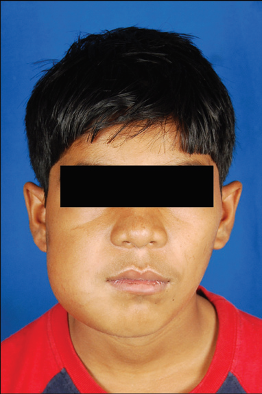
Fig 1: Frontal view, showing swelling over right side of face

Fig 2: Pre operative X-ray OPG

Fig 3: Intra operative view of empty bony cavity
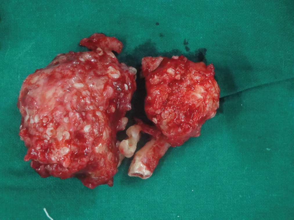
Fig 4: Resected bony mass

Fig 5: Post operative X-ray OPG
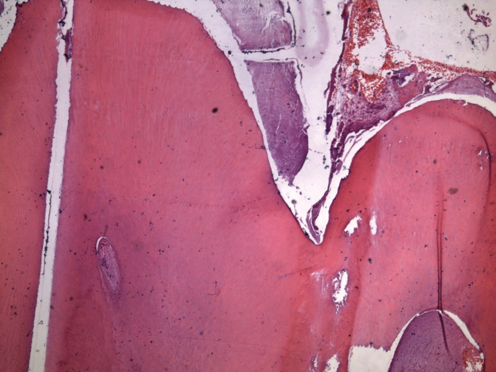
Fig 6: Histopathologic view
DISCUSSION
The term ‘odontoma’ (or odontome) which was firstly described by Paul Broca in 1867, was originally used as a general descriptive for any tumour (in its broadest sense) of odontogenic origin.12 Broca defined the term as ‘tumours which were formed by the overgrowth or the transitory of the complete dental tissue’.13 Odontomas are non-aggressive, hamartomatous developmental malformations or lesions of odontogenic origin appearing as small, solitary or multiple radioopaque lesions found on routine radiographic examinations. They are the most common odontogenic tumours which constitute 22% of all the odontogenic tumours of the jaws14. Although odontomas may be found in any tooth bearing region of the jaws, Compound odontomas show a prediliction in the anterior section of the upper maxilla, while complex odontomas are typically found in the posterior mandibular region 15,16.. In contrast to these data in our case compound odontome located in posterior mandibular region which is quite rare condition. Any trauma during the stage of odontogenesis can seriously affect the morphogenetic stages of dental development, and several developmental alterations can arise in permanent teeth as a consequence of dental injuries and maxillofacial traumas. Odontoma-like malformations and partial or complete arrest of root formation are rare complications developed after trauma. The extent of these malformations depends on the permanent tooth’s developmental stage. Odontomas can also manifest as part of syndromes, such as basal cell nevus syndrome, Gardner syndrome, familial colonic adenomatosis, Tangier disease or Hermann syndrome14,17. Dentigerous cyst arising from the fibrous connective tissue lining of the compound odontome is rare1. Though cases of Complex odontomas associated with dentigerous cyst were reported18. The present case also showed dentigerous cyst arising from fibrous connective tissue lining of the compound odontome. The present case is rare because of posterior mandibular localization, such a huge size and occurrence of surrounding dentigerous cyst in compound odontome.
CONCLUSION
Dentigerous cyst arising from the fibrous connective tissue lining of the odontome is rare. If arise, they are potential for attaining large size, tooth resorption, destruction of jaw bones and some shows neoplastic changes like ameloblastoma. The potential complication justifies radiographic screening, histopathological evaluation and complete enucleation of all the odontomes.
BIBLIOGRAPHY
- Brad W Neville, Douglas D Damm, Carl M Allen, JerryE Bouquot. Oral and Maxillofacial Pathology, 3rd Ed, Elsevier, 2009 Saunders, 2002:724-26.
- Hitchin AD. The etiology of the calcified composite odontoma. Br Dent J 1971; 130: 475-82.
- Kaugers GE, Miller ME, Abbey LM. Odontomas. Oral Surg 1989; 67: 172-6.
- Kaneko M, Fukuda M, Sano T, Ohnishi T, Hosokawa Y. Microradiographic and microscopic investigation of a rare case of complex odontoma. Oral Surg Oral Med Oral Pathol Oral Radiol Endod 1998; 85: 131-4.
- Stajcic ZZ. Odontoma associated with a primary tooth. J Pedodont 1998; 12: 415-20.
- Morning P. Impacted teeth in relation to odontomas. Int J Oral Surg 1980; 9: 81-91.
- Budnick SD. Compound and complex odontomas. Oral Surg 1976; 42: 501-6.
- de Oliveira BH, Campos V, Marcal S. Compound odontoma – diagnosis and treatment: three case reports. Pediatr Dent 2001; 23(2): 151-7.
- Noonan RG. A compound odontoma associated with a deciduous tooth. Oral Surg Oral Med Oral Pathol 1971; 32(5): 740-2.
- 1Martin S.Greenberg, Michael Glick, Jonathan A. Ship. Burkitt’s Oral Medicine. 11th ed. Hamilton: BC Decker Inc, 2008; 151.
- Gulsun Yildirim, Gul Tosun, Dilek Kiziloglu, Ercan Durmus, Yagmur Sener. An unusual association of odontomas with primary teeth. Eur. J Dent 2007; 1: 45- 49.
- Zoremchhingi, Joseph T, Varma B, Mungara J. A compound composite odontoma which was associated with an unerupted permanent incisor. A case report. J Indian Soc Ped Prev Dent 2004; 22(4): 114-7.
- Batra P, Gupta S, Pajan K, Duffal R, Hariprakash. Odontomesdiagnosis and treatment and 4 case reports. JPFA 2003;19:73-6.
- Amado CS, Gargallo AJ, Berini AL, Gay EC. Review of 61 cases of odontoma: Presentation of an erupted complex odontoma. Med Oral 2003;8:366-73.
- Litonjua L, Suresh L, Valderrama L, Neiders M. Erupted complex odontoma: A case report and literature review. Gen Dent 2004;52:248- 51.
- Hidalgo-Sanchez, leco-Berrocal, Martinez-gonzalez. Metanalysis of the Epidemiology and Clinical
- manifestations of odontomas. Medicina oral, Pathologia oral, Oral Y.Cirugia Bucal 2008; 13: 16-17. Patiño Illa C, Berini Aytés L, Sánchez Garcés MA, Gay Escoda C. Odontomas complejos y compuestos: análisis de 47 casos. Arch Odonto Estomatol 1995 Agosto;11(8):423-30.
- P Jeyaraj, S.Murali Mohan, Dentigerous cyst of mandible with multiple complex odontomes, MJAFI 2008;64:382-83
Disclaimer from Guident
The views and opinions expressed in articles published in Guident are solely those of the authors and do not necessarily reflect the views of the editors, the editorial board or the publisher, Ivory India. Guident does not endorse any product, technique or treatment described. Clinical content is intended for qualified dental and medical professionals and is not a substitute for individual clinical judgement. Guident, its editors and the publisher accept no liability for any consequence arising from the use of information published in the journal.
How to cite this article
Dr. Mohit Agrawal, Dr. Balram Garg, Dr. Suket Tailor. A Giant Compound Odontome in Posterior Mandible. Guident. 2013 Jul;6(9). Available from: https://guident.in/articles/oral-surgery/a-giant-compound-odontome-in-posterior-mandible.html