Improving the Appearance of Face with Segmental Bijaw Orthognathic Surgery
Agra, India
INTRODUCTION
Segmental procedure, whether they are in the maxilla or in the mandible, have been recognized as being more stable than ramus2 or total maxillary osteotomies. In 1971, Bell wrote about the correction of anterior open bite, highlighting the stability achieved with segmental osteotomies.
Rosenquist in 1993 followed a series of patients who underwent anterior segmental maxillary ostetomies with the downfracture or the Wunderer method of treatment. Long term stability was found to be acceptable except when osteotomy was used to correct deep overbites. The stability seen following movement of anterior portions of both jaws results from minimal movement of segmentsand muscular attachement..
CASE REPORT
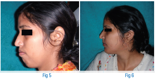
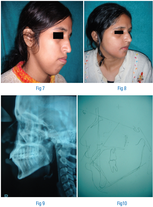
A 23 year old female patient presented with chief complains of protruding lips and bad looks. On intraoral examination both maxillary and mandibular arch were paraboloid in shape, the general dental and periodontal status of the patient was fair. On Extraoralexamination patient had mesocephalic face (fig.3 and fig.5), convex facial profile (fig.3 and fig.5), incompetent lips, hypermentalis activity and loss of labiomental sulcus fig.1 and fig.7).
CEPHALOMETRIC ANALYSIS Refer to figure 9, figure 10 and table.1


TREATMENT PLANNING
After through clinical and cephalometric examination diagnosis of bimaxillary protusion was made and bi -jaw segmental osteotomy was planned. The surgery was first carried on study models with 5mm set back of maxillary segment and 4 mm set back of mandibular segment.
Patient was admitted prior to surgery, routine investigations were done, both maxillay and mandibular first premolars were removed prior to surgery and splint was fabricated.
ANTERIOR MAX ILLA RY OSTEOTOMY
A transverse incision was made in depth of the sulcus from behind the planned osteotomy site to the opposite side. Subperiosteal dissection was done superiorly. The pyriform aperture was exposed. The nasal mucoperiosteum was dissected to expose lateral wall and floor. The cartlaginous nasal septum was elevated from the vomerion groove in the anterior nasal crest of the maxilla. The dissection was then carried out for about 15mm posteriorly. The osteotomy was designed as per model surgery. The osteotomy was
extended to the nasal cavity, protecting the nasal mucoperiosteum with periosteal elevators a 5mm of healthy bone was kept above the apices of the canines1. The osteotomy was done after tunnelling the muperiosteum at the interdental area. In the whole procedure mucoperiosteum in the anterior region was kept intact. Than alveolar osteotomy was performedkeeping the finger in the palatal area to protect palatal tissue (one can feel the bur perforatig the bone). A good margin of alveolar crestal bone was maintained
around the adajacent teeth to the osteotomy site. About 15 mm of the anterior portion of the nasal crest of maxilla was removed for better visulization while doing the transpalatal ostetomy3. Than with help of straight osteotome transpalatal osteotomy was completed, the anterior maxilla was downfractured. All osteotomy sites were inspected for bony protuberances which were removed with the help of round burr6. The segment was then placed on the prefabricated occlusal splint and wired in to the proper position.
ANTERIOR SUBAP ICAL MANDIBULA R OSTEOTOMY
After infiltration of lignocaine with 2% adrenaline in the labial and buccal vestibule, degloving incision made mucoperiosteal flap was reflected. Subperiostel tunneling was done at lingual aspect of planned osteotomy1. Vertical cuts were made through the extractionsockets keeping the finger on lingual aspect to feel rotating burr penetrating lingual cortex, horizontal cut was made joining both vertical cuts keeping a 5mm of safe bone below the root apices7. The osteotomy was completed with help of osteotome. The anterior
segment was mobilized with finger pressure all bony protuberances were inspected and removed with help of round burr. Position of the fragment was checked on the occlusal splint secured to maxillary arch.
Maxillary anterior segment was than stabilized with two L-shaped plate bilaterally while mandibular segment was stabilized with two 4 hole plate bilaterally.
Layer wise suturing with 3-0 vicryl suture, alar cinch suture given, IMF released, patient reversed, extubated and shifted to post op ward for further care.
DISCUSSION AND CONCLUSION
It is well documented that east asians generally have a grater tendency toward bimaxillary protrusion or bialveolar protrusion than caucasions,8,9. Four premolar extraction to retract the anterior teeth was one of the treatment of choices of typical orthodontics. However it has become more complicated in adult patients because of the difficulty in achieving physiologic tooth movement, longer treatment time, relatively worse periodontal status, and increasingly strict social conditions. Considering these inherent limitations, surgery with extraction of premolars and surgical retraction of anterior segments using subapical osteotomy technique is gaining acceptance as a better treatment modality than orthodontic alone.
There have been many reports in the literature of hard and soft tissue changes after orthognathic surgery but very few about anterior segmental surgery of maxilla and mandible. In addition studies of frontal view of face, as compared to profile studies were scarce. However, changes in the frontal view are clearly of substantial clinical importance and are often one of the key criteria by which patient judges the success of treatment10,11.
When orthognathic surgery is contemplated, one wishes to be able to accurately predict the ultimate outcome for the patient (predictability). The other important thing is ability to maintain the desired position (stability)4,5. Predictability depends on ability to position the bony segments in space, soft tissue response to that movement (fig.2, fig.4, fig.6 and fig.8). Stability involves the adaptation of the skeleton and its surrounding soft tissue envelope to a new equlibrium. Stability varies with the type of movement and the technique used. Segmental procedures are considerd to be more predictable and stable as compared to other maxillary and mandibular osteotomies.
ACKNOWLEDGMENTS
The authors thanks Dr. Divya Mehrotra for her valuable advice and guidance throughout the treatment of this case and preparation of this case report.
REFERENCES
1. Epker BN: Va scular considerations in orthognathic surgery. Oral surgery. Oral med, oral path 57: 467-472, 1984.
2. Behrman SJ: Complications of sagittal osteotomy of the mandibular ramus. J maxillofacial surge 30:554, 1972.
3. Bell WH, Phys AM: Biologic basis of maxillary osteotomies, Anthropology 38:279-290, 1979.
4. Storum KA, Bell WH: Hypomobility after maxillary and mandibular oatetomies, oral surg 5:7-12, 1984.
Disclaimer from Guident
The views and opinions expressed in articles published in Guident are solely those of the authors and do not necessarily reflect the views of the editors, the editorial board or the publisher, Ivory India. Guident does not endorse any product, technique or treatment described. Clinical content is intended for qualified dental and medical professionals and is not a substitute for individual clinical judgement. Guident, its editors and the publisher accept no liability for any consequence arising from the use of information published in the journal.
How to cite this article
Dr. Bhupendra Harjani, Dr. Manpreet Singh, Dr. Harjani Dental & Facial Surgery Clinic. Improving the Appearance of Face with Segmental Bijaw Orthognathic Surgery. Guident. 2013 Jul;6(9). Available from: https://guident.in/articles/oral-surgery/improving-the-appearance-of-face-with-segmental-bijaw-orthognathic-surgery.html