
Carcinima Ex Pleomorphic Adenoma
(Prof. & Head)
Abstract
Carcinoma ex pleomorphic adenoma is a relatively rare malignant salivary tumor, especially in the minor salivary gland. We present a case of carcinoma ex pleomorphic adenoma in a recurrent lesion along with a discussion on etiology, clinical presentation, causes, investigation, treatment and management.
Keywords:malignant mixed tumor, carcinoma ex pleomorphic adenoma
INTRODUCTION
Pleomorphic adenoma, also known as benign mixed tumor is the most common salivary gland neoplasm. Malignant salivary tumors represent 3 to 5% of all malignant head and neck tumors[1]. Carcinoma ex pleomorphic adenoma (CXPA) is a relatively rare, highly malignant tumor; accounting for 11.7% of salivary malignancies[2].The development of Carcinoma ex pleomorphic adenoma is from an untreated long-standing benign mixed tumor or from a recurrent benign tumor. Prognosis is usually poor in invasive tumors.
CASE REPORT
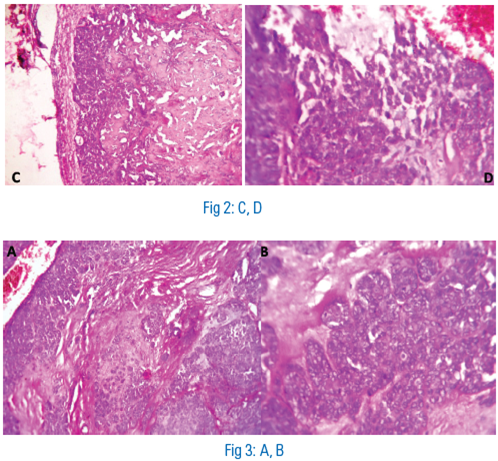
A 74 year old male patient presented to a private clinic with a soft swelling present for the past 5 months (in) on the right side of the hard palate. It was a recurrent swelling, having been earlier diagnosed as palatal pleomorphic adenoma 8 years back, which first recurred and was managed one year ago. The swelling was extending from first premolar to first molar region measuring about 5 cm x 2.5 cm. The swelling presented with no fluid on aspiration and with a perforation. An excisional biopsy was performed and the specimen was sent for histopathological evaluation. Microscopic evaluation showed an encapsulated salivary gland lesion (FIGURE 1, 2) with myxoid, chondroid and hyalinised areas in few foci (FIGURE 3). Mucous acinar cells were seen in the periphery of the lesion. The glandular epithelium exhibited nuclear pleomorphism and was arranged in sheets and islands (FIGURE 4). Keratin formation was visible in few areas. Necrotic areas were also seen (FIGURE 5). In few areas, cells with mucoid differentiation were observed (FIGURE 6). The histopathological features were suggestive of pleomorphic adenoma with features of malignant transformation.


The patient was lost for follow-up and after investigation was reported to have succumbed probably as a result of malignancy.
DISCUSSION
Pleomorphic adenoma is the most common benign neoplasm of the salivary gland. It accounts for 53% to 77% of parotid tumors, 44% to 68% of submandibular tumors and 33% to 43% of minor gland tumors[3]. According to the World Health Organization, malignant derivatives of pleomorphic adenomas of salivary gland origin should be divided into 3 different clinical and histologic entities: 1) carcinoma ex pleomorphic adenoma (carcinoma in pre-existing pleomorphic adenoma), 2) carcinosarcoma (true malignant mixed tumor), and 3) metastasizing pleomorphic adenoma.[4] These are collectively called as malignant mixed tumors.
CXPAs account for 3.6% of all salivary neoplasms and 11.7% of salivary malignancies[5]. Most of the malignant transformation in pleomorphic adenoma occur in the parotid gland and is relatively rare in minor salivary gland[6]. Minor salivary gland tumors account for less than 7% of all cases[7]. The palate is the most common site of occurrence in minor salivary glands as found in our case.
Malignant transformation of pleomorphic adenoma occurs in 5–25% of untreated patients, usually in longstanding cases of 15–20 years[8]. Longstanding lesions PAs or lesions PAs with multiple recurrences have high chance of malignant transformation in pleomorphic adenoma. Studies have shown that 1.6% of malignant transformation occurs in tumor less than 5 years and the risk for over 15 years is 9.5%[ 9, 10]. Exposure to radiation and development of genetic instabilities may also contribute to formation of tumor. Chromosomal abnormalities at 3p, 9p and 17p have been described in cases of CXPA(11).
Most patients report with either an asymptomatic mass or a sudden rapid expansion of the preexisting mass. The age ranges from 34 to 95 years with a mean of 61 years. About 1/3rd of CXPA patients present symptoms such as pain, facial nerve palsy, enlarged lymph nodes, skin ulceration and dysphasia[2,10]. There is a male predilection contrary to female and the predilection of for pleomorphic adenoma with male: female ratio of 1.8:1. Survival rate is largely related to dependent on clinical staging. While the 5-year survival rate for stage I disease is over 80%, the overall 5-year survival rate is only 37%.[2]
Histopathologically, CXPA may be classified based on the extent of capsular infiltration. Non invasive CXPA refers to malignant transformation without violation of the capsule. Minimally invasive CXPA refers to cases where there is less than 1.5 cm penetration of the capsular tissue. Infiltration beyond that limit is classified as frankly invasive. The first two types have excellent prognosis while the invasive type has a guarded prognosis(5).
The commonly reported histologic pattern is poorly differentiated adenocarcinoma. The malignant areas in CXPA usually consist of dysplastic epithelial cells with increased mitosis. Rarely, malignant tissues resembling PLGA(Expand), Mucoepidermoid carcinoma, adenoid cystic carcinoma, epithelial-myoepithelial carcinoma may be present. Malignant mixed tumors metastasize to the lungs, bone, hilar and cervical lymph nodes.[ 3,12, 13]. CXPA is generally an aggressive tumor with high recurrence and metastatic rates, which vary from 25% to 75%[13]. Recurrence and regional and distant metastases are predictive of extremely poor prognosis[14,15]. Diagnostic imaging is done with CT and MRI for providing details about the tumor margins and its extension into the surrounding structures.
Immunohistochemistry has added to our knowledge regarding the processes occurring in CXPA. Amplification and overexpression of HMGIC (high mobility group protein gene) and possibly MDM2 (murine double minute 2) might be important genetic events possibly contributing to malignant transformation of benign pleomorphic adenoma. Immunostaining has revealed the presence of HMGIC protein largely expressed in the cells in the carcinoma part of the tumor.
Ki-67 antigen is present in all the active parts of cell cycle – G1, S, G2, M phase and absent in G0 phase. Ki-67 expression is increased with the cell cycle dysregulation. Ki-67 expression in CxPA was detected predominantly in malignant epithelial component than benign components.[16,17,18]
p53 protein is a product of the tumor suppressor p53 gene. The tumor suppressor gene functions in G1 arrest to allow the repair of DNA damage and to prevent the cell from entering the S phase of the cell cycle or alternatively, to guide the damaged cells to apoptosis.[19] Highest p53 protein expression with high intensity in CXPA suggest mutation of this tumour suppressor gene; this seems to be involved in the transformation of PA to carcinoma CXPA.[16,17]
Surgery and radiation are thought to improve the tumor control. There are controversies on the treatment of CXPA in salivary gland. The presence of positive margins, perineural invasion, facial nerve involvement, and lymph node metastasis are high in CXPA when compared to other malignancies of parotid gland. Post-operative adjuvant radiation could effectively remove residual deposits of the tumor[20].
CONCLUSION
CXPA of the minor salivary gland is very rare and diagnosis of such cases is extremely difficult. Early diagnosis of the tumor is very important for management. The management of the benign tumor should be towards prevention of possible malignancy by completely removing the benign tumor. Treatment of maligngnt lesions consists of surgical excision of the tumor followed by radiotherapy.
REFERENCE
- Laurie SA, Licitra L: Systemic therapy in the palliative management of advanced salivary gland cancers. J Clin Oncol 2006, 24:2673-2678.
- Olsen KD, Lewis JE: Carcinoma ex pleomorphic adenoma: a clinicopathologic review. Head Neck 2001, 23:705-712.
- Neville B, Douglas D, Allen CM and Bouquot. Textbook of Oral and Maxillofacial Pathology. 3rd Ed: WB Saunders Co, 2009: 492-4
- H. Kato,M. Kanematsu,K. Mizuta,Y. Ito,Y. Hirose: Carcinoma Ex Pleomorphic Adenoma of the Parotid Gland: Radiologic-Pathologic Correlation with MR Imaging Including Diffusion-Weighted Imaging. AJNR Am J Neuroradiol 29:865– 67.
- Gnepp DR, Wenig BM. Malignant mixed tumors. In: Ellis G, Anclair P, Gnepp D, editors. Surgical pathology of the salivary glands. Philadelphia: Saunders; 1991. p. 350–68
- Yoshihara T, Tanaka M, Itoh M, Ishii T: Carcinoma ex pleomorphic adenoma of the soft palate. J Laryngol Otol 1995, 109:240-243.
- Hüseyin Akan, Levent Yıldız, Recep Ünal: Carcinoma ex pleomorphic adenoma of the minor salivary gland with pulmonary metastasis. Diagn Interv Radiol 2008; 14:3-5
- Stodulski D, Rzepko R, Kowalska B, Stankiewicz C.Carcinoma ex pleomorphic adenoma of major salivary glands – a clinicopathologic review. Otolaryngol Pol 2007;61:687–93.
- Eneroth CM, Zetterberg A. Malignancy in pleomorphic. adenoma: a clinical and microspectrophotometric study. Acta Otolaryngol (Stockh) 1974;77:289-94.
- Zbären P, Zbären S, Caversaccio MD, Stauffer E: Carcinoma ex pleomorphic adenoma: diagnostic difficulty and outcome. Otolaryngol Head Neck Surg 2008, 138:601-605.
- Fearon ER, Vogelstein B. a genetic model for colorectal tumorogenesis. Cell 1990; 61: 759-67.
- Batsakis JG. Metastatic patterns of salivary gland neoplasms. Ann Otol Rhinol Laryngol 1982; 91:465–466.
- Gnepp DR. Malignant mixed tumor of the salivary glands: a review Pathol Annu 1993; 28:279–328
- Leonetti JP, Marzo SJ, Petruzzelli GJ, Herr B. Recurrent pleomorphic adenoma of the parotid gland. Otolaryngol Head Neck Surg 2005; 133:319–322.
- Beckhardt RN, Weber RS, Zane R, et al.Minor salivary gland tumors of the palate; clinical and pathologic correlates of outcome. Laryngoscope 1995; 105:1155– 1160.
- Freitas LL, Araújo VC, Martins MT, Chone C, Crespo A, Altemani A. Biomarker analysis in carcinoma ex pleomorphic adenoma at an early phase of carcinomatous transformation. Int J Surg Pathol 2005;13:337–42.
- Matsubayashi S, Yoshihara T. Carcinoma ex pleomorphic adenoma of the salivary gland: an immunohistochemical study. Eur Arch Otorhinolaryngol 2007;264:789–95.
- Katori H, Nozawa A, Tsukuda M. Expression of epidermal growth factor receptor, transforming growth factor-alpha and Ki-67 in relationship to malignant transformation of pleomorphic adenoma. Acta Otolaryngol 2007;127:1207–13
- Kumar V, Abbas AK, Fausto N. Robbins and Cotran Pathologic Basis of Diseases, 7th edition. Philadelphia: Saunders, 2004:302–3.
- Chen AM, Garcia J, Bucci MK, Quivey JM, Eisele DW: The role of postoperative radiation therapy in carcinoma ex pleomorphic adenoma of the parotid gland. Int J Radiat Oncol Biol Phys 2007,67:138-143.
Disclaimer from Guident
The views and opinions expressed in articles published in Guident are solely those of the authors and do not necessarily reflect the views of the editors, the editorial board or the publisher, Ivory India. Guident does not endorse any product, technique or treatment described. Clinical content is intended for qualified dental and medical professionals and is not a substitute for individual clinical judgement. Guident, its editors and the publisher accept no liability for any consequence arising from the use of information published in the journal.
How to cite this article
Dr. Sujatha G, Dr. Muruganadhan. J, Dr. Lodd Mahendra Principal. Carcinima Ex Pleomorphic Adenoma. Guident. 2013 Jul;6(9). Available from: https://guident.in/articles/oral-pathology/carcinima-ex-pleomorphic-adenoma.html

