Management of Extra Root and Extra Canals in Mandibular Teeth
Seema Dental College and Hospital, Rishikesh, Uttrakhand, India
INTRODUCTION
A thorough understanding of root canal anatomy and morphology is required for achieving high levels of success in endodontic treatment. Failure to recognize variations in root or root canal anatomy can result in unsuccessful endodontic treatment. Hence, it is imperative that the clinician be well informed and alerted to the commonest possible variations. Hoen and Pink in their analysis on teeth requiring re-treatment, found a 42% incidence of missed roots or canals.1
This article reports a two cases of extra root in mandibular canine and premolar respectively.
CASE 1
A 51 year old male patient reported to the Department of Conservative Dentistry and Endodontics, with the chief complaint of dull aching pain in the lower left front teeth region since one month. History of present illness revealed intermittent pain with hot and cold stimuli for the past three months. Past dental history revealed asymptomatic, endodontically treated right mandibular first premolar. The patient’s medical history was non-contributory.
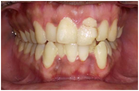
On examination, the lower right canine revealed proximal caries on disto-buccal side. The tooth was not mobile and periodontal probing around the tooth was within physiological limits. Thermal tests were positive and electric pulp testing elicited delayed response with the right mandibular canine. A diagnostic radiograph revealed a coronal disto-occlusal radiolucency involving the pulp space and widening of the periodontal ligament space. The radiograph also revealed an unusual anatomy of involved tooth. It showed presence of two roots and two root canals. For further confirmation of this unusual morphology, multiple pre-operative radiographs were taken at 10-40 degree mesial and distal angulations which confirmed the presence of two roots and two root canals.
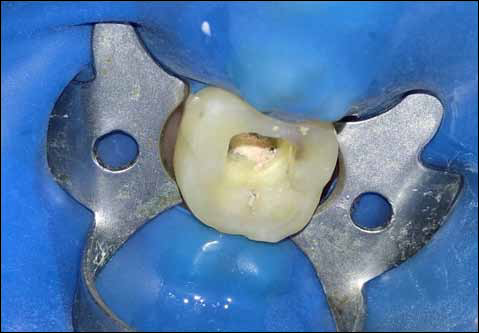
Local anesthesia was administered and access opening was done using endo-access bur under rubber dam isolation. The pulp chamber was opened to facilitate location of buccal and lingual canals. Working length was established using apex locator and confirmed radiographically.
The canals were instrumented using Neoendoflex rotary file till #20/0.06. A 5.2% solution of sodium hypochlorite and 17% EDTA were used alternatively as irrigants, at each change of file. The canals were dried with absorbent paper points and were obturated using #20/0.06 gutta-percha. The final radiograph showed two well obturated canals. After completion of root canal treatment, the tooth was restored using resin composite.

CASE 2
A 45-year-old male patient was referred to our postgraduate endodontic department for management of lower right second premolar. History revealed that the patient reported pain for the past two days. Pain was spontaneous in nature and aggravated on chewing and lying down. On intraoral clinical examination, there was a carious exposure of the pulp and the tooth was tender to percussion. The tooth was subjected to routine clinical tests and a provisional diagnosis of symptomatic irreversible pulpitis with apical periodontitis was made.
On radiographic examination preoperative radiograph, the periodontal ligament outline suggested a rare anatomical feature.
Access was gained to the pulp chamber after administration of local anesthesia, under rubber dam isolation. To gain sufficient access to the canals, the conventional access opening was modified into one that was wider buccolingually as the roots were bucco-lingually oriented. Then the working length determined using CanalPro apex locator and confirmed using #15 K File through radiographic method.
The canal were sequentially irrigated using 5.25% Sodium hypochlorite and 17% EDTA during the cleaning and shaping procedure till #25/0.04. The canals were thoroughly dried and obturation was done using #25/0.04 Gutta-percha and MTA Fillapex sealer. Occlusal access opening was sealed with resin composite. The two roots of the second premolar can be appreciated well in this post-obturation radiograph.

DISCUSSION
The anatomical variations related to teeth must be identified when these teeth are involved in an endodontic diagnosis or treatment planning.The presence of extra roots or canals in mandibular canines and premolars is undoubtedly an endodontic challenge. A collection of previous studies have been tabulated for better understanding [Table 1] and [Table 2].
Mandibular canines having one root with single canal are common, however, two root canals2,3,4,5,6 and in some extraordinary cases, there may be one or two roots with three root canals7,8,9 has been reported.
One mandibular canine and one mandibular premolar were reported in the present case series. The incidence of two root canals in two-rooted mandibular canine teeth has been reported to be up to 14%. Vertucci observed only 6 teeth with Type IV canal configuration (two separate and distinct canals from the pulp chamber to the apex) and of mandibular canine having Type II canals3 in mandibular canine.
In a study at the University of Washington assessing the results of endodontic therapy, the mandibular premolars showed the highest failure rate of all types of teeth.11 Conceivably, these findings could be due to the complex root canal anatomy of a large number of these teeth. A wide range of opinions are reported in the literature regarding the number of root canals, but there are very few reports on the variations in the number of roots that occur in the mandibular second premolars.12
Careful examination of the initial radiograph is extremely important. It allows the identification of multiple roots, root canals and anatomical variations. Failure to recognize the presence of extra root or canals can often lead to acute flare-ups during treatment and subsequent failure of endodontic therapy.
Table 1: Prevalence of lower canine teeth with two roots in specific population groups
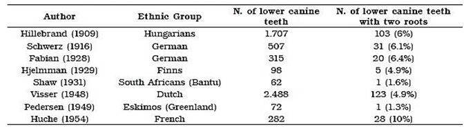
Table 2: Incidence of variations in mandibular second premolar root canal anatomy

CONCLUSION
The clinician should be astute enough to identify the presence of unusual numbers of roots and their morphology. A thorough knowledge of root canal anatomy and its variations, careful interpretation of the radiograph, close clinical inspection of the floor of the chamber and proper modification of access opening are essential for a successful treatment outcome.
REFERENCES
- Hoen MM, Pink FE. Contemporary endodontic retreatments: An analysis based on clinical treatment findings. J Endod 2002;28:834-6.
- Pineda F, Kuttler Y. Mesiodistal and buccolingual roentgenographic investigation of 7,275 root canals. Oral Surg Oral Med Oral Pathol 1972;33:101-10.
- Vertucci FJ. Root canal anatomy of the human permanent teeth. Oral Surg Oral Med Oral Pathol 1984;58:589-99.
- Green D. Double canals in single roots. Oral Surg Oral Med Oral Pathol 1973;35:689-96.
- Sert S, Bayirli GS. Evaluation of the root canal configurations of the mandibular and maxillary permanent teeth by gender in the Turkish population. J Endod 2004;30:391-8.
- Caliskan MK, Pehlivan Y, Sepetçioglu F, Türkün M, Tuncer SS. Root canal morphology of human permanent teeth in a Turkish population. J Endod 1995;21:200-4.
- Arora V, Nikhil V, Gupta J. Mandibular canine with two root canals - An unusual case report. Int J Stomatol Res 2013;2:1-4.
- Heling I, Gottlieb-Dadon I, Chandler N. Mandibular canine with two roots and three root canals. Dent Traumatol 1995;11:301-2.
- Holtzman L. Root canal treatment of a mandibular canine with three root canals. Case report. Int Endod J 1997;30:291-3.
- Orguneser A, Kartal N. Three canals and two foramina in a mandibular canine. J Endod 1998;24:444-5.
- Goswami M, Chandra S, Chandra S, Singh S. Mandibular premolar with two roots. J Endod 1997;23:187.
- Ingle JI, Simon JH, Machatou P, Bogaerts P. Outcome of endodontictreatment and re-treatment - “Textbook of Endodontics”. Ingle JI, Bakland LK, editors. 5th ed. BC Deker Inc: 2002. p. 747-57.
Disclaimer from Guident
The views and opinions expressed in articles published in Guident are solely those of the authors and do not necessarily reflect the views of the editors, the editorial board or the publisher, Ivory India. Guident does not endorse any product, technique or treatment described. Clinical content is intended for qualified dental and medical professionals and is not a substitute for individual clinical judgement. Guident, its editors and the publisher accept no liability for any consequence arising from the use of information published in the journal.
How to cite this article
Dr. Annil Dhingra Dr. Gazal Bisht Dr. Mayuri Kumari Department of Conservative Dentistry and Endodontics. Management of Extra Root and Extra Canals in Mandibular Teeth. Guident. 2018 Jan;11(2). Available from: https://guident.in/articles/endodontics/management-of-extra-root-and-extra-canals-in-mandibular-teeth.html