Reattachment of Maxillary LEFT Central Incisor- a Interdisciplinary Approach
PDM Dental College and Research Institiute, Sector-3A, Village sarai Aurangabad, Bahadurgarh, Haryana, India
ABSTRACT
Introduction: Reattachment of the fractured anterior tooth is a highly conservative and aesthetic treatment that has gained popularity nowadays.The reattachment of the crown fragment to the remaining tooth is a conservative treatmentthat should be considered for young patients. Advancements in the adhesive dentistry has facilitated the restoration of the coronal tooth fractures by minimally invasive procedures when the original tooth fragment is available. Reattachment of fractured fragment offers immediate treatment with improved preponderant aesthetics and restoration of function.
Case Presentation: We reported a case of an adult male who had fractured his maxillary left central incisor in a roadside accident.
Conclusion:The treatment reported for reattachment of the tooth fragment are reasonably easy while providing immediate and lasting results in patients regaining of social confidence and functionality.
Keywords: Composites, Esthetics, Tooth Fractures, Fibre post, Reattachment.
INTRODUCTION
Crown fractures in the anterior region represent 92% of all traumatic injuries of the permanent dentition. 1 Many procedures have been used to restore the fractured teeth, initially including crowns, orthodontic bands, pins and full crowns. 2 Several factors influence the management of coronal tooth fractures, including extent of fracture (biological width violation, endodontic involvement, alveolar bone fracture), pattern of fracture and restorability of fractured tooth (associated root fracture), secondary injuries (soft tissue status), presence/absence of fractured tooth fragment and its condition for use (fit between fragment and the remaining tooth structure), occlusion, aesthetics, finances and prognosis.3-5 One of the treatment modality for managing coronal tooth fracture, especially when there is no or minimal violation of the biological width, is the reattachment of the dental fragment when it is available. Tooth fragment reattachment offers the advantage of being a highly conservative technique that promotes preservation of natural tooth structure and good aesthetics.
The concept of reattachment began in 1964 when Chosak and Eidelman used a cast post and conventional cement to reattach an anterior crown fragment.6 Tennery was the first to use acid etch technique for the reattachment of fractured tooth fragment. 7 Reattachment of natural tooth fragment provides better esthetics, natural wear resistance similar to surrounding dentition, original tooth contour, translucency, and surface texture. Developments in restorative materials, placement techniques, and adhesive advancements allow reattachment using composites as a permanent treatment. Reattachment procedures can be used with a successful outcome on both uncomplicated and complicated fractures.8,9 In the case of extensive loss of tooth structure in the endodonticallytreated tooth a post is required.10 Studies further suggest that a post should be used only whenthere is insufficient tooth structure remaining to support the final restoration11,12.
The present case report describes themanagement of a fractured maxillary left central incisor treated endodontically followed by fragmentreattachment using the glass fiber post. The fractured fragment was intact and available as one single unit,which adapted optimally to the remaining tooth structure.
CASE PRESENTATION
A 22 year old male injured in a road side accident was referred to the Department of Conservative Dentistry and Endodontics, with the chief complaint of broken upper left front tooth associated with pain and sensitivity. On intraoral examination, the maxillaryleft central incisor had a complicated horizontal crown fractureon the buccal side extending 4-5 mm above the gingival margin andoblique in the bucco-palatal direction, extending 3mm below the gingival margin on the palatal side. Clinical examination revealed Ellis class III fracture (involvement of enamel and dentin compromising the pulp) of the maxillary left central incisor (Figure 1). An intraoral periapical radiograph was taken to evaluate the extent of the fracture, the root status and to rule out any alveolar bone fracture (Figure 2). Electric and thermal pulp testing were performed toevaluate the pulp vitality of all the upper anterior teeth, and the result was negative only for tooth21 whereas, all the adjacent teeth displayed a normal response.
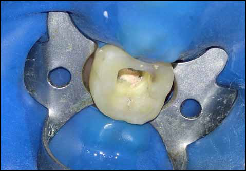
No significant hard or soft tissue injury other than tooth fracture was observed. Local anesthesia was administered, and thefractured coronal fragment was detached withminimal force and immediately soaked in saline solutionto prevent dehydration. After fractured fragment removal the fracture extendingsubgingivally on the palatal surface wasapparent (Figure 3). Upon probing this area, it was determined that the biological width was only minimally invaded and that crown lengthening alone in the palatal aspect would be sufficient for access and isolation during the reattachment procedure. Crown lengthening was done using electro cautery and 2 mm of palatal tissue was excised to expose the subgingivalfracture in order to evaluate the possibilityfor reattachment procedure (Figure 4) and single visitroot canal treatment was planned.
Following isolation using a rubber dam, an endodontic access opening was made using an endo-access bur (Dentsply Maillefer, USA). Patency was checked using 10 K-file (Dentsply Maillefer USA), and the working length was determined by using an electronic apex locator Root ZX II (J. Morita MFG. Corporation, Kyoto, Japan) and radiograph with an endodontic file inserted in the canal was taken to confirm it. The root canal was cleaned and shaped by a Rotary Ni-Ti Protaper system (Dentsply Maillefer, USA) along with Glyde (Dentsply Maillefer, USA), using a crown down technique. The root canal was copiously irrigated with 3% sodium hypochlorite and 17% EDTA solution. The root canal was dried using paper points and before obturation, master cones were selected, and a radiograph was taken. The maxillary left central incisor was obturated with the selected master gutta-percha cone (Protaper, Dentsply Maillefer) and sealer (AH-Plus, Dentsply, Maillefer, USA) (Figure 5). Postspace was created by removing gutta-percha using peeso reamers. Since aesthetics was a concern, the prefabricated glass fiber post was selected. A radiograph of glass fibre post was taken to confirm the post position (Figure 6). Access cavity was sealed and the patient was recalled on next day. The patient reported to the department on next day for the reattachment procedure. Restoration was removed and the prepared root canal was conditioned using self-etching non rinse conditioner (ParaBond-Coltene Whaledent). Placement of the post in the root canal was done using dual cure luting material (ParaCore-ColteneWhaledent). To facilitate polymerization, curing LED light (Woodpecker) was applied through the tip of the post into the root canal for 20 seconds. About 2mm of the post was visible beyond the incisal margin after fixing (Figure 7). The coronal portion of the post was cut to fit into the fractured fragment. Key and lock system was used to attach the fractured fragment with the post, for which an internal retention slot was placed on the dentin of the fractured fragment using a no 4 round bur. The fractured surface of the fragment was then etched with 37% phosphoric acid and the fragment was luted in the correct position using dual cure resin (ParaCore-Coltene Whaledent) with slight pressure. The occlusion was carefully checked and adjusted. Excess of the material was removed using a sharp instrument from the edges and was light cured for 20 seconds (Figure 8). A bevel was prepared on the margins of the approximating surfaces of 21 on the labial side and the margins were sealed with nanocomposite (BrilliantNG-ColteneWhaledent). Using finishing bur the labial surface was finished. Finishing of the lingual surface was done using an egg shaped finishing bur. Polishing of the surface was done with polishing disks which ensured an aesthetic blending of the margins. The margins were properly finished and polished with a series of Sof-Lex disks (3M ESPE) and diamond polishing paste (Figure 9). Post operative radiograph showed the fibre post inside the canal and the fractured fragment reattached with the remaining tooth structure (Figure 10). Gingival tissues had a normal texturewith a normal contouring on palatal aspect (Figure 11). Instructions were given to avoid heavy forces on the tooth and maintaince of the oral hygiene. The patient was kept on recall. The patient came for recall visit at 1, 3 and 6 months and the tooth was found to be intact and functional inside the oral cavity.
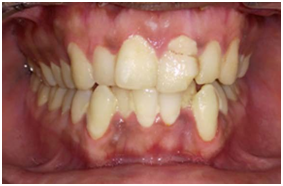
Fig 1:EllisclassIIIfractureofmaxillaryleftcentralincisor
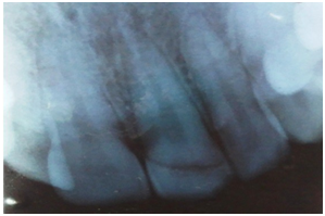
Fig 2:Intraoral periapical radiograph showing coronal fracture
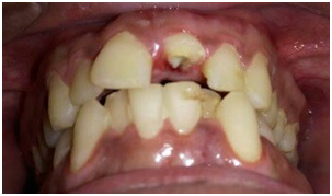
Fig 3:Left maxillary central incisor after removal of fractured fragment
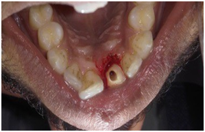
Fig 4:Palatalviewoftooth21 after electrocautery
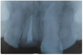
Fig 5:Root canal intervention in tooth 21

Fig 6:Post space preparation and trial post placement radiograph
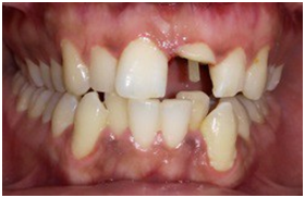
Fig 7:Photograph after post placement
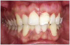
Fig 8:Post treatment reattached toothfragment
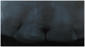
Fig 9:Post treatment radiograph
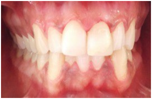
Fig 10:Reattachedtooth21 after finishing and polishing
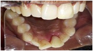
Fig 11:Palatal view of reattached maxillary left central incisor
DISCUSSION
Reattachment of the fractured tooth is an option whenthe broken fragment is intact and available. It has several advantages over conventional methods as itretains the translucency of natural tooth. It is less time consuming and is cost effective. Severalstudies have shown that the impact strength of reattachedtooth is not significantly different from that of intact naturaltooth13,14. Tooth fracture reattachment allows restoration of the tooth with minimal sacrifice of the remaining tooth structure. The natural tooth structure clearly eliminated problems of differential wear of restorative material, unmatched shades and difficulty of contour and texture reproduction associated with other restorative techniques.15
Reattachment procedure is multidisciplinary, mainly dictated by the extension of tooth fracture and trauma to the attachment apparatus. In this case fracture was subgingival on palatal aspect and electrocautery was done to bring the fracture line supragingivally. In the case presented, the pulp was involved and hence, root canal treatment was performed.Post placement and composite resin bonding were used to achieve adequate retention and to resist variousocclusal forces. Fiber posts which have the modulus of elasticity similar to that of dentin are used here to bond with the root and are preferred over metal posts because of less stress concentration on the root and there is low incidence of root fracture16. There is less tooth preparation with a fiber post compared to cast post; thus, the tooth is conserved more. Sometimes failures may occur by debonding of the post and due to root fractures17. When more than 50% of the coronal tooth structure is missing, use of post and core isrecommended prior to restorationv18. Restoration with cast metal posts can cause wedging forces coronallyresulting in failure19. Hence, to meet the patient's esthetic demand, glass fiber post was selected.
Crown-root fractures have immediate implications for endodontic, restorative and periodontal prognosis due to the subgingival line of the fracture. Fracture of the tooth below the gingival attachment presents restorative problems due to difficulty of access20. In such cases, orthodontic or surgical exposure of the fracture is necessary to facilitate further treatment. However, surgical exposure in aesthetic regions has unacceptable results and is best used only for the palatal surface of anterior teeth.21,22 So along with electrosurgical crown lengthening procedure and adequate isolation reattachment can be a logical and justified approach for treating fractured anterior teeth.Several case reports showed that more than80% of fractured anterior teeth separate obliquely fromthe buccal to the palatal side, and the fracture progressestoward the apical area.
Conventional composite restorations can result in less ideal color match,surface texture, contours, and incisor translucency23. Reattachment with composite resin with the help of the acid etch technique is considered to be a highly aesthetic treatment for restoring fractured anterior teeth. It is also necessary topreserve fractured fragment in the hydrated environment as dentin's dehydration results in the collapse ofcollagen fibers and obstruction of adequate resin monomers’ penetration leading to poor adhesion betweendentin and composite material24. There is,however, no synthetic restorative material that can replicate the aesthetic characterization or color stability ofthe natural tooth structure.25 Several case reportsand studies have proved that reattachment of a fracturedtooth fragment is a viable and preferred treatmentwhere indicated, and long-term success can be achieved 26-30. The significant innovation of adhesive methods and resin composites has made reattachment of tooth fragments a procedure that is no longer a provisional restoration, but rather, a restorative treatment offering a promising prognosis.
REFERENCES
- Belcheva A. Reattachment of fractured permanent incisors in school children (review) ASDC J Dent Child. 1998;65:492–4.
- Simonsen R, Thompson VP, Barrark G. Etched cast restorations: clinical and laboratory technique. Chicago: Quintessence;1985;150–1.
- Olsburgh S, Jacoby T, Krejci I. Crown fractures in the permanent dentition: Pulpal and restorative considerations. Dent Traumatol. 2002;18:103–15.
- Reis A, Francci C, Loguercio AD, Carrilho MR, Rodriques Filho LE. Reattachment of anterior fractured teeth: Fracture strength using different techniques. Oper Dent. 2001;26:287-94.
- Andreasen FM, Norén JG, Andreasen JO, Engelhardtsen S, LindhStrömberg U. Long term survival of fragment bonding in the treatment of fractured crowns. Quintessence Int. 1995;26:669–81.
- Chosak A, Eidelman E. Rehabilitation of a fractured incisor using patient's natural crown: Case report. J Dent Child. 1964;31:19–21.
- Tennery NT. The fractured tooth reunited using the acid etch bonding technique. Tex Dent J. 1978;96:16–7.
- Vaz VT, Presoto CD, Jordao KC, Paleari AG, Dantas AA, Segalla JC, et al. Fragment reattachment after atypical crown fracture in maxillary central incisor. Case Rep Dent.2014.
- Taguchi CM, Bernardon JK, Zimmermann G, Baratieri LN. Tooth fragment reattachment: a case report. Oper Dent. 2015.
- Cheung W. A review of the management of endodontically treated teeth post, core and the final restoration. J Am DentAssoc2005;136:611-9.
- Morgano SM. Restoration of pulpless teeth: application of traditional principles in present and future contexts. J ProsthetDent1996;75:375-80.
- Heydecke G, Butz F, Strub JR. Fracture strength and survival rate of endodontically treated maxillary incisors with approximal cavities after restoration with different post and core systems: an in-vitro study. J Dent2001;29:427-33.
- Farik B, Munksgaard EC, Andreasen JO. “Impact strength of teeth restored by fragment-bonding”. Dent Traumatol. 2000;16(4):151–3.
- Farik B, Munksgaard EC. “Fracture strength of intact and fragment-bonded teeth at various velocities of the applied force”. Euro J Oral Sci. 1999;107: 70–3.
- Maia EA, Baratieri LN, de Andrada MA, Monteiro S, de Araújo EM. Tooth fragmentreattachment: Fundamentals of the technique and two case reports. Quintessence Int. 2003;34:99–107.
- Akkayan B, G¨ulmez T. “Resistance to fracture of endodontically treated teeth restored with different post systems,” J Prosthet Dent. 2002;87(4): 431–7.
- Asmussen E, Peutzfeldt A, Heitmann T. “Stiffness, elastic limit, and strength of newer types of endodontic posts,” J Dent. 1999;27(4):275–8.
- Christensen G. When to use fillers, buildups or posts and cores. J Am Dent Assoc.1996;127:1397–8.
- Deutsch AS, Cavallari J, Musikant BL, Silverstein L, Lepley J, Petroni G. Root fracture and the design of prefabricated posts. J Prosthet Dent. 1985;53:637–40.
- Turgut MD, Gonul N, Altay N. Multiple complicated crown root fracture of a permanent incisor. Dent Traumatol. 2004;20:288-92.
- Andreasen JO, Andreasen FM, Andersson L. Crown-root fractures. In; Textbook and color atlas of traumatic injuries to the teeth. 4th edt. Copenhagen: Blackwell Munksgaard;2007:314-36.
- Emerich-Poplatek K, Sawicki L, Bodal M, Adamowicz-Klepalska B. Forced eruption after crown/root fracture crown. Dent Traumatol.2005;21:165-9.
- Mojirade AD, Abiodun Solanke IM, Gbadebo SO. Reattachment of fractured anterior tooth: A 2 year review of a case. Int J Prosthodont Restorative Dent. 2011;1:123–7.
- Perdigão J, Lopes M. Dentin bonding — state of the art 1999. Compend Contin Educ Dent.1999;20:1151–8.
- Terry DA. Pract Proced Aesthet Dent. 2003;15:403–9.
- VamsiKrishna R, Madhusudhana K, Swaroopkumarreddy A, Lavanya A, Suneelkumar C, Kiranmayi G. Shear bond strength evaluation of adhesive and tooth preparation combinations used in reattachment of fractured teeth: an ex-vivo study. J Indian Soc Pedod Prev Dent.2015;33(1):40–3.
- Hegde SG, Tawani GS, Warhadpande MM. Use of quartz fiber post for reattachment of complex crown root fractures: A 4-year follow up. J Conserv Dent. 2014;17(4):389–92.
- Baratieri LN, Monteiro Jr S, Caldeira de Andrada MA. Tooth fracture reattachment: case reports. Quintessence Int. 1990;21(4):261–70.
- Kulkarni VK, Bhusari CP, Sharma DS, Bhusari P, Bansal AV, Deshmukh J. Autogenous tooth fragment reattachment: a multidisciplinary management for complicated crown-root fracture with biologic width violation. J Indian Soc Pedod Prev Dent. 2014;32(2):190–4.
- Patni P, Jain D, Goel G. A holistic approach to management of fractured teeth fragments: a case report. Oral Surg OralMed Oral Pathol Oral Radiol Endod. 2010;109(5):e70–4.
Disclaimer from Guident
The views and opinions expressed in articles published in Guident are solely those of the authors and do not necessarily reflect the views of the editors, the editorial board or the publisher, Ivory India. Guident does not endorse any product, technique or treatment described. Clinical content is intended for qualified dental and medical professionals and is not a substitute for individual clinical judgement. Guident, its editors and the publisher accept no liability for any consequence arising from the use of information published in the journal.
How to cite this article
Dr. Shilpa Arora Dr. Mandeep S.Garewal Dr. Sakshi Bhardwaj Depatment of Conservative Dentistry and Endodontics. Reattachment of Maxillary LEFT Central Incisor- a Interdisciplinary Approach. Guident. 2018 Feb;11(3). Available from: https://guident.in/articles/endodontics/reattachment-of-maxillary-left-central-incisor-a-interdisciplinary-approach.html