PROSTHODONTIC REHABILITATION OF A HEMISECTEDMOLAR: A Case Report
Dr. Himanshu Aeran (Professor & HOD), Dr. Jyotsna Seth (Professor), Dr. Kedar Deole (PG Student), Department of Prosthodontics and Crown & Bridge, Seema Dental College & Hospital, Rishikesh, Uttarakhand, India.
ABSTRACT
Procedural errors like separation of instrument, canal transportation have been the stumbling blocks for effective and perennial endodontic therapy. There are some distinct circumstances where tooth may be indicated for extraction after an endodontic treatment. In such conditions hemisection and root amputation are the modalities which can be exercised for saving the tooth. This case report presents the rehabilitation of a endodontically treated mandibular first molar in a 28 year old female patient who underwent hemisection procedure. A separated instrument in the root canal of mesial root was addressed during the procedure. After the hemisection the mesial portion of tooth was rehabilitated with specifically designed porcelain fused to metal fixed partial denture. This conservative treatment procedure aimed at preserving maximal original tooth structure and an alternative to extraction of the natural tooth.
Key words- Instrument separation, Mandibular first molar, Hemisection, Prosthetic rehabilitation, Fixed partial denture.
INTRODUCTION
The lack of cogent response to endodontic treatment in many instances is primarily attributed to procedural errors, which impede the effective control of endodontic infection.1 Success in endodontic treatment is often roadblocked by mishaps like separation of instrument in the root canal. The retrieval of the separated instrument may not suffice as in some instances fracture of tooth in future is impediment in successful endodontic therapy.2
The terms hemisection and root amputation are known as root resection. Hemisection is the sectioning of multirooted tooth into two halves which is followed by extraction of pathologic root and coronal half of the tooth. The retained root is endodontically treated and restored prosthetically.3
Tooth resection has been indicated in few conditions according to Weine.4
- Severe bone loss along a single root in multirooted tooth.
- Furcation involvement which is through and through.
- Unfavourable root proximity of adjacent teeth.
- Dehiscence of root.
- Prosthetic collapse.
- Irreparable perforation of floor of pulp chamber.
- Vertical root fracture of one root.
Contraindications
- Favourable adjacent abutment teeth for prosthetic restorations
- Inoperable canals to be retained.
- Root fusion.
- Insufficient bone support for remaining root
Thus, this case reports presents prosthetic rehabilitation of a carious hemisected endodontically treated mandibular molar aimed to attain normal structure and functionality.
CASE REPORT
A female aged 28 years set forth to the department of prosthodontics and crown & bridge complaining of pain and food lodgement in the lower right back region of the jaw. The pain was dull in nature, intermittent and occurred in short duration. Patient presented with history of endodontic treatment of right mandibular first molar 5 months back. Intraoral examination revealed carious right mandibular first molar with grade 3 furcation involvement. The tooth was tender on vertical and horizontal percussion. Radiographic investigations were suggestive of a separated instrument in the mesial root of the tooth with a periapical radiolucency (Fig 1). Therefore, the final diagnosis was post treatment apical periodontitis.
Considering awareness and young age of the patient, a conservative approach was planned for the tooth rather than extracting it. The treatment plan was as follows:
- Retreatment of the distal root of #46 (FDI numbering system)
- Hemisection of mesial root of #46
- Prosthetic rehabilitation.
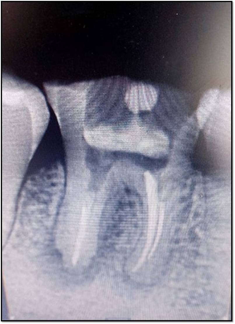
Fig. 1: Preoperative radiographic view of mandibular first molar
TREATMENT PROCEDURES
Retreatment of distal root of #46
The preexisting gutta-percha from the distal root canal was done followed by irrigation and shaping and was obturated using lateral condensation method. Following which post endodontic restoration was given.
Hemisection of mesial root of #46
Under local anaesthesia, a crevicular incision was given to reflect a full thickness flap. Vertical cut method was used to resect the crown. (Fig 2). Separation of mesial and distal halves was checked by passing a probe via the vertical separation. The pathologic root was extracted and the socket was debrided, irrigated and root was planed. Distal root surface was contoured and developmental ridges were removed (Fig 3).
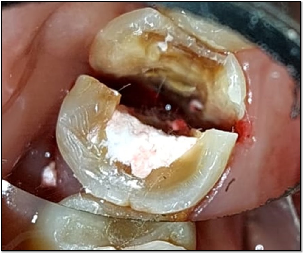
Fig. 2: Vertical cut towards the bifurcation area.
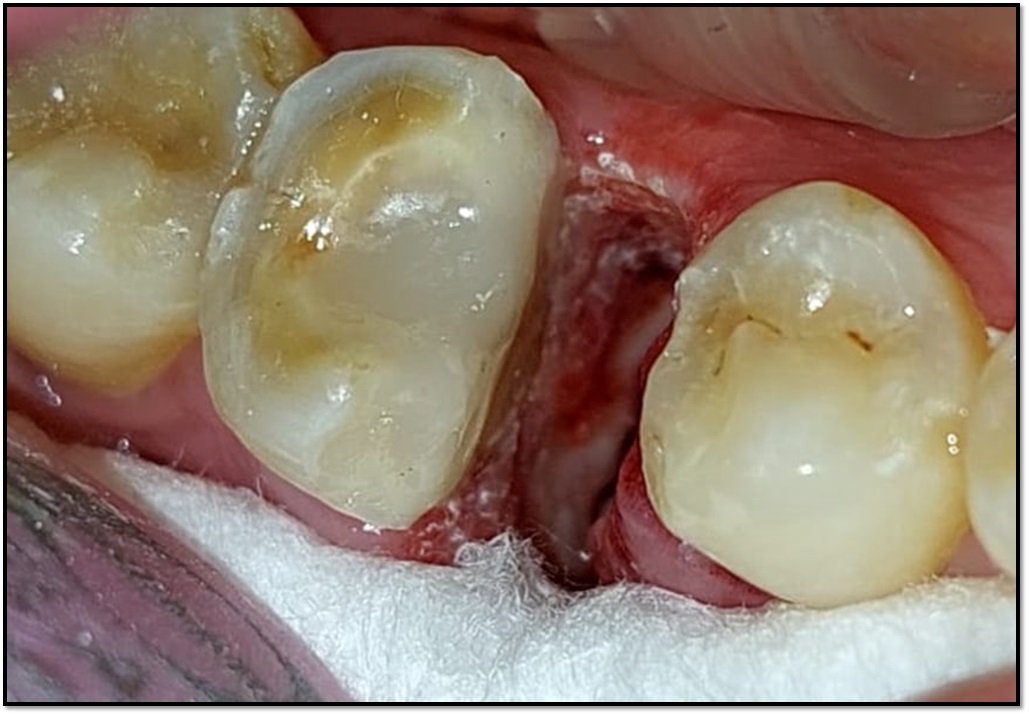
Fig. 3: Hemisection done with mesial root of mandibular first molar.
This was followed by approximation of buccal and lingual flaps which were sutured and dressed with COE pack. This was followed by patient recall after 3 months for prosthetic rehabilitation of hemisected tooth.
Prosthetic rehabilitation
During this phase diagnostic impressions of dentulous maxillary and mandibular arches were made and diagnostic casts were obtained which were then articulated to a semi adjustable articulator following a face bow record. Diagnostic casts were checked for premature occlusal contacts and interferences and were corrected.
This was followed by tooth preparation with 45 followed by distal portion of 46 to receive a porcelain fused to metal prosthesis (Fig 4). After adequate isolation, dual phase impression technique was used to make the impression of preparation. Temporization was done. The master cast was obtained. Wax pattern fabrication was done which was further invested and casted metal framework was obtained which was tried in patient’s mouth. Then the ceramic build up was done followed by cementation of the final prosthesis (Fig 5) on to the prepared teeth using luting glass ionomer cement (GC Fuji I). Post cementation instructions were given and followed by recall periodically was done to constantly assess the rehabilitation.
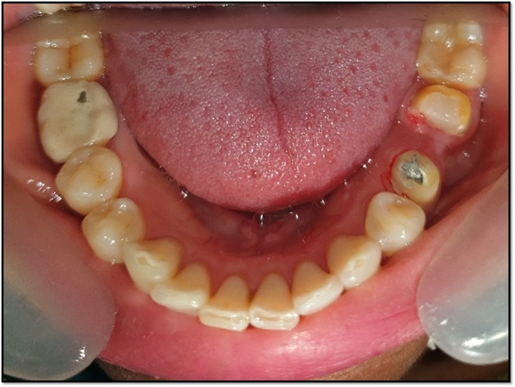
Fig. 4: Tooth preparation done with 45 and 46.
Fig. 5: Final prosthesis cemented.
DISCUSSION
A multirooted tooth can be effectively saved from extraction by hemisection. Endodontic treatment of remaining tooth structure is done followed by splinting the tooth with adjacent tooth and restoring it with a favourable prosthesis.5 Hemisection is a viable alternative to molar extraction in conditions like vertical root fracture of one root, severe vertical bone loss, dehiscence, prosthetic failures of abutments.6 According to Kost WJ et al (1991), hemisection is a conservative treatment option before extraction of any multirooted tooth.7
Singh M et al (2017) presented a case report of severe caries mandibular first molar resection, treated with endodontic treatment and proximal root restoration with a single piece implant with BCS (Cortical Bi-screw).8
Buragohain A et al. (2019) demonstrated management of partial tooth resection and prosthetic rehabilitation in thirty-six patients which followed a conservative approach to preserve maximum tooth structure.9
Therefore, in young patients’ conservative management of grossly carious multirooted teeth preserves the tooth and also the occlusal dysfunction.
CONCLUSION
A conservative approach of preserving a compromised molar is by hemisection and its functional rehabilitation. Success of hemisected tooth is determined by its periodontal support, planned restorative treatment and oral hygiene practices of the patient. Hemisection is an efficient conservative approach over extraction of endodontically or periodontally compromised tooth.
REFERENCES
- Seltzer S, Bender IB, Turkenkopf S. Factors affecting successful repair after root canal therapy. J Am Dent Assoc. 1963;67:651–662.
- Buhler HH. Survival rates of hemisected teeth: an attempt to compare them with the survival rates of alloplatic implant. Int J Periodon Res Dent 1994;14(6):536-543.
- Kost WJ, Stakiw JE. Root amputation and hemisection. J Can Dent Assoc. 1991;57:42–45.
- Weine FS. Endodontic Therapy, 5th Edition.
- Saad MN, Moreno J, Crawford C. Hemisection as an alternative treatment for decayed multirooted terminal abutment: A case report. J Can Dent Assoc 2009;75:387-90.
- Shah S, Modi B, Desai K, Duseja S. Hemisection – A conservative approach for a periodontally compromised tooth – A case report. J Adv Oral Res 2012;3:31-5.
- Kost WJ, Stakiw JE. Root amputation and hemisection. J Can Dent Assoc. 1991;57:42–45.
- Singh M, Batra R, Das D, Verma S, Goel M. A novel approach for restoration of hemisected mandibular first molar with immediately loaded single piece BCS implant: A case report. J Oral Biol Craniofac Res. 2017 May-Aug;7(2):141-146.
- Buragohain A, Nagpal A, Bhatia V, Kaur H. Functional rehabilitation of hemisected mandibular molar: A case report. J Dent Specialities 2019;7(1):38-41.