AESTHETIC REHABILITATION IN MAXILLARY ANTERIORREGION: A Case Report
Dr. Tanya Singh (P.G. Student), Dr. Shubhangi Srivastava (P.G. Student), Dr. Manesh Lahori (Principal & HOD), Department of Prosthodontics and Crown & Bridge, K.D. Dental College & Hospital, Mathura, UP, India.
ABSTRACT
Aesthetic rehabilitation is a specialty of dentistry dedicated to the restoration and maintenance of oral functions in terms of chewing, phonetics and aesthetics, through the repair of damaged teeth and the placement of fixed, partial or total dental prostheses. Aesthetic rehabilitation has been glorified with the advent of all ceramic restorations. In order to get increasingly beautiful, harmonic and natural results, the search for development of rehabilitation, indirect materials is constant, such as ceramic crowns (metal free).This article presents variety of cases for Maxillary anterior rehabilitation using all ceramic restorations.
INTRODUCTION
The aesthetic enhancement is one of the most frequent reasons for patients to seek prosthetic rehabilitation of the smile, especially when discrepancies are found in maxilloanterior region.1,2
The required harmony and consistency are accomplished when there is a proper link between dental and gingival components, as well as those with individual facial patterns. Thus, characteristics such as: tooth shape and size, interdental proportion, gingival contour, presence of interdental papillae, among others, are essential elements to be evaluated during an aesthetic analysis.3
In the last 30 years, dentistry has undergone a revolution, not only in terms of the introduction of new materials and techniques, but also in terms of the scientific data backing their clinical applications. As ceramic materials for dentistry improve and patient demand for aesthetic restorations grows, practitioners must stay up with both research and patient demand.4 Proper practitioner assistance is essential in selecting the optimal system for crowns, as well as understanding of the optical qualities of different ceramic systems, which will allow the clinician to make suitable choices when faced with varied aesthetic issues.
Recent advancements in the strengthening of dental ceramics have resulted in the creation of novel ceramic restorative systems that use porcelain directly on an opaque ceramic foundation rather than a metal framework. The various new ceramic restorative systems generally can withstand relatively high compressive forces and offer a range of flexural strengths.5-7
Full ceramic crowns with opaque cores are superior in strength and aesthetics, and can be used for both posterior and anterior teeth with significant discolouration. For fixed partial dentures, crowns with a zirconia core are advised. Because the colour of the cement has no effect on the shade of the crown, resin or conventional luting agent can be used for cementation. When rebuilding anterior teeth with these crowns, it is best to finish the margin subgingivally to avoid a shade mismatch between the tooth margin and the restoration. The strength of these restorations is dependent on the ceramic material used, the core- veneer bond strength, the crown thickness, and the design of restoration.8
This article presents case series of maxillary anterior region rehabilitation.
CASE REPORT 1
A 25 yr old man reported to the Department of Prosthodontics, crown and bridge, K. D. Dental Colege, for dental aesthetics treatment with the chief complaint of spacing in the maxillary anterior region.[Figure 1]
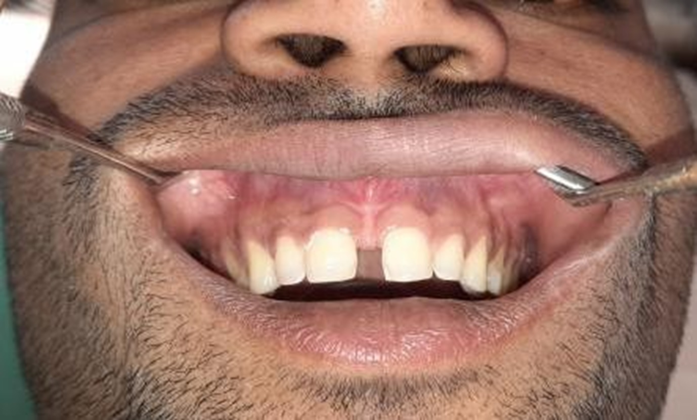
FIGURE-1
On clinical examination, 4mm of space was found between the maxillary central incisors and 2mm of space between lateral incisor and canine of both sides of maxillary arch. Patient was offered the treatment option of Zirconia crowns on 11.12. 21.22.
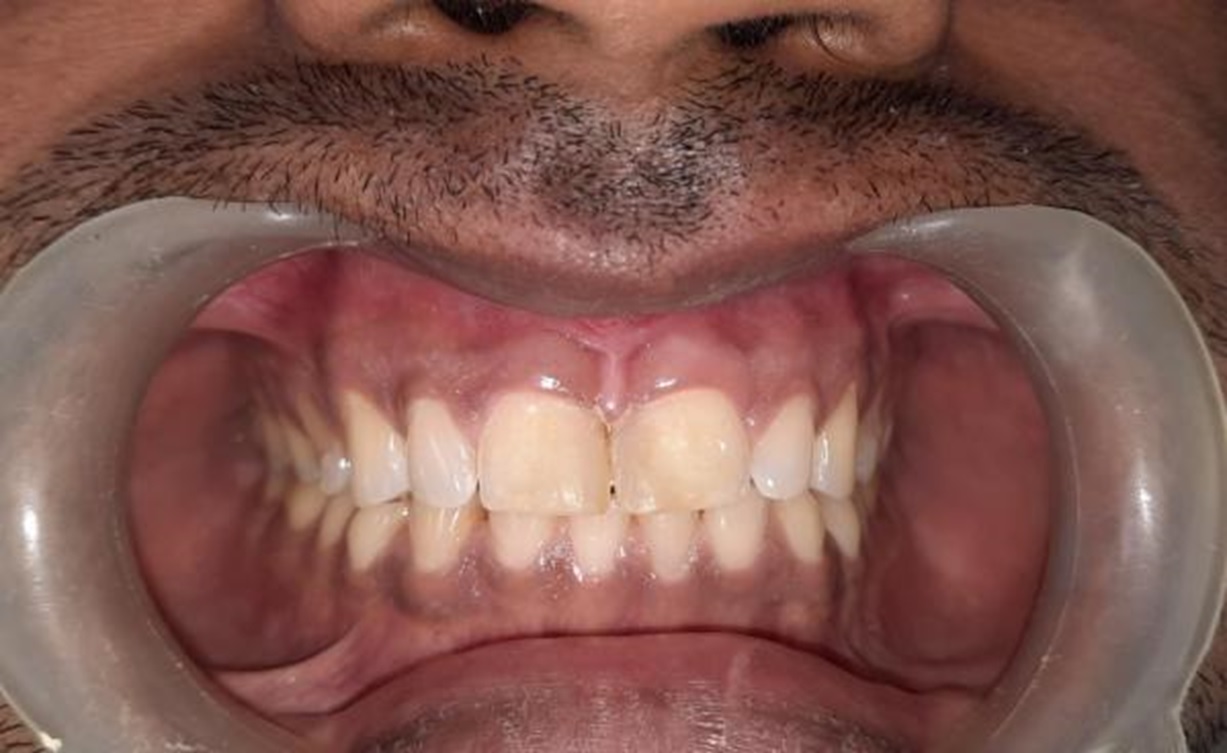
FIGURE-2
Gingival health is of prime importance before beginning with any aesthetic procedure. Hence, oral prophylaxis was carried out before the inception of the prosthodontic treatment.
In order to address the patient’s chief esthetic concerns, the plan included the following elements: Development of a simulation or mock- up to evaluate proper tooth morphology and tooth length for better esthetics and proper gingival contours [Figure 2]. This was presented to the patient to assist in determining the course of treatment.It was achieved using composite restorative material. After being satisfied with the mock-up, the patient authorised the final treatment plan.
After 1 week, the patient was recalled for the treatment and composite resin was removed. Teeth preparation was done in relation to 11,12, 21 and 22 for all ceramic restoration [Figure 3].
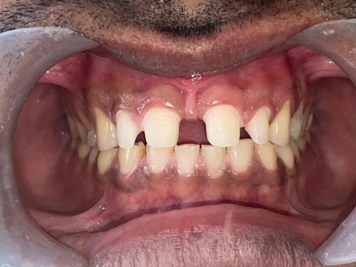
FIGURE-3
For the impression, gingival retraction was achieved using 3M ESPE gingival retraction paste, followed by making impressions using vinyl poly siloxane impression material of putty and light body consistency using double mix impression technique for upper arch.Impression was washed and inspected for any voids, followed by disinfection with 2% glutardlehyde solution. Shade selection was done with VITA classical shade guide.The final impression, mock-up, along with pictures of aesthetic pre-evaluative intra oral mock up done with composite resin was sent to the laboratory for the crowns fabrication.
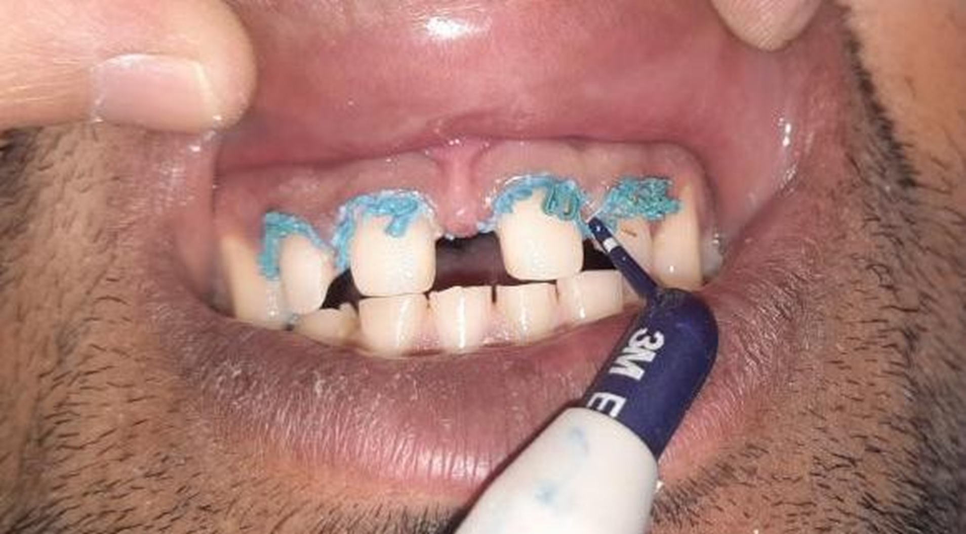
FIGURE-4
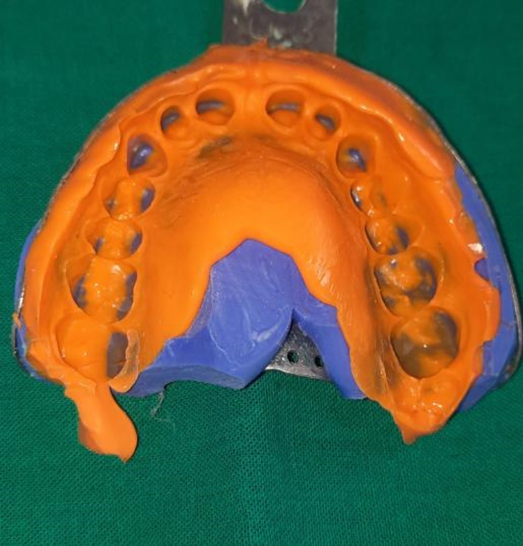
FIGURE-5
Provisional restorations were fabricated with a tooth colored auto polymerizing acrylic resin and cemented with noneugenol temporary cement.
After one week, temporary crowns were removed and the fit of all- ceramic restorations was evaluated intraorally alongwith occlusion assessment. The crowns were cemented with Glass Ionomer cement(GC) luting consistency. Patient was satisfied with the crown length, width and esthetics which was improved and equal to adjacent natural teeth [Figure 7].
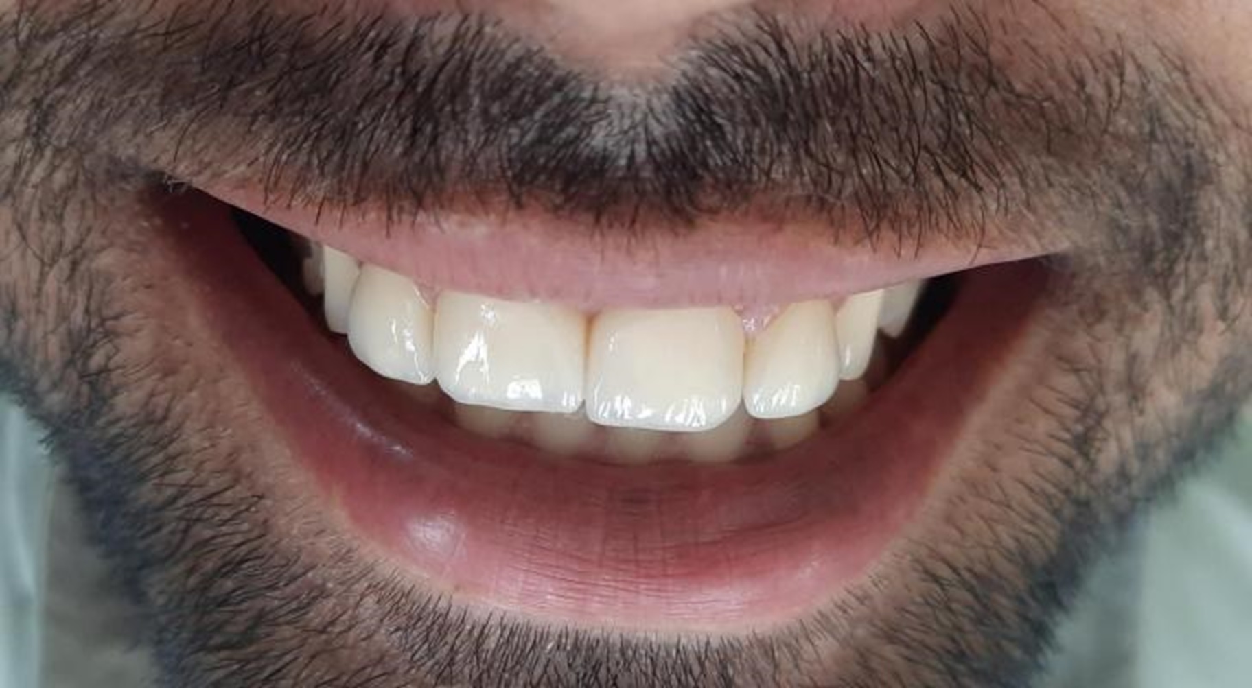
FIGURE-6
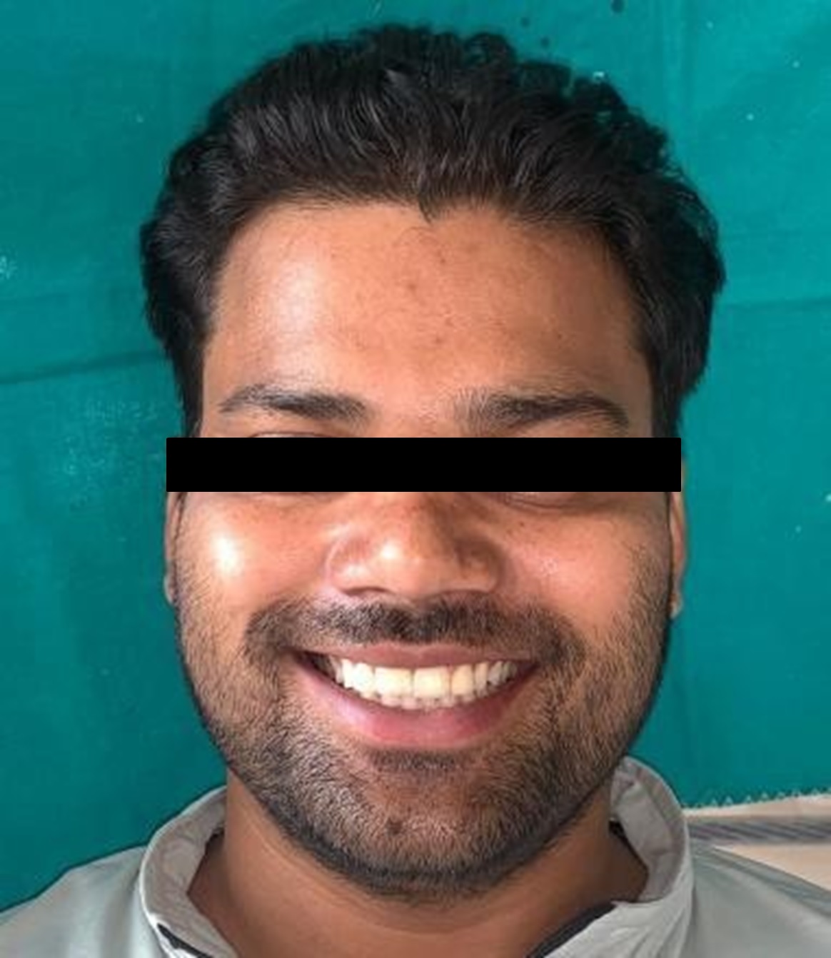
FIGURE-7
CASE REPORT 2
A 24 yr old man reported to the Department of Prosthodontics, crown and bridge, K.D.Dental College,for dental aesthetics treatment with the chief complaint of missing teeth in the maxillary anterior region.[Figure 8]
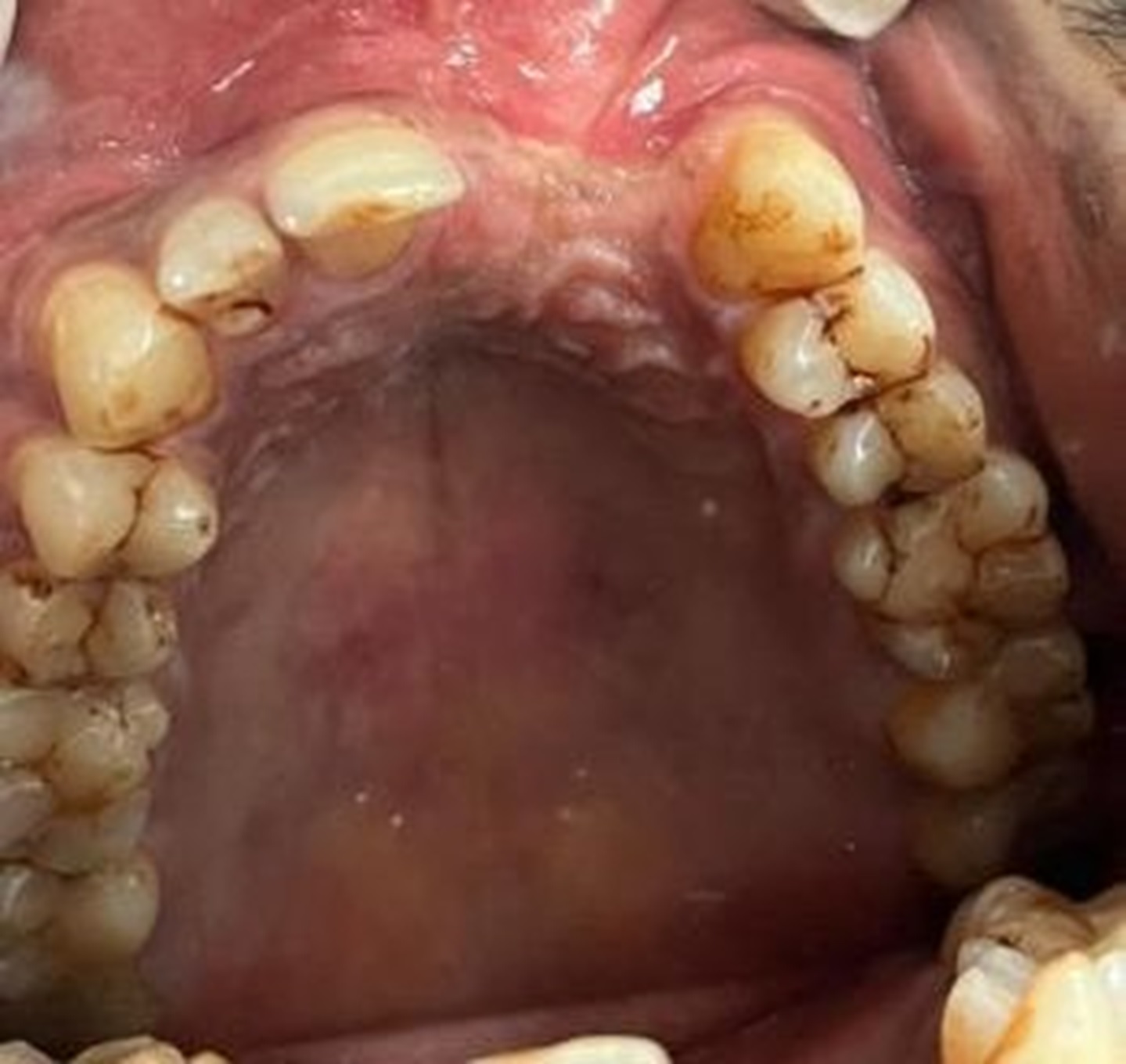
FIGURE-8
On clinical examination,the teeth missing were 21, and 22, and the patient presented with Siebert’s II ridge defect(Maxillary). The teeth were extracted 4 years back alongwith cleft lip surgery post accident.Patient was educated about all the treatment options available and approved for the treatment option of Fixed dental prosthesis(Zirconia) alongwith pink porcelain.
Oral prophylaxis was carried out first.Teeth preparation was done in relation to 11, 12 and 23 for all ceramic restoration [Figure 9].
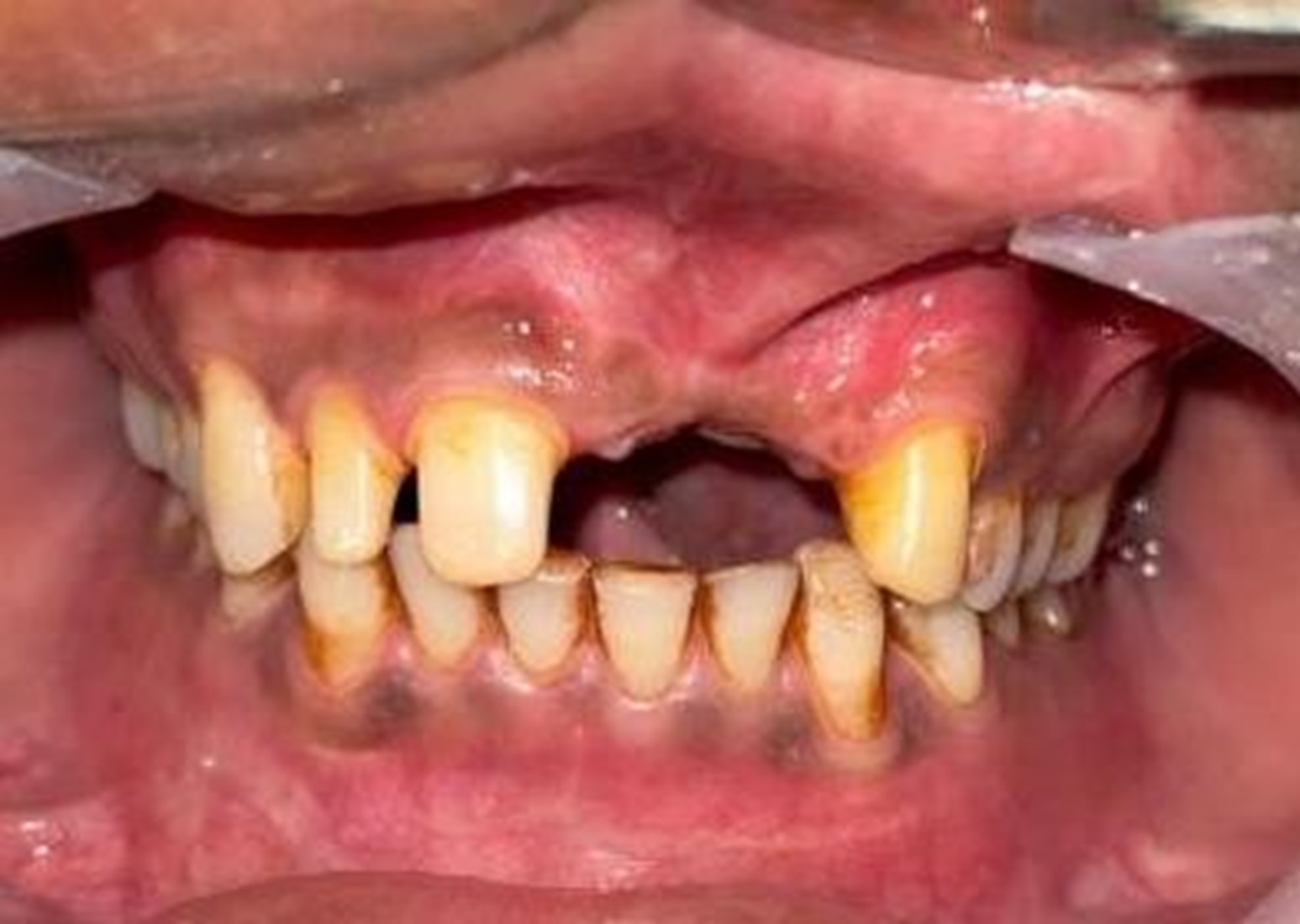
FIGURE-9
Final impression was made using vinyl poly siloxane impression material of putty and light body consistency using double mix impression technique for upper arch.Impression was washed and inspected for any voids, followed by disinfection with 2% glutarlehyde solution. Shade selection was done with VITA classical shade guide.
Provisional restorations were fabricated with a tooth colored auto polymerizing acrylic resin and cemented with noneugenol temporary cement [Figure 10].
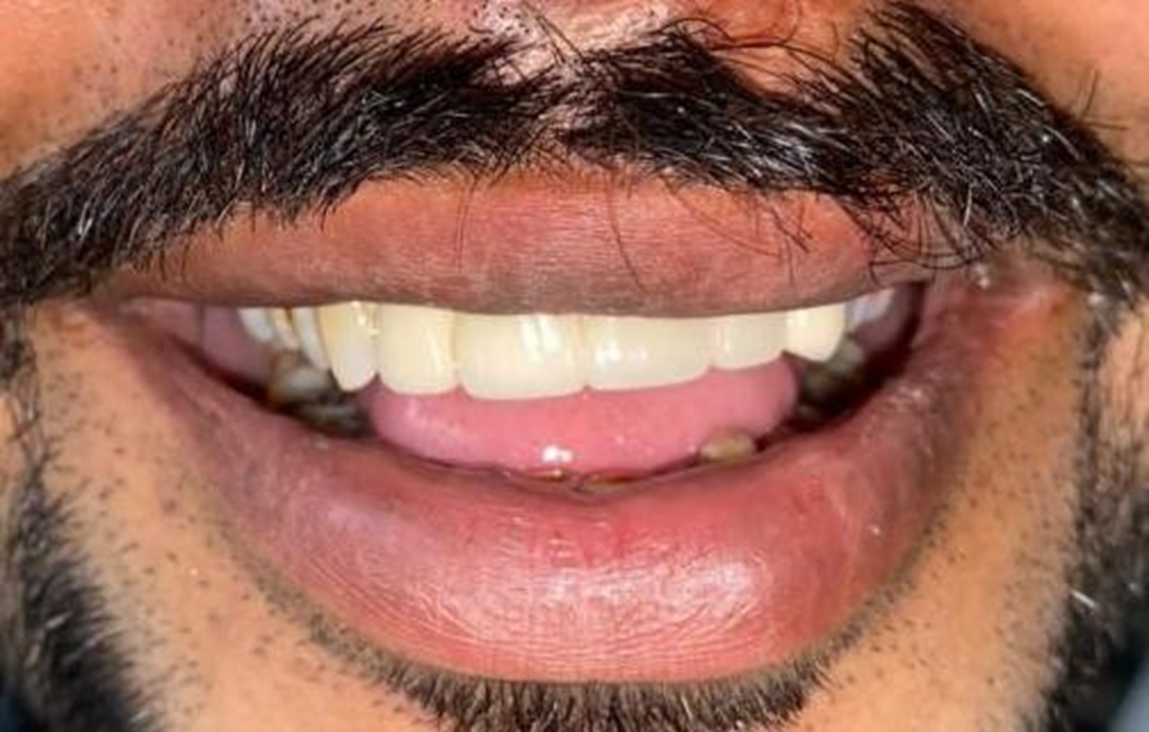
FIGURE-10
After one week, temporary crowns were removed and the fit of all- ceramic FDP was evaluated intraorally alongwith occlusion assessment. The crowns were cemented with Glass Ionomer cement GC luting consistency. Patient was satisfied with the smile and esthetics. [Figure 11, 12]
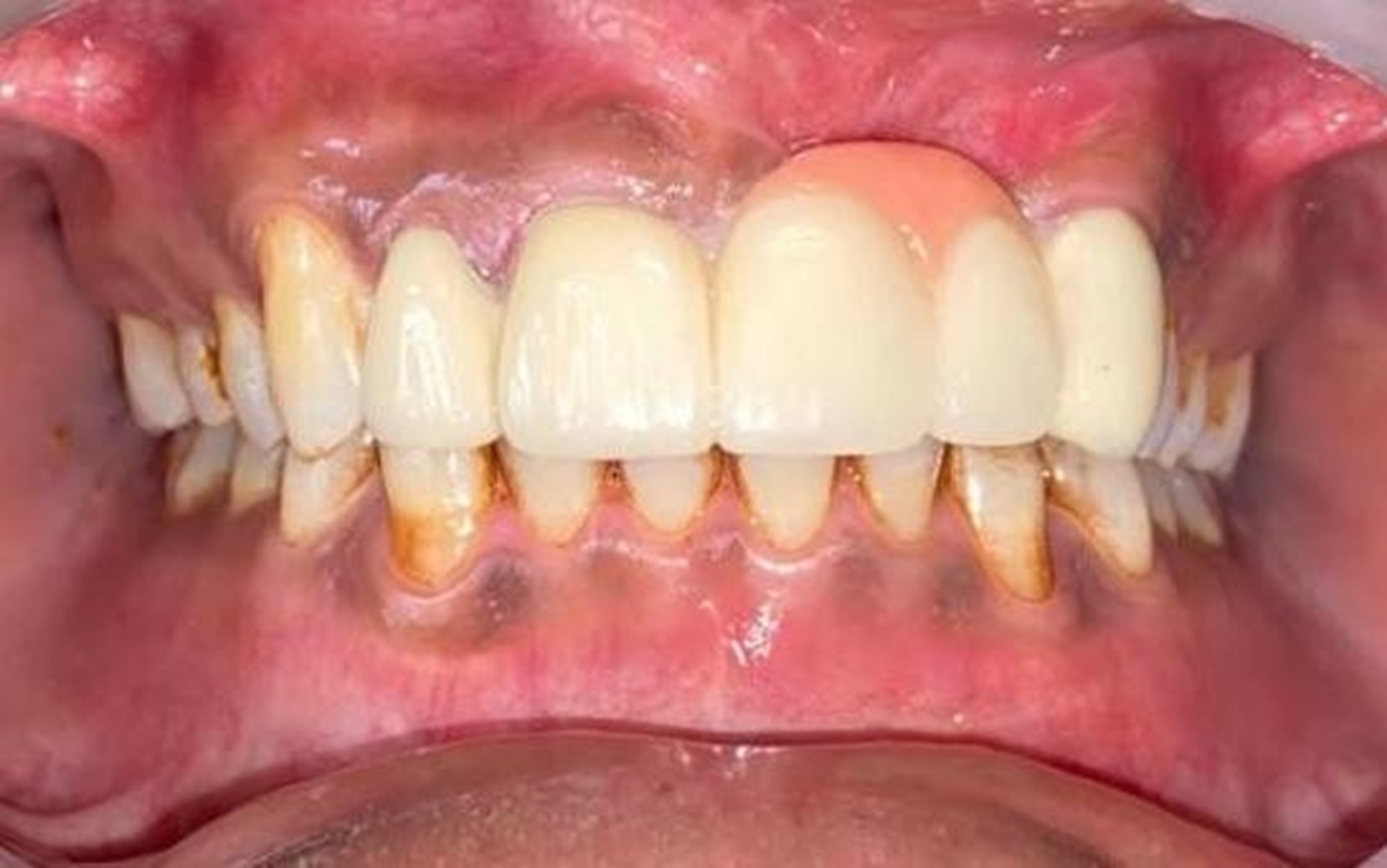
FIGURE-11
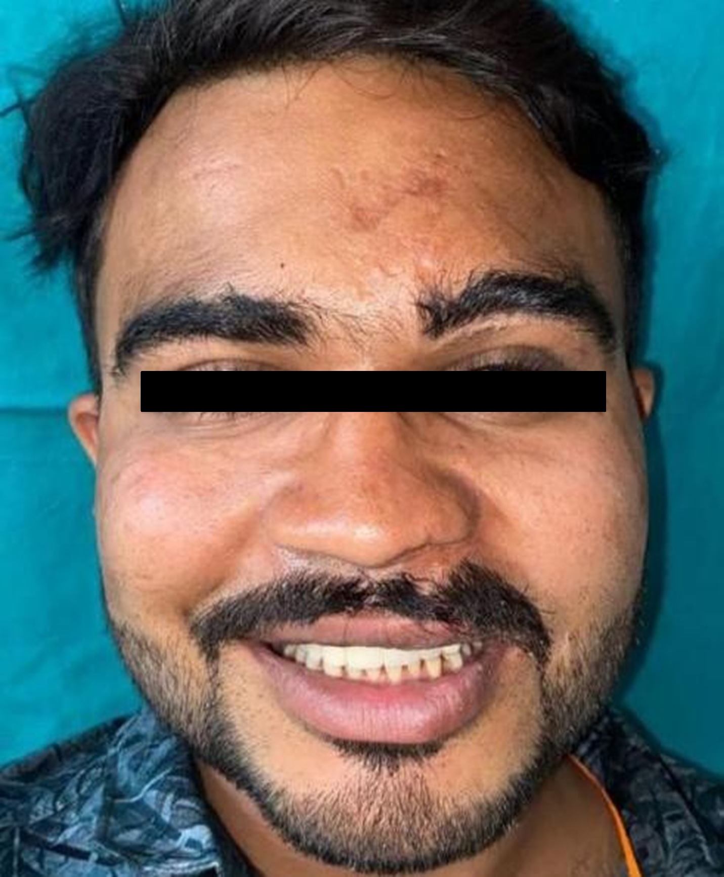
FIGURE-12
CASE REPORT 3
A 38 yr old woman reported to the Department of Prosthodontics, crown and bridge, K.D.Dental College, for dental aesthetics treatment with the chief complaint of missing teeth in the maxillary anterior region. On clinical examination, both maxillary central incisors were found to be missing and all ceramic FDP was planned from 12 to 22.
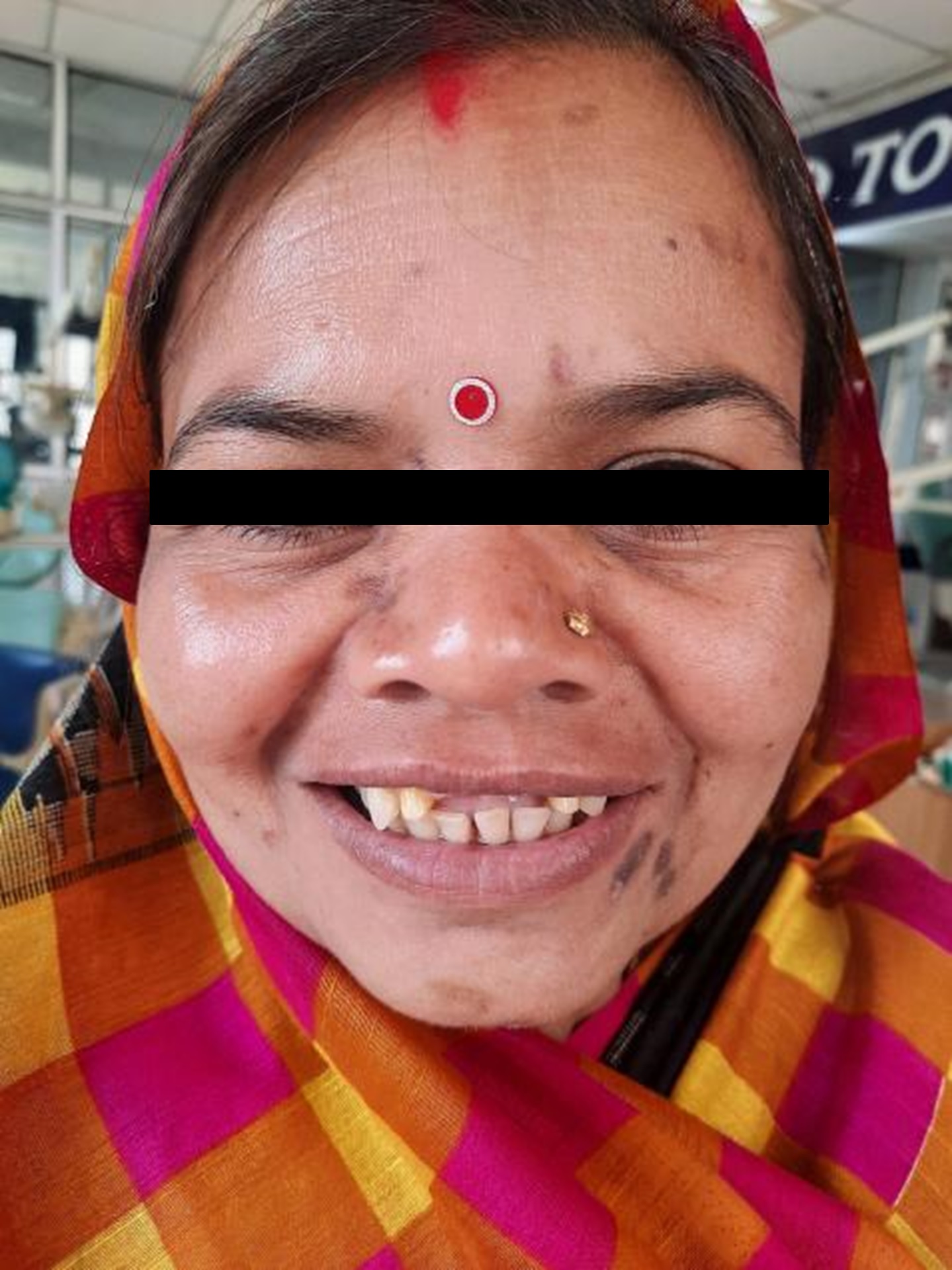
FIGURE-13
Oral prophylaxis was carried out before the inception of the prosthodontic treatment.
Tooth preparation was done in relation to12 and 22 for all ceramic restoration [Figure 14]. Final impression was made using vinyl poly siloxane impression material of putty and light body consistency using double mix impression technique for upper arch. Shade selection was done with VITA classical shade guide.
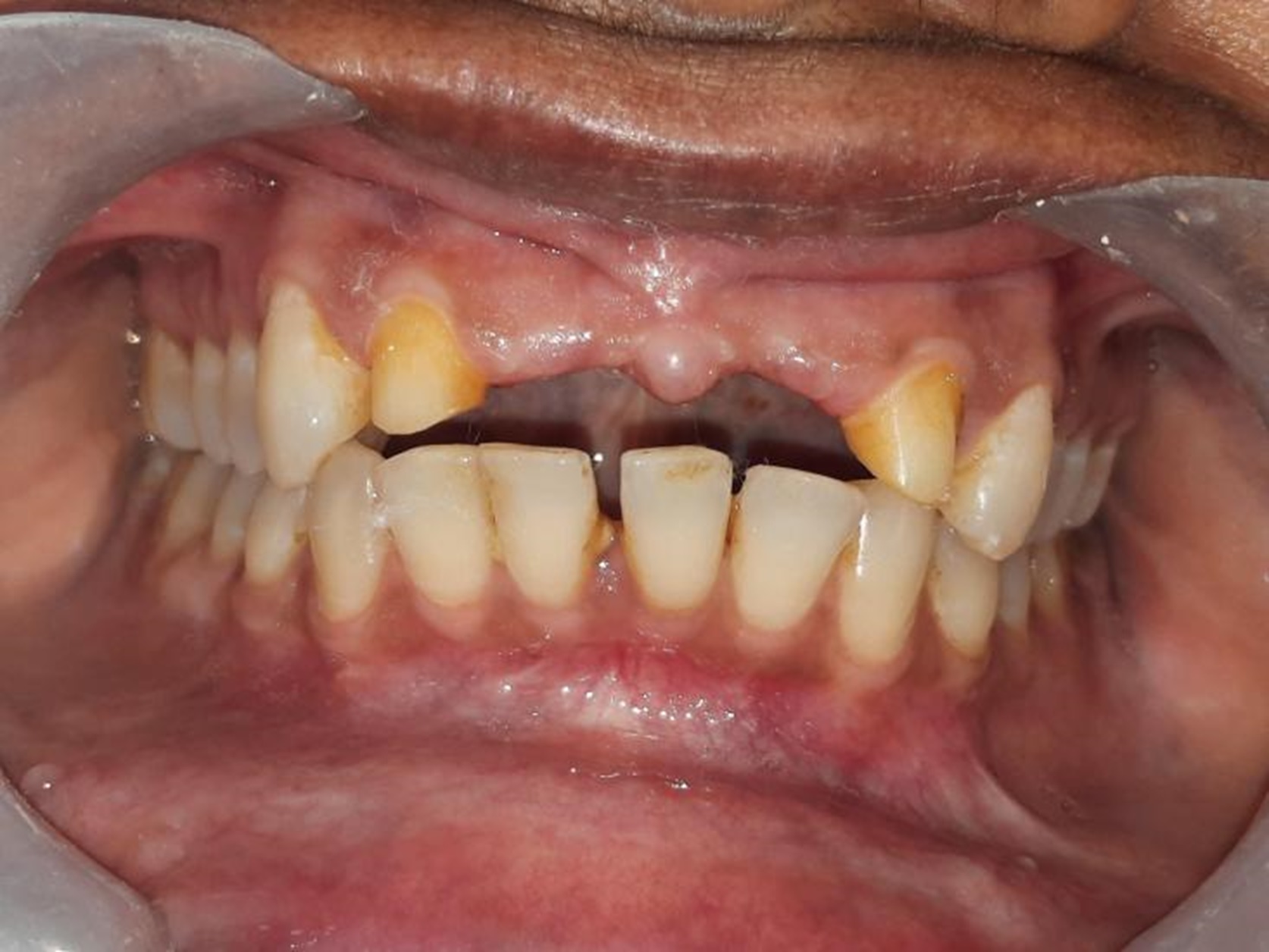
FIGURE-14
Provisional restorations were fabricated with a tooth colored auto polymerizing acrylic resin and cemented with noneugenol temporary cement.
After one week, temporary restoration was removed and the fit of all- ceramic restorations was evaluated. The crowns were cemented with Glass Ionomer cement
(GC)luting consistency. Patient was satisfied with the aesthetic smile [Figure 15,16].
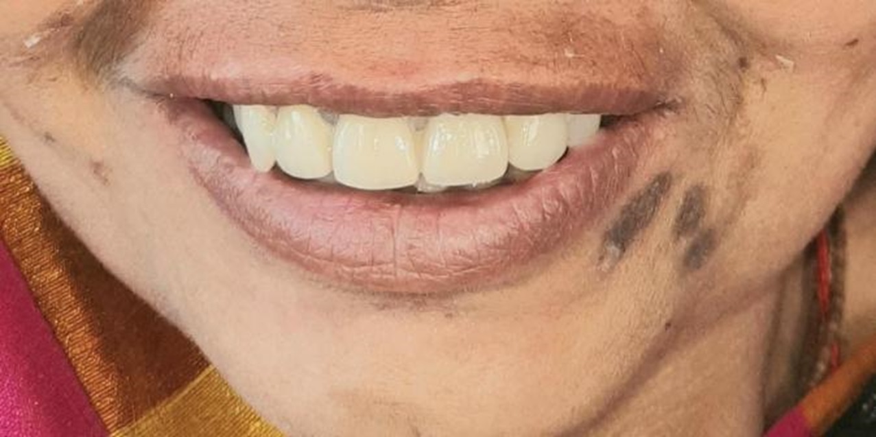
FIGURE-15
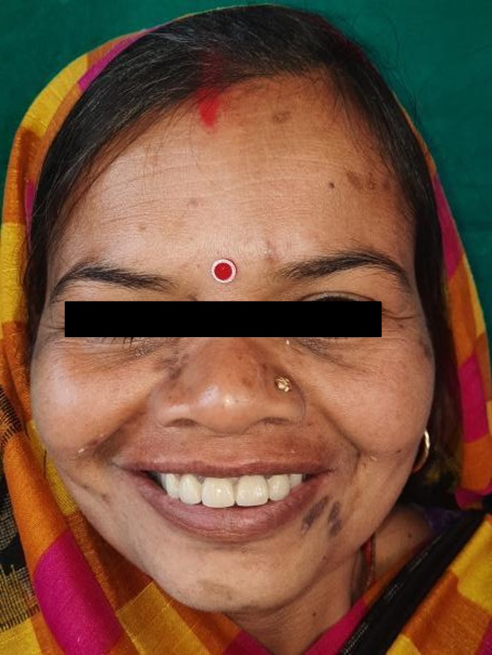
FIGURE-16
DISCUSSION
A good face is a letter of recommendation.For many years, it has been assumed that the initial impression a person creates is due to his appearance, which lasts a long time. The media's projected flawless appearance has a huge influence on our beauty- conscious society's behaviour and thoughts .The dental look is an important aspect of face beauty. Dental appearance can influence an individual's judgements about the personal attributes of others.
This article presents three different cases for aesthetic rehabilitaion in maxillary anterior region.
Diastema is a term that simply describes a gap between teeth. It can be the result of a wide range of causes from hereditary reasons to tooth damage. Regardless of the cause, there are various treatment options available for those who suffer from diastema which include: Dental Veneers, Dental Bonding, Braces and Aligners and Dental crowns. The modality opted here for diastema closure was Dental crowns (All ceramic).
In the second case described here,the patient presents with ridge defect alongwith missing anterior teeth in maxillary anterior region. Hence, we have used pink porcelain to mask the defect coupled with All-ceramic FDP. From an aesthetic point of view, the dentogingival prosthesis is an important treatment option in cases of previous bone defects associated with tooth loss.
The third case presents maxillary anterior region rehabilitation with All-ceramic FDP.
CONCLUSION
Happiness is a mental state. It is brought about by a sense of wellbeing, security, and self-confidence. The ability of the dentist to replace missing teeth, both in contour and colour, especially for the anterior teeth, is critical for the formation of a good oral and facial expression for the patient.
REFERENCES
- Hasanreisoglu U, Berksun S, Aras K, Arslan I. An analysis of maxillary anterior teeth: Facial and dental proportions. J Prosthet Dent 2005; 94:530-38. https://doi.org/10.1016/j.prosdent.2005.10.007
- Seibert JS. Reconstruction of deformed, partially edentulous ridges, using full thickness onlay grafts. Part I. Technique and wound healing. Compend Contin Educ Dent 1983;4:43753.
- Rotundo R, Nieri M, Bonaccini D, Mori M, Lamberti E, Massironi D, Giachetti L, Franchi L, Venezia P, Cavalcanti R, Bondi E, Farneti M, Pinchi V, Buti J. The Smile Esthetic Index (SEI): A method to measure the esthetics of the smile. An intra-rater and inter-rater agreement study. Eur J Oral Implantol 2015;8(4):397-403
- Spear F, Holloway J. Which all ceramic system is optimal for anterior esthetics? J Am Dent Assoc 2008;139:19- 2
- Cho GC, Donovan TE, Chee WW. Rational use of contemporary all- ceramic crown systems. J Calif Dent Assoc 1998;26:113- 20.
- Lee S. All- ceramic crowns. Dent Asia 2001;2:20- 3.
- van Dijken JW. All- ceramic restorations: Classification and clinical evaluations. Compend Contin Educ Dent 1999;20:1115- 24, 6.
- Mizrahi B. The anterior all- ceramic crown: A rationale for the choice of ceramic and cement. Br Dent J 2008;205:251- 5.