Prosthodontic Rehabilitation in Ectodermal Dysplasia
Department of Prosthodontics Dr. Ziauddin Ahmad Dental College Amu, Aligarh, India
Abstract
Ectodermal dysplasia (ED) is a rare, inherited disorder, the manifestation of which could be seen in more than one ectodermal derivative. The most common syndrome within this group are hypohydrotic (anhydrotic) and hidrotic ED. The tissues which are primarily affected are skin, hair, nails eccrine glands and teeth. Oral manifestations of ectodermal dysplasia are anodontia or oligodontia, complete or partial absence of teeth with frequent malformation of any teeth present, both deciduous and permanent dentitions. Teeth are commonly truncated or cone shaped. Several ectodermal dysplasia syndromes may manifest in association with midfacial defects, mainly cleft lip and palate.No treatment for the condition is available. However, affected individuals with dental defects could be subjected to early dental evaluations and intervention beginning withdenture as early as two years.This paper presents a case report of a 09 year old patient diagnosed with ectodermal dysplasia and with clinical and radiographic evaluation it was found that the patient had only four maxillary teeth. A maxillary removable partial denture and a complete mandibular denture were fabricated for prosthetic rehabilitation after considering her growth and condition of her present teeth. No major complications occurred during the 3 month follow up and the patient was fully satisfied.
Key words: Eccrine glands, anodontia, oligodontia
INTRODUCTION
Ectodermal dysplasias are a heterogeneous group of disorders characterized by developmental dystrophies of ectodermal structures. It is an X linked recessive disorder. It is characterized by the triad of signs which comprises of sparse hair (atrichosis or hypotrichosis), abnormal or missing teeth (anodontia or hypodontia)and inability to sweat due to lack of sweat glands (anhidrosis orhypohidrosis). The etiology of ectodermal dysplasia appears to be genetic in nature[1]
A definitive classification is complicated because of the overlapping features of the syndrome .
Lamartine in 2003 has described various well defined ectodermal dysplasia as Hypohidrotic (anhidrotic), Hidrotic (Clouston’syndrome), Ectrodactyly-ectodermal dysplasia-cleft syndrome (EEC), Rapp- Hodgkin Syndrome, Hay-Wells syndrome or ankyloblepharon ectodermal dysplasia.Usually the ectodermal dysplasia is divided into two types based on the number and function of sweat glands. (Viera et al, 2007) as mentioned below[2]:
A. Hypohidrotic (anhidrotic) Ectodermal Dysplasia (Christ-Siemens- Tourine Syndrome)
In this form sweat glands are absent or significantly decreased. This disorder is usually inherited as either autosomal dominant / recessive or X-linked recessive trait and the gene locus is X q13- q21. It is commonly X-linked recessive with full expression in males. Female carriers have a minimal expression. 60- 70% of cases usually show manifestations restricted to minimal hypodontia, aplastic or hypoplastic mammary glands, impaired lacrimal gland function,
glaucoma and increased susceptibility to allergic disorders such as asthma or eczema. Typical general mental development, frontal bossing with characteristic reduction in amount of hair (hypotrichosis), absence of sweat glands (anhidrosis) resulting in temperature elevation, absence of sebaceous glands (asteatosis) resulting indry skin, depressed nasal bridge, protuberant lips, prominent supra orbital ridges , sunken cheeks, wrinkled hyperpigmented skin around the eyes and large low set ears (Crawford et al ,1991).
The oral manifestations include conical or peg shaped teeth, hypodontia (partial absence of teeth) or complete anodontia (complete absence of teeth) of both the deciduous and the permanent dentition with malformation of any teeth that may bepresent, generalized spacing, underdeveloped alveolar ridges and delayed eruption of permanent teeth but the growth of the jaw is never impaired. This would imply that the development of the jaws except for the alveolar process is not dependent upon the presence of teeth. However, since the alveolar process does not develop in the absence of teeth, there is a reduction in the normal vertical dimension resulting in the protuberant lips. In addition, in oropharynx the defect may be manifested as a high palatal arch or even a cleft palate. The salivary glands including the intraoral accessory glands are sometimes hypoplastic in this disease. This result in xerostomia, and the protuberant lips may be dry and cracked.
B. Hidrotic Ectodermal Dysplasia (Clouston Syndrome)
Here the clinical features include nail dystrophy, hair defects and palmoplantar dyskeratoris. The patients have normal facies, normal sweating and no specific defect is seen.

CASE REPORT
A 09 year old female patient reported to the department of pedodontics with the complaints of absence of teeth. The history from the patient also reveals the complete absence of primary dentition.
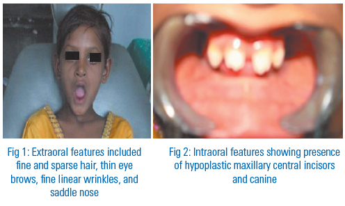
On general examination the patient exhibited typical characteristics of ectodermal dysplasia; Extraoral features included fine and sparse hair, thin eye brows, fine linear wrinkles, and saddle nose. (Fig.1)
Intraoral features included absence of several teeth and hypoplastic maxillary central incisors and canine. These were the only teeth present in the patient’s mouth (fig. 2). Also loss of vertical dimension on occlusion, and underdeveloped alveolar ridges were detected on oral examination.
It was found that there was absence of sweating with increased body temperature along with increased frequency of stools especially after meals. These complaints may be related to the absence of teeth and indigestion of food. Patient also reported the history of chronic pharyngitis and associated problems of airways which were probably due to abnormal mucosal thickening (presented in ectodermal dysplasia).
In the clinical and radiographic evaluation, it was confirmed that she had only maxillary central incisors and canine in her mouth and no other succadaneous teeth were present. (Fig. 3)
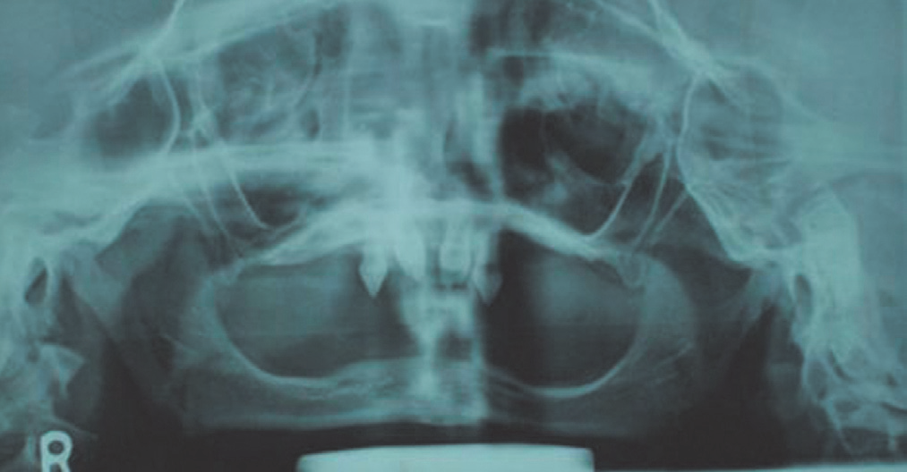
Fig 3: OPG confirming the absence of teeth other than maxillary central incisors and canine
MANAGEMENT
A maxillary removable partial denture and a complete mandibular denture were planned for prosthodontic rehabilitation after considering her growth and the number, and condition of her present teeth.
Impressions for the maxillary removable partial denture and mandibular complete denture were made with custom trays and medium viscosity additional silicone impression material.
A bilateral balanced occlusion was developed using 33 degree anatomic acrylic resin teeth. The maxillary removable partial denture and the mandibular complete denture were processed with flexible denture base acrylic resin. (Fig. 4)
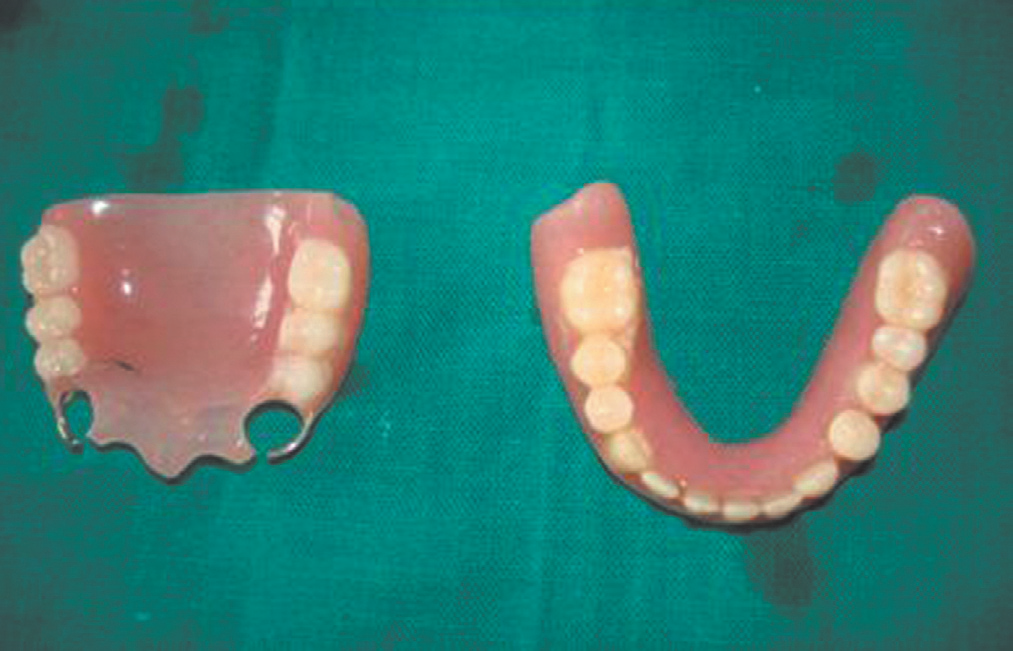
Fig 4: maxillary and mandibular removable prosthesis given to the patient
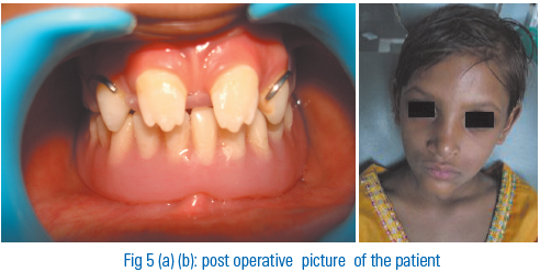
The patient was examined after 48 hrs for post insertion adjustments, and then followed on a monthly basis till 3 months. She had no notable complains about the dentures, good esthetic and functional results were achieved.
Follow up pictures: fig. 5 (a) and (b)
DISCUSSION
The Ectodermal dysplasias are a group of inherited disorder that share in common developmental defects involving at least two ofthe major structure classically hold to derive from the embryogenic ectoderms hair, teeth, nails, sweat glands[4]. The incidence of ectodermal dysplasia in males is estimated at 1 in 100,000 births[5] .Thurman first described this entity in 1848[6].
Dental defects represent a core clinical feature of many EDs: anodontia, polydontia, dysplastic teeth, retained primary teeth, deficient enamel development (amelogenesis imperfecta), dentine
deficiency (dentinogenesis imperfecta), and underdevelopment of the alveolar ridge[7].
Prenatal diagnosis of ectodermal dysplasia has occasionally been reported which has been diagnosed by foetal skin biopsy, obtained by fetoscopy by 20 weeks gestation[8].The characteristic facies is pathognomonic but may not be recognized in infancy. In partial forms, the pointed conical teeth provide the most valuable indication and should suggest the need for sweat test and a skin biopsy[9]. The disorder was assigned to chromosome 11q23 by linkage mapping[10].
Dental treatment is often necessary in patients with some forms of ED and some children may need dentures as early as 2 years of age [11]. It is important to seek dental advice early as maintenance of the alveolar ridge is important for later dental intervention[12,13,14].Prosthetic teeth are implanted in adults for mastication and speech. Importantly, aesthetic dental interventions in patients with ED and malformed teeth and malocclusion helps with the development of positive self image and over all oral health[15].
Prosthetic rehabilitation of patients with ectodermal dysplasia is an ordinary process. Early and extensive dental treatment is needed throughout childhood because of the absence of most of the deciduous and permanent teeth. A multi- disciplinary team approach to management of these patients is recommended. Osseo-integrated implants should be an alternative treatment in older patients with ectodermal dysplasia.
Considering the poor economical situation of the present case, it was deemed better to postpone Osseo-integrated implants.
This is commonly a difficult condition to manage the patients with prosthodontics because of the typical oral deficiencies and because the afflicted individuals are quite young when they are evaluatedfor treatment. It is important that these individuals receive dental treatment at an early age for physiologic and psychosocial reasons. Adaptation to the prosthesis depends on various factors as capabilityof the specialist, age of the patient, and cooperation in a growing child. A removable partial denture or an overdenture is often suitable treatment of choice, because of the need to easily modify the intra oral prosthesis during rapid growth periods. Also these treatment options are easy, affordable, and reversible rehabilitation methods.
The treatment goals for this patients were establish a functional occlusion with prosthetic rehabilitation and, to obtain an esthetic smile with adult sized teeth. Nutrition of the patient is improved and self confident appearance is provided.
REFERENCES
1. Mortier K, Wackens G. Ectodermal Dysplasia anhidrotic. Orphanet Encyclopedia. 2004:1-6..
Disclaimer from Guident
The views and opinions expressed in articles published in Guident are solely those of the authors and do not necessarily reflect the views of the editors, the editorial board or the publisher, Ivory India. Guident does not endorse any product, technique or treatment described. Clinical content is intended for qualified dental and medical professionals and is not a substitute for individual clinical judgement. Guident, its editors and the publisher accept no liability for any consequence arising from the use of information published in the journal.
How to cite this article
Dr. Geeta Rajput, Dr. Vasundhara Bhatt, Dr. Himangi Srivastava. Prosthodontic Rehabilitation in Ectodermal Dysplasia. Guident. 2013 Jun;6(8). Available from: https://guident.in/articles/prosthodontics/prosthodontic-rehabilitation-in-ectodermal-dysplasia.html