Key Factors in Osseointegrated Supported Prosthesis
A.B.Shetty Memorial Institute of Dental Sciences, Nitte University Deralakatte, Mangalore, India
Abstract
Osseointegrated supported prosthesis has become a scientifically accepted and predictable treatment for completely and partial edentulous patients. The osseointegration is a complex process that can be influenced by many factors such as loading conditions, surgical techniques, as well as restorative conditions, which will play an important role in osseointegration. Diagnostic imaging plays an important role in evaluating the dental implant patient. Planning and executing optimal occlusion schemes is an integral part of osseointegrated supported prosthesis. This article makes an attempt to identify the important key factors for the success of osseointegrated supported prosthesis.
Key words: Osseointegrated supported prosthesis, Key factors, Implants
INTRODUCTION
The goal of modern advanced dentistry is to restore the patients to normal contour, function, comfort, esthetics speech and health, regardless of the atrophy, disease or injury of the stomatognathic system. Since the introduction of the concept of osseointegration, the success of dental implants has increased dramatically because of better understanding of bone response and the improvement in bone loading concepts. However, in spite of high success rate, occurrence of implants failure even though rare has been reported.
The treatment plan should begin with a clear idea of the desired end result of treatment, which should fulfill the functional and esthetic requirements of the patient. Evaluation begins with a patient consultation and assessment of the esthetic and functional requirements, and proceeds to more detailed planning with intraoral examination, diagnostic mock up and appropriate radiographic examination. Successfully achieving osseointegrated supported prosthesis involves detailed pre surgical planning, optimal three-dimensional implant placement, meticulous soft tissue management, the use of bone grafting techniques when required, and use of various prosthetic components.
It is important to establish success criteria for implant systems, and for implants to be tested
in well controlled clinical trials. The minimum success criteria proposed by Albrektsson et al.,1 are as follows:
MINIMUM SUCCESS CRITERIA FOR DENTAL IMPLANTS
1. An individual, unattached implant is immobile when tested clinically
2. Radiographic examination does not reveal any periimplant radiolucency
3. After the first year in function, radiographic vertical bone loss is less than 0.2 mm per annum
4. The individual implant performance is characterized by an absence of signs and symptoms such as pain, infections, neuropathies, paresthesia, or violation of the inferior dental canal.
5. As a minimum, the implant should fulfill the above criteria with a success rate of 85% at the end of a 5 year observation period and 80% at the end of a 10 year period.
Key Factors may be identified under the following headings as related to Osseointegrated supported prosthesis:
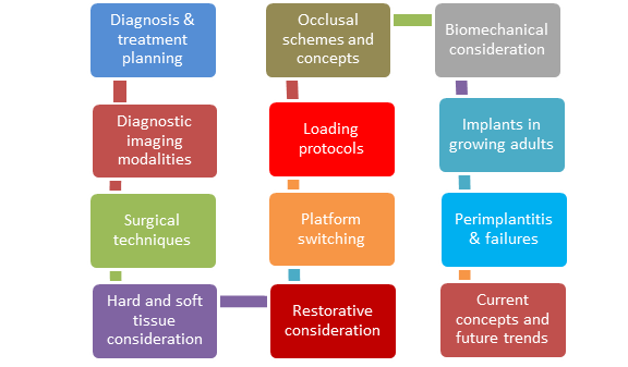
The treatment plan should begin with a clear idea of the end result which should fulfill the functional and esthetic needs of the patient. It is important that these goals are realistic, predictable and readily maintainable.
The phase of diagnosis and treatment planning should include a careful and detailed examination of the surgical site to identify any variations from normal in the anatomic structures of the maxilla and the mandible. One should make use of the available medical imaging modalities like CT and MRI, as they provide accurate information regarding the topography and location of the anatomical structures. They also provide information regarding the quality and quantity of the available alveolar bone which is essential to gauge the required implant dimensions, number as well as the position. Appropriate implant selection ensures achievement of predictable results.
Mandibular endosseous implant, if impinges on the neurovascular bundle that lies in inferior alveolar canal, can cause pain, paresthesia, numbness and excessive bleeding in case of injury to the artery. In the posterior maxilla, perforation of the sinus lining may occur, if the implant length is more than desired, leading to implant failure. The length and diameter of the implant must be selected keeping in mind the available height and width of the bone. If the available bone is too little, advanced surgical procedures such as ridge augmentation, bone grafting, sinus elevation and nerve repositioning may be done. Shorter implants are a tangible option in clinical situations with insufficient available bone and should be considered whenever suitable.
KEY FACTORS RELATED TO DIAGNOSTIC IMAGING
There are many imaging modalities that have been employed for implant imaging, including devices developed specifically for dental implant imaging. These modalities can be described as either analog or digital and two- dimensional or three-dimensional.
Intraoral periapical and occlusal radiography provide images of perhaps the greatest details of any imaging technique. They are used in initial phase of patient evaluation to detect the presence of pathosis, the approximate location of anatomic structures such as maxillary sinus and also estimate the quality of the trabecular bone.The objectives of diagnostic imaging depend on a number of factors, including the amount and type of information required and the time period of the treatment rendered. The decision to image and determine which imaging modality to use depends on the phase of the treatment.
Peripaical radiographs (PA), panoramic radiographs (Pan), lateral cephalographs (Ceph), conventional/computed tomography (Tomo/CT) are the recommended imaging modalities for treatment planning. During the surgical placement of implants, PA, Ceph or CT imaging may be used in order to analyse the placement of the implants and carry out any corrections in the angulations or depth of the implant. PA, Pan, Tomo, CT or Ceph are the recommended treatment modalities to analyse how well the healing and osseointegration of the implant has taken place following the first 3 months of implant placement. If no complications are encountered during the first 3 months, Periapical radiograph or Panoramic radiograph may suffice during the remodelling phase of 4-12 months and the maintenance phase i.e., follow up approximately every 3 years post implant placement. However, in case of any complications, CT imaging may be indicated.2
KEY FACTORS RELATED TO SURGICAL TECHNIQUE
Evaluation of soft tissue includes:
- Gingival recession and biotype
- Interdental papilla
Gingival recession and biotype: Two different biotypes have been described in relation to the morphology of the interdental papilla and the osseous architecture: The thin scalloped biotype and the thick flat biotype. The biotype should be assessed because such an assessment will partly determine the risk for postsurgical recession. A thin, highly scalloped gingival biotype is much less resistant to trauma from surgical or restorative procedures and, consequently, is more prone to recession in comparison with a thick, flat gingival biotype. A thin gingival biotype dictates placement of the implant in a slightly more palatal position to reduce the chance of recession and prevent a titanium ‘‘shadow’’ from showing through the thin gingival tissue. Similarly, the implant should be placed somewhat more apically to achieve a proper emergence profile and avoid a ridge lap restoration.
Interdental papilla: The distance between the underlying interproximal bone height on the adjacent natural teeth and the final prosthetic contact point dictates the formation and spontaneous regeneration of the interdental papillae associated with the osseointegrated supported prosthesis.
EVALUATION OF HARD TISSUES
An implant placement yields ideal esthetics only when it conforms to a prosthetically driven position, the orientation of the desired emergence profile equals the path of implant insertion there are specific ridge dimensions required supporting the implant initially and withstanding future resorption from occlusal forces. Primary stability of an implant calls for at least 1 mm of buccal and of lingual bone. The prevention of facial recession, however, requires a minimum of 2 mm of buccal support. Inevitably, some level of alveolar turnover occurs after tooth extraction, with buccolingual resorption exceeding apicocoronal loss.
A diagnostic wax-up is often required, especially in cases involving placement of multiple fixtures. The wax-up previews the future restoration and potential difficulties and can be used to educate the patient during the informed consent process. A duplicate cast can be fabricated from an impression of the wax-up and be used to create a surgical template, to serve as a guide for the surgeon during implant placement.3
SURGICAL PHASE
:
In the age of prosthetic-driven implant treatment, grafting procedures may be indicated not exclusively due to lack of bone volume, but to ensure favourable biomechanics and long-term esthetic outcome.4
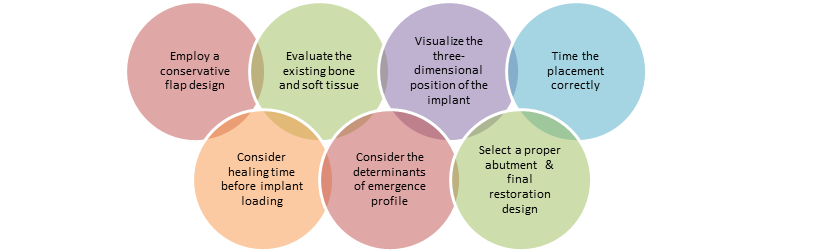
The surgical approach must be carefully planned and executed.
KEY FACTORS RELATED TO RESTORATIVE PHASE
It comprises of two phases: provisional restoration and final restoration.
Provisional restoration: Provisional restoration is an important step in obtaining treatment esthetics because it creates an adequate emergence profile and enhances the formation of papilla between the adjacent implants that will be maintained until the insertion of the final restoration.
Final restoration: The adequate selection of prosthetic components is important for esthetics with implant-supported prostheses. When esthetics was compromised during previous steps, some prosthetic adjustments can be carried out to minimize this limitation, such as selection of angulated abutments, fabrication of restorations with greater contact area, and use of gingival ceramic and ceramic abutments.
As the need for functional and esthetic restorations has increased in the present times, the need to use angled abutments to achieve these goals has also been widely accepted in implant dentistry. The use of angled abutments has simplified the management of clinical situations when implant placement is suboptimal.
KEY FACTORS RELATED TO LOADING PROTOCOLS
In carefully selected patients and sites where there are few risk factors for osseointegration and primary implant stability can be achieved and maintained during the healing phase, immediate and early functional loading protocols are recommended. Modification in implant design, the use of longer implants and altered surgical techniques can contribute to a better implant primary stability. In situations where primary stability is questionable or where there is compromised bone quality, parafunction, or systemic risk factors, delayed loading is strongly recommended.
KEY FACTORS RELATED TO PLATFORM SWITCHING
Platform switching is the use of smaller diameter abutments on wider diameter implants. It is used as a method of reducing crestal

Clinical relevance of platform switching
bone loss and maintaining the gingival papillae. Platform switching appears to limit crestal resorption and seems to preserve peri-implant bone levels.
KEY FACTORS RELATED TO OCCLUSAL SCHEMES & CONCEPTS:
Occlusal scheme for an implant-supported restoration should be designed to decrease cuspal interferences, centralize forces along the long axis, and minimize lateral forces; i.e., it should be like that of a similar restoration supported by a natural dentition.5

KEY FACTORS RELATED TO BIOMECHANICAL CONSIDERATION
Occlusal overload is often regarded as one of the main causes for peri-implant bone loss and implant or implant supported prosthesis failure. Many clinical complications such as prosthesis fracture, screw loosening or fracture, implant fracture, continuing marginal bone loss, and implant loss may be attributed to implant overload.
These complications can be prevented by application of biomechanical principles such as the passive fitting of prostheses, reducing the cantilever length, narrowing the buccolingual and mesiodistal dimensions of the prosthesis, reducing cusp inclination, eliminating excursive contacts, and centering occlusal contacts.
KEY FACTORS RELATED TO IMPLANT PLACEMENT IN GROWING INDIVIDUALS
The dynamic relationship of dental and craniofacial development to the use of dental implants in a growing patient must be understood before the treatment is initiated. Maxillary and mandibular skeletal and dental growth results in dramatic changes in all three dimensions during active growth.
Growth changes may result in the burying or loss of implants. Hence, implants placed in the early mixed dentition may lack the proper occlusion and lead to unaesthetic situations particularly in the anterior segment. They may disturb the normal growth or may have to be replaced. Implants placed during late puberty or early adulthood has the best chance for long-term usefulness.6
KEY FACTORS RELATED TO PERIMPLANTITIS AND FAILURES

Periimplantitis is an inflammatory process that affects both the hard and soft tissues around a functional implant and results in marginal bone loss, which may eventually lead to loss of osseointegration.
Diagnosis of periimplant tissue breakdown was done by periimplant probing, mobility, suppuration, clinical indices, periapical radiography and microbiology. Periimplant lesions may develop after several years.
CURRENT CONCEPTS AND FUTURE DIRECTIONS IN OSSEOINTEGRATED SUPPORTED PROSTHESIS
Knowledge about current concepts and recent advances with respect to the materials used in implant manufacturing and the variations in the implant surface morphology will help in achieving even better results.
Esthetics being the need of the hour, implants with ceramic coatings, ceramic abutments and zirconia implants has come into being for particularly restoring the high esthetic zones. Further efforts are constantly being made to maximize the strength of zirconia implants to efficiently withstand the masticatory load.
However, the future of implantology may be ruled by further research and advancement in Stem Cell Technology wherein stem cells may be used to create living dental implants. Stem Cell dental implants could well be a promising future of implant dentistry.
DISCUSSION
When discussing the impact of various medical conditions on osseointegration, it is necessary to keep in mind that recorded data may be interrelated. Potential risk factors, particularly those found more frequently in older adults in general like systemic chronic diseases, medications taken on a long-term basis, reduced salivary flow etc. may not be independent of each other. On the other hand, one single factor alone may not influence the risk measurably, whereas a combination of multiple independent factors may have a significant impact.
The quality and quantity of keratinized mucosa has demonstrated moderate influences on bone loss, with the risk of tissue recession greater in thin, scalloped biotypes. A minimum of 2 mm of thickness at the buccal walls can prevent further bone loss. The use of modern imaging techniques provides some information regarding quality and density, which cannot be related to any resorption pattern.
Placement of endosseous implants in fresh extraction wounds or small anatomic jawbone defects may often require preceding grafting procedures. Autogenous bone blocks remain the gold standard of grafting material, especially in the rehabilitation of major jawbone defects or advanced resorption. The introduction of the distraction Osteogenesis is an alternative treatment modality in cases with limited bone volumes. Barrier membranes are used in bone augmentation procedures, sometimes to reduce resorption of bone grafts. Membranes have often been used for lateral alveolar ridge enlargement and also used to alleviate bone dehiscences.
Cantilever extensions with one to two units are common in implant restorations, seemingly without controversy. In contrast, the cantilevers on restorations on natural teeth may lead to lower survival rate than for fixed dental prostheses with end abutments.
If an implant is placed before dentoalveolar growth is complete, the implant will become submerged relative to the adjacent teeth because implant behaves like an ankylosed tooth, resulting in poor esthetics and poor implant to crown ratio.
In this present article an attempt has been made to identify the important key factors for the success of osseointegrated supported prosthesis. The identification of the factors to be considered has to be done depending on the individual clinical situations.
CONCLUSION
Significant advances have been introduced including techniques to develop or regenerate implant recipient sites by stimulating both hard and soft tissues and to reproduce healthy periimplant tissue contours that resist mechanical forces and masticatory trauma. Patients with increased time of osseointegrated supported prosthesis in function should be monitored carefully and should have more frequent maintenance visits to prevent or control peri-implant bone loss. The clinical skills and updated knowledge on various aspects of osseointegrated supported prosthesis is very essential and evaluation of the patient based on important key factors along with the anticipation of the probable difficulties during the clinical procedures and management depending on the situation at various stages of implant prosthesis fabrication will result in a successful treatment outcome of osseointegrated supported prosthesis as a advanced treatment modality of rehabilitation.
REFERENCES
- Richard P. Introduction to dental implants. Part 1. Br Dent Jour.1999; 187:127-132.
- Allan B Reiskin. Implant imaging status, controversies and new developments. Dental Clinics of North America 1998;42(1): 47-56.
- Al-Sabbagh M. Implants in the esthetic zone. Dent Cin North Am.2006; 50:391-407.
- Prasad DK, Shetty M, Mehra DR. Anatomical Considerations in Implant Selection and Positioning. Int J Oral Implantol Clin Res 2013; 4(1):24-29.
- Rilo B, da Silva JL, Mora MJ, Santana U Guidelines for occlusion strategy in implant-borne prostheses. A review. Int Dent J. 2008 Jun; 58(3):139-45.
- Prasad DA, Prasad DK. Effect of implant placement in growing adults on craniofacial development: a literature review. J Dent Implant 2012; 2:97-102.
Disclaimer from Guident
The views and opinions expressed in articles published in Guident are solely those of the authors and do not necessarily reflect the views of the editors, the editorial board or the publisher, Ivory India. Guident does not endorse any product, technique or treatment described. Clinical content is intended for qualified dental and medical professionals and is not a substitute for individual clinical judgement. Guident, its editors and the publisher accept no liability for any consequence arising from the use of information published in the journal.
How to cite this article
Dr. Krishna Prasad, Dr. Anshul Bardia, Dr. Anupama Prasad. Key Factors in Osseointegrated Supported Prosthesis. Guident. 2014 Mar;7(4). Available from: https://guident.in/articles/prosthodontics/key-factors-in-osseointegrated-supported-prosthesis.html