Dental Implant Complicationspredict & Plan for a Better Prognosis
Department of Prosthodontics, Crown & Bridge and Oral Implantology Seema Dental College and Hospital, Rishikesh
Abstract
Dental implants over the years have developed as one of the successful treatment option for replacement of missing teeth. Inspite of high success rates there have been a considerable amount of failures have also been seen commonly in dental practice. A well planned dental implant treatment will provide a long term success rate while a small negligence at any stage of treatment may lead to complications and hamper with the long term success rates. Implant complications can be avoided by careful patient evaluation, selection of proper biomaterial and a sterile surgical treatment protocol.
Key Words: Implant Complications, Comfort Zone, Danger Zone, Peri-implantitis, Retrograde Periimplantitis, Osseointegration
INTRODUCTION
The proper diagnosis will help in assessment of various factors that may lead to a successful or a compromised treatment. Diagnosis preceeds the treatment plan and a well planned treatment will help us in the prediction of the treatment outcome. The role of diagnosis in the patients planned for implant placement is to assess various factors such as bone quality, bone quantity, and patient’s ability to fight against possible infections that might occur during or after implant treatment.
The use of dental implants has not only simplified the treatment for tooth loss but also has reduced the discomfort to overcome the difficulty obtained with conventional prosthesis. Failures of dental implants has a multifactorial dimension as many factors come together to cause the failures of dental implants. The proper placement of dental implants has enabled the fabrication of highly functional and esthetic restorations and also improves the predictability of treatment. Although dental implants are successful treatment modality for loss of teeth but it is not without risk of complications and failures.
Failure may be difficult to predict and after failure the cause of it may be difficult to identify. Complications may be due to aging, changing health conditions, long term wear and tear, poor home care and inadequate professional maintenance. Prosthesis instability, implant mobility, occlusal trauma may lead to various symptoms like pain, inflammation and infection.1,2
The total number of implants being placed has increased significantly over the past 10–15 years. The increased numbers of implants and implantrelated procedures being performed would have in itself resulted in a greater number of complications. Nowadays dentists are placing implants in compromised sites using more aggressive protocols including implantsplaced at the same visit of tooth extraction, immediate provisionalization of the implant following placement, and in many cases the occlusal loading of an implant on the day of placement which may result in unesthetic, biomechanically unsound prosthesis. Various factors may be responsible for the treatment outcome that includes the patient related biological and anatomic risk factors, factors related to the dentist’s experience and skills his ability to formulate the best treatment plan and mechanical risks related to the treatment and factors related to the materials and its properties that are being used to restore the lost tooth. (Fig.1) As a prosthodontist meticulous attention must be provided to the treatment planning. Subsequently medical and dental history, intraoral and extraoral examination and radiographs, diagnostic casts should be made for proper treatment planning and designing of dental prosthesis. Clinician placing or restoring implants must be prepared for the possibility of potential complications. These may be minor or major, reversible or irreversible in nature. So a better understanding of the factors associated with implant complications helps us in making correct decision for implant success.
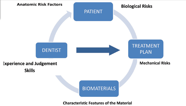
Fig. 1 Interrelationship between principal factors that influence the outcome of implant therapy
PRECAUTIONS IN PATIENTS WITH SYSTEMIC DISORDERS AND RELATED COMPLICATIONS
The number of patients suffering from systemic disorders are constantly growing. The evaluation of patient’s general health becomes an important factor in medically compromised patient. The patient’s medical history plays a key role in treatment planning of an implant patient. A proper communication between the patient’s physician and a prior knowledge of the physiological condition of the patient are basic requirements for selection, treatment planning and follow-up for the implant case. The dentist’s however holds the sole responsibility to make the final decisions for the betterment of treatment planning. A sound knowledge of the various implications of the systemic diseases are also important criteria.
CARDIO VASCULAR DISEASE
Cardio vascular disease directly affects the blood supply of the tissues which leads to impaired wound healing and a reduced supply of oxygen to the tissues that is carried out by the blood. This in turn affects the process of osseointegration. Osseointegration is best facilitated by proper implant bone contact, a good supply of oxygenated blood and the absence of debilitating factors like poor oral hygiene and improper occlusion. proper monitoring of blood pressure and patient stress related factors is important for such patients. the appointments should be short, relaxed and efficient. the vital signs of the patients should be monitored and the support staff should be trained to handle complications that might occur.1, 2
OSTEOPOROSIS
Osteoporosis is a skeletal condition characterized by decreased mineral density (mass/volume unit) of normally mineralized bone. The concern that osteoporosis is a risk factor for dental implants is grounded in the assumption that the bones of the mandible and maxilla are affected similarly as the other bones in the body by impaired bone metabolism. Impaired bone metabolism can also affect the osseointegration of implants.3,4
PAGET’S DISEASE
Osteitis Deformans or Paget’s disease of bone (PDB) is a chronic disorder of the adult skeleton in which localized areas of bone becomes hyperactive, resulting in replacement of the normal bony matrix with a highly vascular, softened and enlarged bone. The most common clinical features of Paget’s disease are malocclusion, tooth mobility, root resorption, hypercementosis, excessive bleeding on extraction, signs of osteomyelitis. The denture wearer suffering from Paget’s disease reports with poorly fitting surfaces.Patients also have compromised bone density and may be contraindicatedfor dental implant surgery.5
DIABETES
Diabetes is one of the most common systemic diseases that is prevalent throughout the world. Though diabetes is not an absolute contraindication for implant placement, the susceptibility of failure of an implant is higher in diabetic patient. Diabetes negatively impacts bone metabolism, with decreased osteoblast differentiation and proliferation, decreased collagen production, and increased osteoblast apoptosis having been demonstrated in hyperglycemic environments. Trabecular bone volume around implants is also decreased in diabetic patients. The subjects suffering from diabetes mellitus have altered wound healing, increased susceptibility to infection and a higher incidence of postsurgical complications following implants. The diabetic subjects show a higher incidence of periodontal disease which may lead to implant failure. Though diabetes is not an absolute contraindication for implant placement, a diabetic patient should have controlled levels of blood sugar which should be maintained even after implant placement. It has been reported that a patient with moderately controlled diabetes, with HbA1c in the range 8.5–9.3% presented with suppuration which was noted upon probing, with 6–7 mm peri-implant probing depths. When flap was reflected, bone loss around coronal 3–7 threads of implants was seen in patient with diabetes.2,6,7,8
PRECAUTIONS FOR PATIENTS ON MEDICATION
A proper treatment planning is necessary for placement of dental implants. A well planned implant treatment ensures a long term success. The implant treatment plan may be modified based on identified systemic conditions or in response to the medications being taken.5,6,9
In case history all the medications that the patient is taking should be recorded and evaluated with emphasis on how they might affect implant surgery. For example, Patients with osteoporosis are often prescribed medications such as bisphosphonates which reduce osteoclastic activity. These medications have names such as ibandronate (Boniva), alendronate (Fosomax), and risedronate (Actonel). The problem that has been associated with this medication is its link to bisphosphonate-related osteonecrosis of the jaw. The bone necrosis and delayed healing may manifest itself following surgical procedures which include tooth extraction, implant placement, bone augmentation, or other surgeries that affects the bone.The patient’s habits (e.g. Smoking, parafunction, recreational drugs), compliance with instructions, homecare, and psychologic issues should be assessed following the patient interview.1,10,11
A history of surgical complications is a warning that the patient may have unknown medical problems. A history of frequent dental infections or previous implant failure should alert the clinician that a thorough medical examination and blood work-up may be necessary before treatment planning begins.
PRECAUTIONS WHILE EVALUATIN DIAGNOSTIC IMAGES
An essential part of the surgical planning phase is performed in conjunction with a radiographic examination. Two and three-dimensional diagnostic images like Orthopantomograph, Dentascan, Cone Beam Computed Tomography can be used to precisely locate various anatomic structures that should be considered while planning a dental implant treatment. One of the most critical steps in the surgical planning phase is a comprehensive evaluation of the local anatomy before treatment. Surgical implant failures
and complications can arise from overlooking a relevant pre-existing anatomic condition. Anatomic variations can lead to perforations of the alveolar bone during placement of implant.12,6,9
PRECAUTIONS WITH IMPLANT SITE EVALUATION
Numerous factors should be considered during implant site evaluation. If the ridge appears atrophic, then bone grafting may be necessary to allow implant placement. The proper number of implants to be placed must be determined in relation to the number of teeth being replaced. Potentialimplant sites should be evaluated for the amount of keratinized tissues present. If an area is lacking adequate keratinized tissue, a soft-tissue graft can be placed before or at the time of implant placement.13,14,15 Precautions during implant positioning The “concept of comfort and danger zones” was introduced in 2003 for the placement of dental implants in the esthetic zone. These comfort and danger zones have been defined in three directions:5,12 (Fig 2)
a. Mesiodistally
b. Coronoapically
c. Orofacially
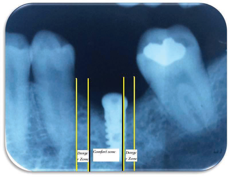
Figure 2: Comfort zone and danger zone for implant placement
MESIODISTAL MALPOSITION
An implant is positioned inside the mesiodistal danger zone when it is placed too close to an adjacent tooth. This complication is mainly caused by the development of a crestal bone modeling process during healing and after implant restoration.
The concept of “comfort” and “danger” zones for the positioning of implants should be considered precisely while performing a dental implant placement. In a mesiodistal dimension, the implant should be positioned within the comfort zone. The danger zone is 1.0–1.5 mm wide. If an implant is placed too close to a root surface it results into a reduced papilla height since there is not enough space for the soft tissues to develop.
CORONOAPICAL MALPOSITION
Apicocoronally, the implant shoulder should be positioned about 1 mm apical to the cemantoenamel junction (CEJ) of the contralateral tooth in patients without gingival recession. The danger zone is entered when the implant shoulder is placed too deeply or too coronally in relation to the comfort zone.
If Implant is not inserted deep enough, metal shoulder can be visible which will lead to unpleasant esthetic outcome. The more common complication arises when implant is placed too deep into the tissues. This apical malposition can cause recession of the facial mucosa, if the implant only has a thin facial bone wall at implant placement.
OROFACIAL MALPOSITION
In the orofacial plane, The facial extent of the implant shoulder is about 1 mm orally to the point of emergence of the adjacent teeth . The implant enters into the danger zone when the shoulder is placed too facially: which increases the risk of mucosal recession. The implant should not be placed too far orally as an orofacial malposition of an implant can also cause two different complications.
The first complication occurs if the implant is positioned too far palatally. This will often lead to a ridge-lap design of the implant crown. If the palatal malposition is combined with deep placement, it can sometimes be difficult to seat the abutment because of the thick facial and palatal mucosa. The second complication is a recession of the facial mucosa. If the implant is clearly positioned too far facially, This can cause severe esthetic complications, since the harmonious gingival course is significantly disturbed and often requires the removal of the implant.
A surgical guide stent should be considered if it is anticipated that there may be difficulty in correctly positioning the implant. Surgical stents are highly recommended in sites with multiple missing teeth. If at the time of surgery there are any doubts about the position of the implant, it is better to abort the procedure than to place the implant into an incorrect position. Reassess the site, and determine the reason for the incorrect position. If necessary, augment the site to optimize the hard- and soft-tissue conditions before returning to place the implant(s). Esthetic complications due to implant malposition are unforgiving and difficult to treat, and it is recommended that the clinician is always on the side of caution.9, 12
PRECAUTIONS WHILE PLACING IMPLANTS AT IMMEDIATE EXTRACTION SITES
Length and cost of treatment are two most common objections to implant dentistry. Poor implant positioning could occur owing to the failure of the clinician to initiate the osteotomy in the correct position. The ideal position is along the lingual incline in a maxillary anterior tooth, at the apex of the socket for a premolar, and in the area of the interdental septum for a molar.5,6,9,16 Implant failure can occur with the immediate implant placement. Studies demonstrated that the survival rate of implants is similar with immediate implant placement and delayed placement protocol. If an implant fails it can be replaced, either immediately by placing a wider implant orin a staged protocol after removal and ridge augmentation. Complicationsmay also occur in implants when implants are placed at sites which have been infected previously.
COMPLICATIONS ASSOCIATED WITH ESTHETICS AND PRECAUTIONS DURING TREATMENT
Esthetics is the prime need of any treatment outcome. Inability to restore a missing tooth in an unaesthetic manner will lead to a compromised treatment. While replacing a missing tooth with dental implants a proper care of both hard and soft tissue should be taken. A strong insight of the future treatment outcome will help restoring the patient’s esthetics in the best possible way.
Implant placement too far to the labial surface or the use of very wide implants tend to increase the potential for gingival recession around implants. Adequate space labial to the implant is essential for long-term maintenance of the implant.17,18 The most common esthetic complication arises from placement of two adjacent implants in the esthetic zone leading to loss of height of the interdental papilla. A deficient papilla leads to a “black triangle” in the interproximal area between two adjacent teeth or implant crowns. The average height of interproximal tissue from the interimplant bone crest to the height of the papilla has been reported approximately 3.5 mm which is 2 mm shorter than the average papilla height that is normally seen between two adjacent teeth. This difference in the height becomes a major esthetic complication for a patient with a high smile line.
COMPLICATIONS WITH POOR IMPLANT MAINTENANCE AND PRECAUTIONS IN POST TREATMENT PHASE
a. Peri-implantitis
The formation of plaque biofilm leads to the development of peri-implant microbiota. This peri-implant microbiota may lead to peri-implant mucositis or peri-implantitis. Peri-implantitis is defined as an inflammatory process that affects the tissues around an osseointegrated implant in function and resulting in loss of supporting bone. Radiographically peri-implantitis appears to be a saucer-shaped circumferential bony defect.6,12( Fig.3) Proper maintenance of oral hygiene and treatment of preexisting oral
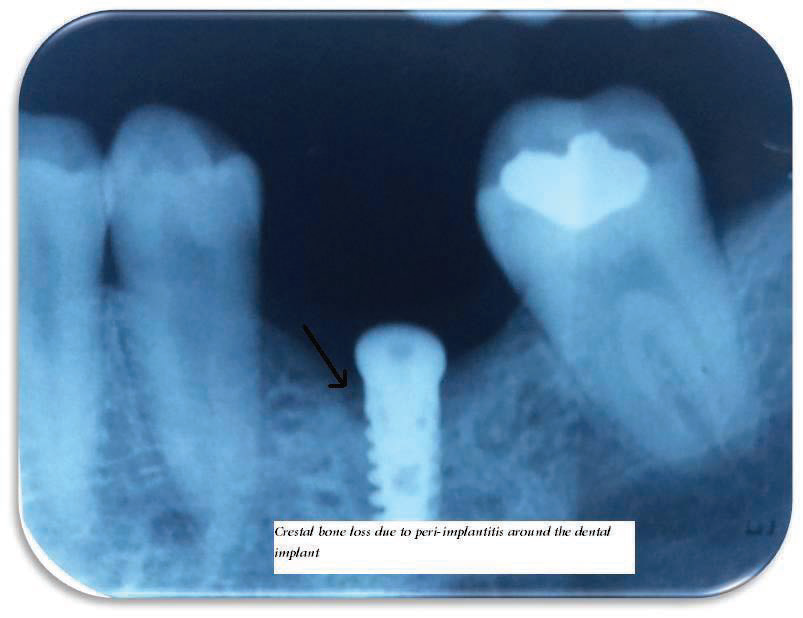
Figure 3: Crestal bone loss around dental implant due to peri-implantitis
infections such as periodontal infections should be treated before implant therapy. Prevention of plaque accumulation plays a key role for preventing progression of periodontal diseases.
b. Retrograde peri-implantitis
A few months post implant placement, a radiolucent area may be visible at the apical region of the implant. The implant may be asymptomatic in mostof the situations. The etiology of retrograde peri-implantitis is generally of unknown origin.19
COMPLICATIONS AND PRECAUTIONS ASSOCIATED WITH PROSTHETIC COMPONENTS
The following factors are most often responsible for such problems. 5,6,20
i. Manufacturing defects in the implant itself
ii. Ill-fitting superstructures
iii. Unfavorable occlusal loading
iv. Parafunctions by the patient
v. Expansive bone resorption in the peri-implant osseous bed.
LOOSENING AND FRACTURE OF OCCLUSAL SCREWS
If loosening or breakage occurs with transmucosal elements and occlusal screws, it is necessary to check the precise fit, positional stability and occlusal relationships of the superstructure , as well as the solid anchoring of implant in the bone.5,6,16 Sealing the screw access holes with acrylic resin can prevent loosening of occlusal screws.
FRAMEWORK FRACTURE
If the strength of the metal framework is inadequate or distal extension segment of the bridge is too expensive, there is a risk of fracture of framework distal to end-standing implant in implant-borne fixed bridgework. The maximum extension from the mid-point of end-standing implant must not exceed 15 mm in the mandible or 12mm in the maxilla . Several authors have reported increased incidence of framework breakage if distal extension in the mandible exceeds 20mm .6
CONCLUSION
As a prosthodontist our aim should be to identify the cause rather than to treat the present condition as a learning experience for future treatment. Understanding the etiology of a complication is fundamental to developing a strategy for corrective intervention and preventing progressive and recurrent problems. Despite strict monitoring and standard protocols to enhance the long-term prognosis of implant dentistry, certain systemic issues do not lend themselves to definitive solutions. In these cases the clinician, together with the patient and his or her physician, must work to minimize the occurrence of complications.
REFERENCES
- Balshi TJ: Preventing and resolving complications with osseointegrated implants. Dental Clinics of North America 1989: 33: 821-868.
- Valerie V. Woo, Nancy E. McDermott, Complications of dental implants: identification, frequency, and associated risk factors, Int J Oral Maxillo Fac. Implants 2003;18:848–855.
- Becker W., Hujoel P.P., Becker B.E., Willingham H.: Osteoporosis and implant failure. An exploratory case control study. J. Periodontol 2000: 71(4): 625-631.
- Dao TT, Anderson JD, Zarb GA. Is osteoporosis a risk factor for osseointegration of dental implants? Int J Oral Maxillofac Implants 1993;8:137-44.
- Stuart J. Froum, dental Implant Complications: etiology, Prevention And Treatment, Wiley Blackwell Publishers, 1st Edition, 9-46
- Carl E. Misch, Contemporary Implant Dentistry, Mosby’s publication, 3rd edition, 720-39
- Fiorellini JP, Chen PK, Nevins M, Nevins ML. A retrospective study of dental implants in diabetic patients. Int J Periodontics Restorative Dent 2000;20:366-73.
- American D. Sones: Complications with osseointegrated dental implants. J. Prosthet Dent. 1989: 62: 581.
- Principles and Practice of Implant Dentistry – Charles M. Weiss, Adam Weiss. 1st edition, chapter 17.
- El Askary A.S., Meffert R.M., Griffin T.: Why do dental implants fail? Part I. Implant Dentistry 1999: 8: 173.
- E1 Askary A.S., Meffert R.M., Griffin T.: Why do dental implants fail? Part II. Implant Dentistry 1999: 8: 265.
- Charles A. Babbush, Dental Implants: Art and Science, 2nd Edition,467-89
- Block M.S, Kent J.N: Factors associated with soft and hard tissue compromise of endosseous implants. J. Oral Maxillo Fac. Surg. 1990:48: 1153.
- Michael S. Block, Kent J.N.: Factors associated with soft and hard tissue compromise of endosseous implants. J. Oral Maxillo Fac. Surg. 1990:48: 1153.
- Zarb G.A., Schmitt A.: The longitudinal clinical effectiveness of osseointegrated dental implants: The Toronto stud III: Problems andcomplications encountered. J. Prosthet. Dent. 1990: 64: 185.
- Ross Beirne O., Philip Worthington: Problems and complicationsin implant surgery. Oral and Maxillofacial Surgery Clinics of North America 1991: 344: 993.
- Clinical Periodontics & Implantology, 4th edition, Jan Lindhe, 1016-26
- Soft tissue and Esthetic consideration in Implant Therapy – Anthony G. Sclar.
- Quirynen M. et al, Predisposing conditions for retrograde periimplantitis and treatment suggestions. Clin. Oral Implants Res. 2005 Oct, 16(5): 599-608
- Thomas D. Taylor: Prosthodontic complications associated with implant therapy. Oral and Maxillofacial Surgery Clinics of North America. 1991: 3 (4):
Disclaimer from Guident
The views and opinions expressed in articles published in Guident are solely those of the authors and do not necessarily reflect the views of the editors, the editorial board or the publisher, Ivory India. Guident does not endorse any product, technique or treatment described. Clinical content is intended for qualified dental and medical professionals and is not a substitute for individual clinical judgement. Guident, its editors and the publisher accept no liability for any consequence arising from the use of information published in the journal.
How to cite this article
Dr. Himanshu Aeran Director, Dr. Varun Kumar, Dr. Abhishek Sharma. Dental Implant Complicationspredict & Plan for a Better Prognosis. Guident. 2014 Mar;7(4). Available from: https://guident.in/articles/implantology/dental-implant-complicationspredict-plan-for-a-better-prognosis.html