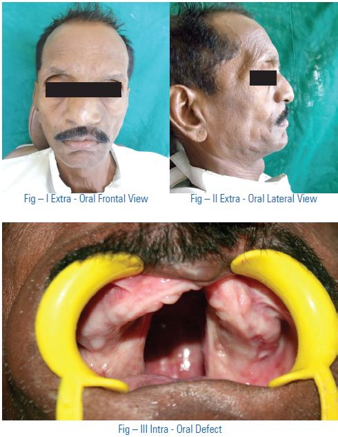
Rehabilitation of an Acquired Maxillary Defect with Close Bulb Obturator - a Simplified Prosthodontic Approach
ABSTRACT
Maxillofacial Prosthodontic Rehabilitation of total/partial maxillectomy patients is a great challenge to the prosthodontist, as patients experiences difficulties in mastication, phonetics, and deglutition. Although each patient has a unique combination of problems there are certain generalities that apply to the fabrication of most obturators. In this case report a simplified treatment procedure was described for the fabrication of light weight one piece closed hollow obturator prostheses that was closely adapted to the Armany class I defect area for a patient who has undergone partial maxillectomy five years back.
Key words: maxillary palatal defect, definitive obturator, maxillectomy.
INTRODUCTION
The prosthodontic rehabilitation of patients with acquired defects of the maxilla after surgical resection is sole responsibility of a maxillofacial prosthodontist. The maxillary/soft palatal defects cause oro nasal/ oro antral communication with difficulties in mastication, speech, deglutition, nasal regurgitation and also in esthetics. It results diminished quality of life and self esteem for the patients, who create a challenging situation to the maxillofacial prosthodontist.1
Basic prosthodontic principles should be followed during the fabrication of maxillofacial prosthesis, while taking extreme care of resilient and unsupported tissues. The defect, in conjunction with the remaining structures should be used to provide retention, stability and support to the prosthesis. Obturator is a prosthesis used to close a congenital or acquired tissue opening, primarily of hard palate and/or contiguous alveolar/soft tissue structures. For acquired defect obturator may be of three types include: immediate temporary obturator, temporary obturator, and the permanent/definitive obturator.
This case report describes the clinical and laboratory management of a patient of partial maxillectomy using one piece hollow close bulb obturator.
CASE REPORT
A 63 years old male patient reported in our department for post surgical evaluation in upper back region of mouth. The patient had undergone surgery (partial maxillectomy) for surgical treatment of sinus problem in the palate 5 years back. The patient was wearing surgical obturator in conjunction with denture to replace the gap formed due to surgical treatment. The patient was not using the prosthesis for past one year as it was not fitting properly.
The Problems encountered after one year of surgery were nasal regurgitation of fluids, impaired functions of speech & mastication, poor esthetics, resonance of voice & psychologically depression. Intraoral examination revealed class 1 defect (Aramany 1978)2 that oro-nasal & oro-antral communication was present after partial maxillectomy(fig 1).
The patient was planned for prosthodontic rehabilitation with obturator. For which he was first provided with interim obturator with minimal extension into the defect area and later on permanent obturator. It solved many of the problems of the patient to some extent. Considering the feasibility & ease of manipulation, one piece closed hollow bulb obturator3,4 , was planned & subsequently
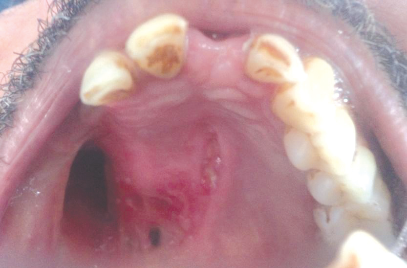
Figure 1: Intra Oral Defect
fabricated for the patient. Considering all materials available for fabrication of maxillofacial prosthesis, silicone rubber5 material has proved to produce the best results in function. But in our case considering the feasibility & ease of manipulation, heat activated acrylic resin was used for this particular patient for rehabilitation.
CLINICAL PROCEDURE
The Preliminary impression of maxillary and mandibular arch were made with irreversible hydrocolloid material (Plastigin,Septodont Inc, USA)(fig2) and was poured in dental stone(Ultrastone, Kalabhai,India). Undercuts were blocked out (fig 3) and after that wax spacer was adapted on maxillary cast. A custom tray was fabricated using auto-polymerizing resin (Pyrax,Rapid repair, India)(fig 4)and the extension was checked in patient’s mouth. After this border-
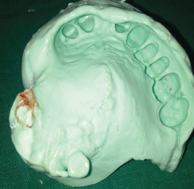
Figure 2: Preliminary Impression
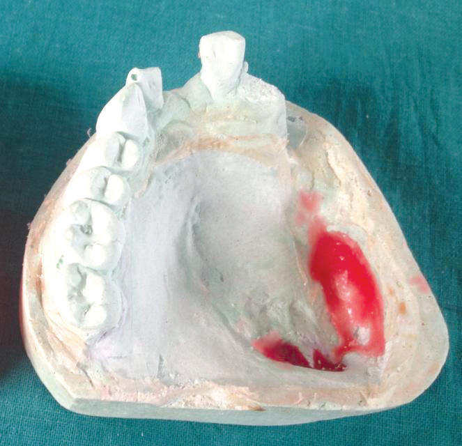
Figure 3: Undercuts Blocked
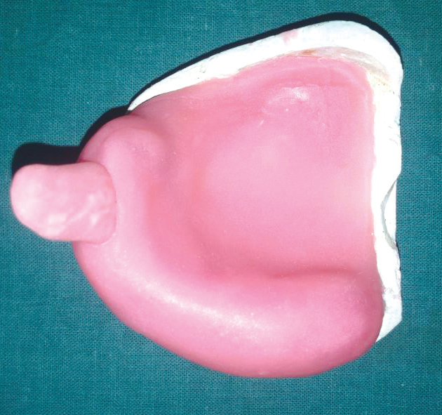
Figure 4: Custom Tray made
molding was performed with low fusing compound (Hiflex,Tracing Sticks,India), then final impression was made using medium bodied addition silicone impression material and a wash impression was made. Final Impression was poured using type IV die stone (Ultra Stone,Kalabhai,India) and master cast was obtained. Wax occlusal rim was constructed over the master cast (fig 5) and jaw relations were recorded in patient’s mouth and then transferred onto the articulator. After completion of teeth arrangement complete try in was done (fig 6).
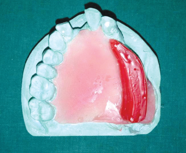
Figure 5 : Record base for jaw relation
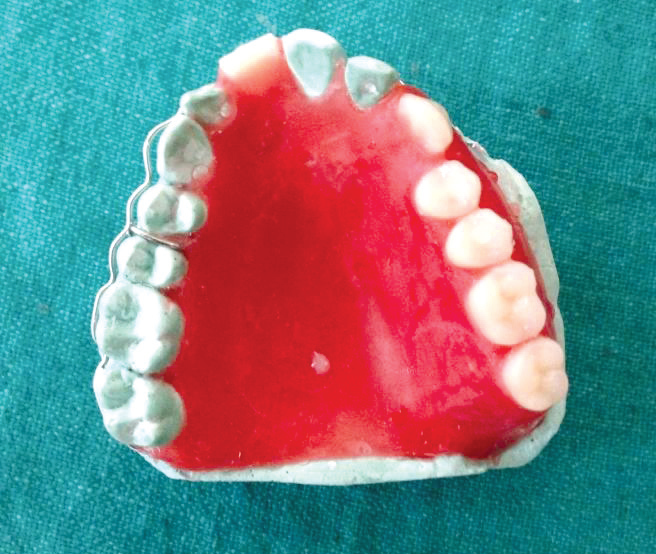
Figure 6: Teeth Arrangement
FABRICATION OF ONE PIECE HOLLOW BULB OBTURATOR
Wax-up was done after the try in. Entire defect area was relieved with one thickness of base plate wax which should follow the contour of the defect. Investing was done in the conventional flask followed by flasking and deflasking in the usual manner(fig 7). After cooling, separating medium was applied and packing of prosthesis was done in heat cure acrylic resin and placed it for short curing cycle. After finishing and polishing put salt (fig 8), or pumice in the depression closer to the outer surface with the auto-polymerizing resin shim, prepared two bur holes on the shim of 0.5mm diameter and used syringe and water to flush out the salt then bur hole were closed with auto-polymerizing resin. After finishing (fig 9) obturator prosthesis was inserted into the patient mouth (fig 10).

Figure 7: Flasking and dewaxing

Figure 8: Defect filled with salt
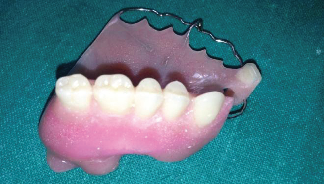
Figure 9: Finished and polished obturator

Figure 10 : Patient with Prosthesis
Patient was trained and educated for proper oral hygiene maintenance and insertion /removal of prosthesis Patient was reviewed after two days, it showed greater improvement in deglutition, mastication and speech also it was found to be more comfortable due to weight reduction. Post operative insertion adjustments were scheduled for 6 months at regular intervals of time.
DISCUSSION
Maxillary defects usually results in severe limitations of patient’s speech, mastication, and swallowing. The loss of supporting structures may also cause considerable facial collapse. Immediate and long-term prosthodontic treatment facilitates physical and psychological recovery and can restore some of the functions and esthetics. Rehabilitation of maxillectomy patient is a challenge to the maxillofacial prosthodontist1. Although each patient has a unique combination of problems, there are certain general rules that apply to the fabrication of most obturators.
This article describes the use of a flexible impression tray and construction of hollow obturator prosthesis. Patients with large maxillary defects have special problems in addition to the usual discomfort in deglutition following surgery. They must continue nasal-gastric feeding until the palate can be closed either prosthodontically or by surgical reconstruction. Speech is usually unintelligible; the mucous, general nasal, sinus & oral fluids tend to dry and crust upon already tender tissues in the defect. An impression of the defect should be obtained as soon as the patient can tolerate the procedure.
Obturator is a maxillofacial prosthesis used to close a congenital or a acquired tissue opening, primarily of hard palate or contiguous alveolar structures. The obturator have numerous functions including : Reduces psychological impact of surgery, keeps the defective area clean, enhance the healing of traumatic or post surgical defects and also help to reshape or reconstruct the defect. It also improves or in some instances makes speech possible and can be used to correct lip and cheek position. When deglutition and mastication are impaired, it can be used to improve functions. It also reduces the flow of exudates into the mouth3.
An interim obturator, that will permit removal of the nasogastric tube and restore intelligible speech, is constructed from this impression. Periodic impressions and additional obturators will be required to accommodate tissue changes during healing until a definitive prosthesis can be made. Patient accommodation is usually slow and difficult, after the initial obturator has been provided. Additional procedures should be spaced weeks apart. The patient should be told that prosthesis is a functional and esthetic compromise that has many limitations2.
It is proved that silicone prosthesis provides best result when used in function as it offers maximum potential for gaining retention from within the defect due to its flexible nature5,6. But in our case considering the feasibility & ease of manipulation, economic status of the patient heat activated acrylic resin was selected to restore the defect.
The technique described is easy, time saving & inexpensive. After insertion of prostheses mastication, deglutition and phonetics were improved. Breathing problems were resolved and esthetics was improved. Bulb portion was hollow & made of heat cure resin, so weight was less & less chances of tissue irritation. Hence there was rebuilding the self confidence in patient.
CONCLUSION
A simplified technical approach for the treatment of a patient with palatal defect of and other supportive structure has been presented. The technique presented offers a method of obtaining a detailed impression of the defect and promptly provides the patient with a light weight, easy-to-use and flexible tissue- tolerant obturator. The procedure was carried out giving utmost importance to functional, psychological & esthetic needs of the patient.
REFERENCES
- Desjardins R.P. Obturator prosthesis design for acquired maxillary defects. J Prosthet Dent 1978; 39: 424.
- Matalon J.W. et al – A simplified method for making a hollow obturator. J Prosthet Dent 1976; 36:580-82.
- Aramany M.A: Basic principles of obturator design for partially edentulous patients. Part I : Classification. J Prosthet Dent 1978; 40:351.
- Aaron Schmider: Method of fabricating a hollow obturator. J Prosthet Dent 1978;40:351.
- Robert h. wood: Hollow silicone obturators for patient after total maxillectomy. J Prosthet Dent 1977;38: 643-651.
- Russell R. Wang – Refilling hollow obturator base using light activated resin J Prosthet Dent. 78:327, 1997
- Beumer J, Curtis TA, Marunick MT. Maxillofacial Rehabilitation Posthodontic and surgical considerations.St. Louis, Ishiyaku EuroAmerica, 1996
Disclaimer from Guident
The views and opinions expressed in articles published in Guident are solely those of the authors and do not necessarily reflect the views of the editors, the editorial board or the publisher, Ivory India. Guident does not endorse any product, technique or treatment described. Clinical content is intended for qualified dental and medical professionals and is not a substitute for individual clinical judgement. Guident, its editors and the publisher accept no liability for any consequence arising from the use of information published in the journal.
How to cite this article
Dr. Himanshu Aeran, Dr. Cheshta Saxena, Dr. Rahul Dhami. Rehabilitation of an Acquired Maxillary Defect with Close Bulb Obturator - a Simplified Prosthodontic Approach. Guident. 2014 Apr;7(5). Available from: https://guident.in/articles/prosthodontics/rehabilitation-of-an-acquired-maxillary-defect-with-close-bulb-obturator-a-simplified-prosthodontic-approach.html