Breathless Nights and Dental Insights: A Prosthodontic Approach to Obstructive Sleep Apnea
Department of Prosthodontics and Crown & Bridge, Seema Dental College & Hospital, Rishikesh, Uttarakhand, India
ABSTRACT
Obstructive sleep apnea (OSA) is a prevalent sleep disorder characterized by recurrent episodes of partial or complete upper airway obstruction during sleep, leading to intermittent hypoxia and disrupted sleep architecture. This article provides an overview of the pathophysiology, clinical manifestations, diagnostic approaches, and current treatment modalities for OSA. It discussed the role of continuous positive airway pressure (CPAP) therapy, alternative treatment options such as oral appliances and surgical interventions, and emerging research trends. The importance of early recognition, personalized management strategies, and multidisciplinary care approaches is emphasized to optimize outcomes and improve quality of life for individuals with OSA.
Keywords:- Obstructive Sleep Apnea, Apnea, Obesity, Prosthodontic management.
INTRODUCTION
Sleep is a complex physiological process involving both the brain and the body. During this state, consciousness undergoes alterations, leading to a partially inhibited response to sensory stimuli. This phase is characterized by changes in muscle activity, with almost all voluntary muscles experiencing inhibition during the Rapid Eye Movement (REM) stage of sleep.1 Additionally, there is an overall diminished response. Contrasting wakefulness with sleep reveals that the latter exhibits a reduced reaction to stimuli, albeit more pronounced than that observed in a comatose state or in cases of altered consciousness, as evidenced by distinct patterns of brain activity.2
Sleep apnea, a prevalent sleep disorder, disrupts the rhythmic breathing patterns during sleep, leading to intermittent pauses in breathing or significantly shallow breaths. These interruptions, termed apneas, can last from a few seconds to minutes and may occur multiple times throughout the night. Shortened sleep duration and compromised sleep quality, prevalent issues in contemporary society, significantly impact overall health. Among various sleep disorders, Obstructive Sleep Apnea (OSA) stands out as the most frequently diagnosed condition.3
Obstructive Sleep Apnea, is characterized by interruptions in breathing during sleep. These pauses in respiration arise from either partial or complete obstructions in the upper airway, disrupting the natural sleep cycle. Such apneas transpire when the muscles in the upper respiratory tract, crucial for breathing, lose their tone during sleep, leading to the collapse of the airway and a halt in oxygen intake.4 Sleep apnea can manifest in various forms, including Central Sleep Apnea, Obstructive Sleep Apnea, and Complex Sleep Apnea. The frequent drops in blood oxygen saturation levels prompt the individual to experience intermittent awakenings, gasping for air as they emerge from deep sleep to resume breathing. As the frequency of these episodes escalates over time, they begin to impede daily life, necessitating further medical intervention.
In recent times, the role of prosthodontists has become increasingly pivotal in addressing snoring and managing mild-to-moderate cases of OSA.5 Notably, OSA exhibits a higher prevalence among males compared to females, with its incidence escalating with age. Globally, its prevalence spans from 0.3% to 5.1% across the general populace. Specifically, among Indian males, the prevalence ranges between 4.4% and 19.7%, while for females, it varies from 2.5% to 7.4%, as indicated by several studies.6
CLASSIFICATION
The International Classification of Sleep Disorders (ICSD-3) delineates Sleep Disordered Breathing into two primary categories: Adult Obstructive Sleep Apnea (OSA) and Pediatric OSA.7 These disorders encompass various breathing irregularities during sleep, including apnea, wherein breathing halts or diminishes due to an obstruction in the upper airway. Conversely, Central Sleep Apnea is characterized by a lack of respiratory effort, indicating a pause in breathing without any corresponding attempt to breathe. The specificity of diaphragmatic activity serves as a crucial metric to discern the nature of respiratory effort, determining whether inspiratory efforts remain consistent or intensify during periods of interrupted airflow.
The term Obstructive Sleep Apnea-Hypopnea Syndrome encapsulates a spectrum of sleep disturbances, wherein an individual frequently experiences daytime sleepiness alongside nocturnal episodes of both Hypopnea (shallow breathing) and Apnea. Diagnostic criteria for obstructive sleep apnea typically encompass one or more of the following observations8:
- Audible snorting or choking sounds during sleep.
- Diminished airflow in both the oropharyngeal and nasopharyngeal regions.
- Paradoxical movements of the chest and abdomen, indicative of thoraco-abdominal paradoxical breathing during episodes.
To quantify the severity of sleep apnea, the medical field utilizes the Apnea-Hypopnea Index (AHI). This metric is derived from the cumulative count of apnea and hypopnea episodes occurring per hour of sleep, with each event lasting a minimum of 10 seconds. Obstructive Sleep Apnea (OSA) is typically categorized into three distinct levels based on its severity, as determined by the Apnea-Hypopnea Index (AHI)9:
- Mild AHI = ≥5 events /hr.
- Moderate AHI = 15-30 events/hr.
- Severe AHI = >30 events/ hr
ETIOLOGY
Obesity significantly amplifies the likelihood of developing obstructive sleep apnea (OSA). The incidence of OSA escalates markedly among those with obesity and aligns with a rising body mass index (BMI). In the category of severely obese individuals, OSA's prevalence can vary between 55% and a full 100%. Moreover, those grappling with obesity frequently exhibit a more pronounced form of the condition, evidenced by elevated apnea-hypopnea index (AHI) values and reduced low points in overnight pulse oximetry readings.
RISK FACTORS FOR OBSTRUCTIVE SLEEP APNEA
- Anatomical Factors: Certain anatomical features can predispose individuals to OSA, such as:
- Enlarged tonsils or adenoids
- Deviated nasal septum
- Small jaw or recessed chin
- Large tongue or soft palate
- Age: While there is a notable rise in the prevalence of Sleep Disordered Breathing as individuals age, this trend seems to stabilize after the age of 65. Several mechanisms have been suggested to explain the heightened occurrence of sleep apnea in older adults. These include a greater accumulation of fat in the parapharyngeal region, elongation of the soft palate, and alterations in the anatomical structures surrounding the pharynx.
- Gender: OSA is more prevalent in males than in females. Additionally, postmenopausal women have a higher likelihood of OSA compared to their premenopausal counterparts. In the context of Neuromuscular Disorders, individuals with Postpolio syndrome often experience compromised upper airway function due to insufficient neuromuscular control.
- Smoking: Smoking can increase inflammation and fluid retention in the upper airway, leading to narrowing and increased susceptibility to collapse during sleep.
- Alcohol and Sedative Use: Consuming alcohol or sedatives can relax the muscles of the throat and interfere with the brain's ability to signal the muscles that control breathing, increasing the risk of airway collapse.
- Medical Conditions: Certain medical conditions are associated with an increased risk of OSA, including:
- Hypothyroidism
- Acromegaly
- Down syndrome
- Neuromuscular disorder
INFLUENCE OF TOOTH LOSS ON OBSTRUCTIVE SLEEP APNOEA
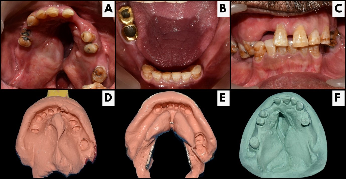
Edentulism, or tooth loss, is known to induce structural alterations in craniofacial anatomy, which are believed to elevate the risk of OSA. The absence of teeth leads to several anatomical modifications, including:
- Reduction in the vertical dimension of occlusion
- Shift in the positioning of the mandible
- Alteration in the placement of the hyoid bone
- Compromised functionality of oropharyngeal muscles, marked by diminished tone in the soft palate and pharynx, as well as conditions like macroglossia.4
PATHOPHYSIOLOGY
The pathophysiological mechanisms underlying snoring and OSA, while not entirely elucidated, are often described through two primary theories: the obstacle theory and the Bernoulli theory.
Per the obstacle theory, during inspiration, an augmented negative pressure causes the pharyngeal structures to retract. This retraction, coupled with the airflow, induces vibration, resulting in the characteristic sound of snoring. In severe cases, this vibration may lead to a partial or complete obstruction, giving rise to OSA.
The Bernoulli theory, rooted in the principles articulated by Bernoulli in 1738, posits that within a constricted tube, the velocity of the airflow increases while the pressure decreases. Applying this to the context of the airway, such conditions can lead to a suction effect on the pharyngeal structures. As these structures get pulled inward due to the decreased pressure, the ensuing vibrations of the walls produce the audible sound known as snoring.10
SYMPTOMS
Obstructive sleep apnea (OSA) can manifest with a variety of symptoms, some of which may be subtle and overlooked. Recognizing these symptoms is crucial for timely diagnosis and management. Here are some common symptoms associated with OSA11:
- Breathing heavily or panting during nighttime hours
- Impaired memory recall and cognitive function.
- Persistent and overwhelming daytime drowsiness.
- Difficulty maintaining focus and concentration.
- Excessive salivation or drooling during sleep.
- Feelings of sadness or hopelessness (despondency).
- Heightened irritability or impatience with others.
- Persistent dryness and discomfort in the mouth.
- Decline in work performance and productivity.
- Increased risk of workplace accidents due to reduced alertness.
- Limited participation in social activities or reduced social interactions.
DIAGNOSIS
The diagnosis of obstructive sleep apnea (OSA) typically involves a combination of clinical evaluation, patient history, and diagnostic testing. Proper diagnosis is crucial for effective management and treatment of OSA. Here are the common steps and investigations involved in diagnosing OSA6:
CLINICAL EVALUATION AND PATIENT HISTORY
- Waking up multiple times during the night.
- Challenges in initiating or falling asleep.
- Feeling unrested or not rejuvenated after a night's sleep.
- Persistent drowsiness or excessive sleepiness during waking hours.
- Difficulties with attention span, maintaining focus, and concentration.
- Impaired memory recall or short-term memory issues.
- Morning headaches
- Excessive nocturia
PHYSICAL EXAMINATION
- Obesity
- Retrognathia
- Neck circumference in males >42cm
- Neck circumference in females >37cm
- Underdeveloped jaw
- Constricted air passage
- Swollen tonsillar tissue
- Elevated palate structure
- Alterations in nasal anatomy.
INVESTIGATIONS
Currently, the most reliable method for conclusively diagnosing OSAHS is through laboratory polysomnography.12
Polysomnography: Polysomnography (PSG) stands as the benchmark examination for identifying OSA. This procedure captures a comprehensive view of sleep, encompassing breathing patterns and oxygen levels throughout the night. The assessment delves into factors like apnea occurrences, oxygen levels, variations in heart rate, snoring tendencies, oxygen saturation rates, and the various stages of sleep. Key measurements within PSG encompass electroencephalography, electro-oculography, electromyography, and electrocardiography.
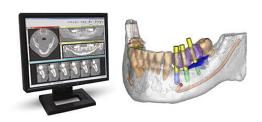
Computed tomography Scan: CT scanning plays a pivotal role in constructing three-dimensional representations and evaluating the volume of the upper airway, offering intricate cross-sectional views for analysis.
Lateral cephalograms: serve as tools to examine both the skeletal framework and soft tissue attributes in individuals diagnosed with OSA.
Magnetic resonance imaging: In compare to lateral X-ray cephalometry or CT scans, MRI provides several benefits, including superior visualization of soft tissue, three-dimensional analysis of tissue constructs, and the absence of ionizing radiation.
Acoustic reflection test: serves as a method to identify airway blockages and assess the impact of mandibular advancement and protrusion on the upper airway. During this examination, a sound wave is emitted into the airway and subsequently reflected back via a tube to a computer system. This computer then generates a graph pinpointing the exact location of the obstruction.
Spirometry: It is a pulmonary function test, is employed to examine pulmonary ventilation, meticulously documenting the airflow dynamics throughout both the inhalation and exhalation phases.
TREATMENT
The treatment of obstructive sleep apnea (OSA) aims to alleviate symptoms, improve sleep quality, and reduce associated health risks. The choice of treatment depends on the severity of OSA, individual patient factors, and preferences. Treatment options can be divided into 4 categories :
Lifestyle Modifications
- Weight Loss: For overweight or obese individuals with OSA, weight loss can significantly reduce the severity of OSA. Even a modest weight loss can lead to improvement in symptoms.
- Positional Therapy: Some individuals experience more pronounced apnea when sleeping on their back (supine position). Using positional devices or techniques to encourage side sleeping can help reduce apnea.
Continuous Positive Airway Pressure (CPAP) Therapy
- CPAP: CPAP is the most common and effective treatment for moderate to severe OSA. It involves wearing a mask over the nose and/or mouth during sleep, which delivers a continuous flow of air to keep the airway open.
- Auto-CPAP (APAP): APAP devices automatically adjust the air pressure throughout the night based on the patient's needs, offering more personalized treatment.
Oral Appliances
- Soft Palate Lifting Appliance: This device elevates the soft palate during sleep, mitigating airflow disruptions by minimizing turbulence.12
- Tongue Retention Appliance: This mechanism activates tongue musculature, concurrently shifting the mandible downwards.15
- Mandibular Repositioning (MRAs): As illustrated in, MRAs adjust the mandible in an antero-inferior direction, propelling the tongue forward and expanding the posterior airway. This adjustment augments the available superior airway volume.
- Mandibular Advancement Device: it is crafted from plastic and features tooth impressions. By facilitating movement of the mandibular teeth, it promotes the forward adjustment of the mandible, ensuring airway clearance during sleep. Notably, the achieved advancement is reversible.16 Titanium precision attachments are integrated into the design, positioned at the incisor level. These attachments enable gradual movement ranging from 2 mm to 8 mm and allow lateral shifts of up to 6 mm.
- Tongue Retaining Device: Constructed from soft polyvinyl material, its design features a distinctive bubble. The appliance is meticulously crafted with custom-fit grooves surrounding the bubble, providing a resting place for the teeth. The tongue protrudes through and adheres to the bubble via suction, aided by the lips. By positioning the tongue forward, the device effectively prevents the airway collapse often caused by the base of the tongue.17
- Thornton Adjustable Positioner (TAP): it operates through a screw mechanism situated anteriorly on the labial side of the maxillary splint. This design allows for incremental advancements of up to 9 mm for the jaw. Notably, the positioner features distinct attachments for both the mandibular and maxillary splints.
- Elastic Mandibular Advancement (EMA): it stands out as the most streamlined and least cumbersome among its counterparts. Resembling clear acrylic orthodontic retainers, it shifts the jaw forward in noticeable increments, although it may pose challenges in terms of comfort.
- DUOBLOC: it represents a custom-crafted adjustable mandibular advancement device (MAD) specifically tailored for managing obstructive sleep apnea (OSA). This MAD integrates attachments near the frontal teeth, facilitating gradual advancements of the mandible.
- Palatal Lift Prosthesis: notably enhances the dimensions of the upper airway passage. Consequently, it addresses issues of snoring and airway blockages, significantly enhancing the overall quality of life for the patient.
- Modified Herbst Appliance: it features a unique design that connects the upper and lower splints using a piston-post and sleeve adjustable telescopic mechanism on either side. This configuration effectively restricts lateral movements. However, due to the mandible's proximity maintained by petite orthodontic rubber bands, opening the jaws remains relatively effortless.
- ProSomnus® Sleep and Snore Device: it represents a custom-made anti-snoring appliance crafted using CAD/CAM technology. Its bifurcated design ensures ample intraoral space, enabling the tongue to find a comfortable resting position during sleep.1
Surgical
- Uvulopalatopharyngoplasty (UPPP): This surgical procedure involves removing excess tissue from the throat to widen the airway.
- Tonsillectomy and/or Adenoidectomy: Surgical removal of enlarged tonsils and/or adenoids can be beneficial, especially in children with OSA.
- Nasal Surgery: Procedures such as septoplasty or turbinate reduction may be considered if nasal obstruction contributes to OSA.18
Offer a non-invasive therapeutic approach for individuals diagnosed with sleep apnea.13 Within dental appliances, there are three foundational concepts designed to enhance the airway.14 These principles can be applied singularly or synergistically to bolster airway openness during episodes. Few of the oral appliances used are:-
RECENT ADVANCEMENT
Following the initiation of CPAP, certain patients previously diagnosed with OSA may transition to central sleep apnea. This condition is termed Complex Sleep Apnea Syndrome (CompSAS), with its prevalence ranging between 10% to 20%. Individuals afflicted with CompSAS often struggle to adapt to CPAP therapy due to heightened sleep disruptions caused by central sleep apnea occurrences. For their management, they are often prescribed an Adaptive Servo-Ventilator, a specialized type of PAP device.1
The Proven device operates on the principle of a 1-way valve, ensuring consistent pressure within the posterior region of the pharynx. Its standout feature is its user-friendly design. Positioned over the nostrils akin to a tape, this appliance has been introduced in the European market specifically for the treatment of OSA.19
CONCLUSION
Obstructive sleep apnea (OSA) is a common sleep disorder characterized by repetitive episodes of upper airway collapse during sleep, leading to breathing pauses and disrupted sleep patterns. It is associated with significant health risks, including cardiovascular disease, hypertension, and impaired quality of life. Early recognition, diagnosis, and management of OSA are crucial to mitigate complications and improve outcomes. Treatment options range from lifestyle modifications and positional therapy to continuous positive airway pressure (CPAP) therapy, oral appliances, and surgical interventions. Personalized approaches tailored to individual patient needs, ongoing research, and technological advancements are key to optimizing the management of OSA and enhancing long-term outcomes for affected individuals.
REFERENCES
- M Juneja, S S Phukela, B Yadav, A Nagpal, O Shetty, P Ritwal. Prosthodontic Management of Obstructive Sleep Apnea . Bull. Env.Pharmacol. Life Sci., Spl Issue [2]: 2022: 550-560
- Brain Basics: (2019). Understanding Sleep” National institute of Neurological Disorders and stroke.p90.
- Methani S, Aras M, Chitre V. Obstructive sleep apnea and its prosthodontic management. J Indian Prosthodont Soc. 2004;4:8-13.
- Garbarino S, Bardwell WA, GugliemiO,Chioori C, Bonanni E, Magnavita N. (2018). “Association of Anxiety and Depression in Obstructive Sleep Apnea Patients: A systematic Review and Meta-Analysis”. Behavioral Sleep Medicine.;18(1): 35-57.
- Ralls, F., & Cutchen, L. (2019). A contemporary review of obstructive sleep apnea. Current opinion in pulmonary medicine, 25(6), 578-593.
- Chandra, S. D., & Raj, K. S. (2018). Obstructive sleep apnea and its prosthodontic management-an overview. Int J Health Sci Res, 8(3), 259-265.
- Sateia M. (2014). International classification of sleep disorders-third edition: highlights and modifications Chest. ;146(5): 1387-1394.
- American Academy of Sleep Medicine. (2019)."AASM Practice Guidelines".
- Vijaya Laxmi Murty, Gulab Chand Baid, Saumya Sharma, Sanjeev Singh, Vivek Lath, Priyabrata Jena. Obstructive Sleep Apnea: A Prosthodontist’s Viewpoint. Sch J Dent Sci, 2023 May 10(5): 102-107.
- Meyer J B, Knudson C R, Hall W: The sleep apnea syndrome. Part I: Diagnosis. J Prosthet Dent 1989 ;2:675-9.
- Chander, N. G. (2020). Sleep apnea and prosthodontic implications. The Journal of the Indian Prosthodontic Society, 20(4), 335.
- Y. K. Tan, P. R. Lestrange, Y. M. Luo, C. Smitha, H. R. Grant, A. K. Simonds, S. G. Spiro. Mandibular advancement splints and cpapin patients with obstructive sleep apnea: a randomized cross over trial. Eur. J. Orth.2002. 24:239-49.
- Qureshi A, Ballard RD.(2003). Obstructive sleep apnea: J Allergy Clin Immunol. 112(4):643- 50.
- Lam B, Sam K, Mok WYW, Cheung MT, Fong DYT, Lam JCM, Lam D, Lorett YC, Yam, Mary SM. (2007). Randomised study of three non-surgical treatments in mild to moderate obstructive sleep apnoea. Thorax; 62:354–9.
- Yoshida K. (1994). Prosthetic therapy for sleep apnea syndrome. J. Prost. Dent. 72(3): 296–302.
- F. Gagnadoux, B. Fleury, B. Vielle, B. Pe´Telle, N. Meslier, X.L. N?guyen, W. Trzepizur And J.L. Racineux. (2009). Titrated Mandibular Advancement Versus Positive Airway Pressure For Sleep Apnoea. Eur Respir J. 34:914–20.
- LS Bennett, RJ Davies and JR Stradling. (1998). Oral appliances for the management of snoring and obstructive sleep apnoea; Thorax, (53): 58-64.
- Kline CE, Crowley EP, Ewing GB, Burch JB, Blair SN, Durstine JL, Davis JM, Youngstedt SD. (2011). The Effect of Exercise Training on Obstructive Sleep Apnea and Sleep Quality: A Randomized Controlled Trial. SLEEP. 34(12): 1631-40.
- Reichmuth KJ, Austin D, Skatrud JB, Young B. (2005). Association of Sleep Apnea and Type II Diabetes - A Population-based Study. Am J Respir Crit Care Med. 172(12): 1590-5.
Disclaimer from Guident
The views and opinions expressed in articles published in Guident are solely those of the authors and do not necessarily reflect the views of the editors, the editorial board or the publisher, Ivory India. Guident does not endorse any product, technique or treatment described. Clinical content is intended for qualified dental and medical professionals and is not a substitute for individual clinical judgement. Guident, its editors and the publisher accept no liability for any consequence arising from the use of information published in the journal.
How to cite this article
Dr. Varun Kumar, Dr. Jyotsna Seth, Dr. Manas Aeran. Breathless Nights and Dental Insights: A Prosthodontic Approach to Obstructive Sleep Apnea. Guident. 2024 Apr;17(5). Available from: https://guident.in/articles/prosthodontics/breathless-nights-and-dental-insights-a-prosthodontic-approach-to-obstructive-sleep-apnea.html