Digital Dentistry in Prosthodontics: Transforming Precision and Efficiency in Restorative Dentistry
Department of Prosthodontics and Crown & Bridge, K.D. Dental College & Hospital, Mathura, UP, India
ABSTRACT
The advent of digital technology has revolutionized the field of prosthodontics, ushering in an era of unprecedented precision, efficiency, and patient-centered care. This abstract explores the impact of digital dentistry on prosthodontic practices, focusing on the advancements that have transformed traditional approaches to restorative dentistry.
Digital impressions, aided by intraoral scanners, have replaced conventional impression materials, offering improved accuracy and patient comfort. Computer-aided design (CAD) and computer-aided manufacturing (CAM) technologies have streamlined the prosthetic fabrication process, allowing for precise and reproducible restorations. The utilization of three-dimensional (3D) printing in prosthodontics further extends the possibilities, enabling the fabrication of complex and patient-specific prostheses with enhanced material options.
Virtual articulation and dynamic occlusal analysis have empowered prosthodontists to refine treatment planning, ensuring optimal function and esthetics in the final prosthetic outcomes. The integration of digital smile design tools has enhanced communication between clinicians and patients, fostering a collaborative approach in treatment decision-making.
Additionally, the incorporation of digital workflows facilitates seamless interdisciplinary collaboration among dental professionals, promoting a comprehensive and integrated approach to patient care. Teledentistry platforms further expand accessibility to prosthodontic consultations, enhancing patient engagement and satisfaction.
Despite these advancements, challenges such as initial setup costs, learning curves, and ongoing technological developments persist. Nevertheless, the undeniable benefits of digital dentistry in prosthodontics underscore its potential to redefine the standard of care in restorative dentistry, ultimately improving patient outcomes and shaping the future of prosthodontic practice.
KEYWORDS: Digital dentistry, Intraoral scanner, CAD-CAM, Virtual Articulator
INTRODUCTION
The rich tapestry of dentistry, with its roots stretching back to the eighteenth century, unveils a captivating journey marked by remarkable transformations in techniques, materials, and equipment. In those early days, the impressions were crafted with waxes and plaster of Paris, and the dental equipment was characterized by hand-driven and subsequently water-driven motors. The evolution from these rudimentary tools to the contemporary paraphernalia of dentistry reflects a profound trajectory of progress and innovation.
During the nascent stages, dentistry grappled with limited materials and equipment, leading to selective treatment options. However, with the relentless march of time and an exponential growth in research, a myriad of options emerged, reshaping the landscape of dental practice. The modern dental arena now boasts an array of choices for preserving oral health, offering aesthetics that closely mimic natural teeth, all delivered through an enhanced approach that minimizes treatment time, reduces error potential, and ensures superior quality assurance.
One of the pivotal transformations in contemporary prosthodontics is the assimilation of digitalization into its core. The digital era has seamlessly woven itself into the fabric of dentistry, offering efficiency, speed, precision, and error-free production of fixed prosthodontic appliances. The advent of computer-aided design/computer-aided manufacturing (CAD-CAM) technology in the early 1980s marked a significant milestone. This technology, along with other digital techniques such as stereolithography, rapid prototyping, virtual articulators, digital face bows, digital radiographs, and a plethora of dental software applications, has revolutionized the field.
The scope of digitalization and technology in prosthodontics extends beyond clinical and lab procedures. It encompasses training, education, and research, with innovations like virtual patient programs, optoelectronic recording of jaw motion, retention testing devices, and audio technology. The possibilities are seemingly endless, continually pushing the boundaries of what can be achieved in the pursuit of excellence in dental care.
Looking ahead, the future promises even more integration of digital imaging in dentistry. The potential for online testing and upgrades of x-ray equipment and software hints at a dynamic and interconnected future. Furthermore, ongoing research into the development of 'smart cards,' credit card-sized devices carrying a patient's medical and dental records along with radiographic images, could revolutionize the way patient information is managed and accessed.
In navigating this era of rapid technological advancement, it becomes imperative for the dental community to embrace these innovations. Acceptance and utilization of technological advances are crucial to ensuring that clinical practice and patient care continually evolve and improve. The journey of dentistry, from its humble beginnings to the digital frontier, serves as a testament to the unyielding spirit of progress and the unwavering commitment to enhancing the well-being of patients through the marvels of science and technology.
VARIOUS DIGITALIZATION TECHNIQUES USED IN PROSTHODONTICS
Dentistry, like many fields, is undergoing a profound transformation due to the increasing influence of digitalization. This technological wave is poised to revolutionize every facet of dental practice, potentially reshaping the conventional paths of treatment.
The interconnected workflow among dentists, surgeons, and dental laboratories stands at the forefront of this digital evolution, promising significant alterations. It is widely recognized that digitalization will fundamentally alter dental workflows, marking a paradigm shift in how dental professionals collaborate and deliver care.
Esteemed figures in the dental community, such as Professor Daniel Wismeijer from the Academic Center for Dentistry Amsterdam, are keenly aware of the transformative power digitalization holds for their profession. Professor Wismeijer eloquently describes digital dentistry as a journey where the patient remains anchored in the analog realm while the tools and processes in between become seamlessly digital.
In his vision, digital dentistry opens doors to a collaborative landscape where practices, laboratories, and beyond can collectively optimize the value chain, thereby enhancing patient care and outcomes.
STRAUMANN DIGITAL SOLUTIONS
In the early 2010s, Straumann unveiled a groundbreaking suite of integrated computer-driven technologies dubbed 'Straumann Digital Solutions'. This innovative portfolio encompasses three distinct categories: computer-guided surgery, intra-oral scanning, and CAD/CAM prosthetics.
At the heart of Straumann's Digital Solutions lies a vision where the patient and their dental restoration retain their analog essence, while the intricate processes that bridge them transition into the digital realm. This vision underscores Straumann's commitment to leveraging digital advancements to enhance patient care and streamline dental procedures.
COMPUTER GUIDED SURGERY
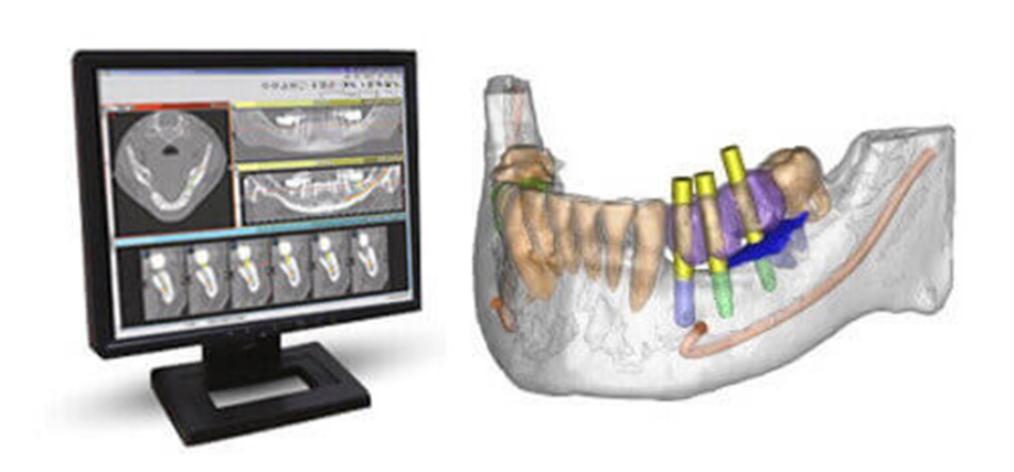
Computer Guided Implant Surgery
Straumann's computer-guided surgery product comprises three primary components:
- the coDiagnostiX planning software,
- the gonyX surgical template production tool,
- and the Guided Surgery Kit and Implants.
The guided surgery process commences with a master model derived from an impression of the patient's current dental condition.
This master model serves as the foundation for generating both the scan and surgical template. Utilizing this model, a scan prosthesis is crafted to emulate a provisional teeth set-up, facilitating precise planning and execution of the guided surgery procedure.
co-DIAGNOSTIX PLANNING SOFTWARE
Within coDiagnostiX, all pertinent data can be seamlessly integrated – from the patient's CT scan to the STL file of the impression. This comprehensive approach allows for a holistic examination of the patient's condition, empowering dental professionals to devise more effective treatment plans. By amalgamating these datasets, clinicians gain a thorough understanding of the patient's oral landscape, enabling them to deliver superior treatment outcomes tailored to each individual.
DELIVER HIGH-QUALITY AESTHETIC CARE WITH DIGITAL TREATMENT PLANNING
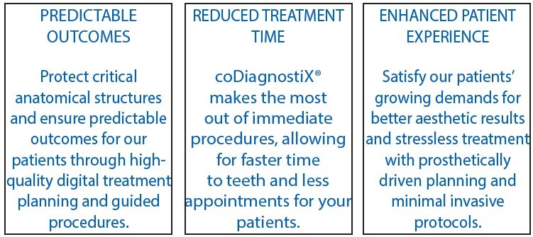
In our fast-paced and ever-evolving world, patients rightfully expect top-notch, aesthetically pleasing dental care with minimal disruptions to their busy schedules. Embracing digital treatment planning is swiftly becoming the norm, empowering clinicians to fulfill these escalating expectations while enhancing patient involvement. Enter coDiagnostiX®, our cutting-edge dental treatment planning software poised to propel us forward on this transformative journey. With its advanced capabilities, coDiagnostiX® stands as a reliable ally, enabling us to deliver unparalleled care and forge deeper connections with our patients.
goNYX SURGICAL TEMPLATE PRODUCTION TOOL
- The gonyX coordinate table is an integral part of the coDiagnostiX system.
- It is utilized for the production of scan and surgical templates.
- These templates are crucial for guiding dental procedures accurately.
- Pins are incorporated into the templates to establish connectivity between the gonyX table and the coDiagnostiX dataset.
- This integration ensures seamless coordination between physical and digital elements.
- Ultimately, it enables clinicians to execute treatments with enhanced precision and efficiency.
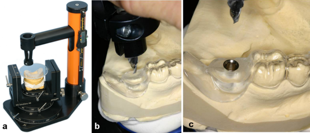
Creation of drill guides using a gonyX table (IVS Solutions AG, Chemnitz, Germany) on the basis of virtual planning a) Transfer of computer-assisted implant planning to the implantation template using the gonyX table; b) Drilling into the template; c) Template with guide sleeve.
THE GUIDED SURGERY KIT AND IMPLANTS
- Fully guided placement of 3.1 mm implant and implants in sizes 3.7 mm, 4.1 mm and 4.7 mm
- Compact kit, which is useful for space-constrained dental offices
- No handles or keys are necessary while preparing the osteotomy
- DLC (Diamond-like Carbon) coating on the drills provide wear resistance characteristic
Drilling System

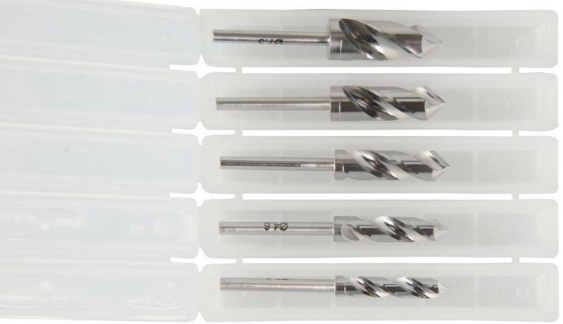
The RealGUIDE Z3D Guided Surgery Kit boasts drills crafted from resilient AISI 420B stainless steel, meticulously treated with DLC (Diamond-like Carbon) coating. This innovative enhancement not only enhances surface durability but also mitigates friction forces encountered during operation, ensuring smoother and more precise surgical procedures.
Implant Mounts

Place the implant vial on the dedicated implant staging pedestal corresponding to the implant length.
Screwdriver and Ratchet
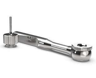
They can be bundled with the kit purchase, or use the instruments from our existing standard.
Intra-oral scanning
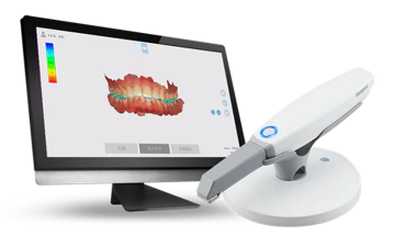
The advent of intra-oral scanning technology empowers dentists to generate three-dimensional representations of a patient's teeth using a digital scanner positioned within the oral cavity.
Revolutionizing traditional methods, the intra-oral scanner supersedes the need for cumbersome impression-taking and model casting in dental practices and laboratories, respectively.
These three-dimensional images seamlessly integrate into computer-aided design (CAD) software, facilitating streamlined restorative procedures.
The advantages of the intra-oral scanner are manifold:
- Diagnostic Precision: Serving as diagnostic aids, they allow dentists to visually and interactively educate patients about their oral health status.
- Time Efficiency: Digital impression-making significantly reduces time consumption, enabling dentists to attend to more patients efficiently.
- Rapid Impressions: Compared to conventional techniques, digital impressions are swift and seamless.
- Accuracy and Precision: The technology ensures high levels of accuracy and precision in impression-making.
- Versatility: With the broadest range of applications, intra-oral scanners cater to diverse dental needs.
- Cost Savings: By eliminating the need for traditional impression materials, this technology helps in saving costs.
- Enhanced Patient Comfort: Patients benefit from increased comfort during procedures, while reduced chair time improves overall experience.
In summary, intra-oral scanning represents a transformative leap forward in dental practice, optimizing efficiency, precision, and patient comfort.
DIFFERENT TYPE OF INTRAORAL SCANNER
Trios 5 intraoral scanners
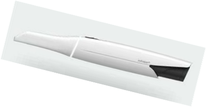
Crafted to deliver a seamlessly intuitive scanning encounter, unparalleled ergonomic design, and elevated standards of cleanliness. This scanner epitomizes a revolutionary benchmark in safeguarding patients and enhancing infection control measures.
Trios 4 intraoral scanner
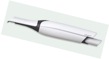
Embracing wireless technology, this intraoral scanner integrates advanced diagnostic aids and monitoring tools. Renowned for its rapidity and comfort, it also excels in delivering remarkable precision, particularly for individual units.
Trios 3 colour pod intraoral scanner
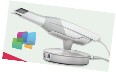
Compact and feather-light, this device boasts ultrafast optical sectioning technology, facilitating vibrant intraoral scanning. Not only does it capture intricate details in color, but it also accurately measures tooth shade, enriching the patient journey while streamlining chair-side procedures with heightened precision.
Trios 3 Move + intraoral scanner
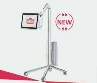
Featuring a multi-touch screen and an integrated high-performance computer tailored for 3D scanning, this device heralds a new era of dental technology. Its revolutionary capabilities ignite patient enthusiasm and foster advanced case acceptance, marking a significant leap forward in dental practice.
Aeralscan 3 intraoral scanner
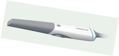
Introducing the groundbreaking Aoralscan 3 by Shining 3D, a cutting-edge design in intraoral scanning. This swift scanner marries high accuracy with state-of-the-art features, elevating our digital scanning experience to unprecedented levels of excellence.
CAD/CAM TECHNOLOGY
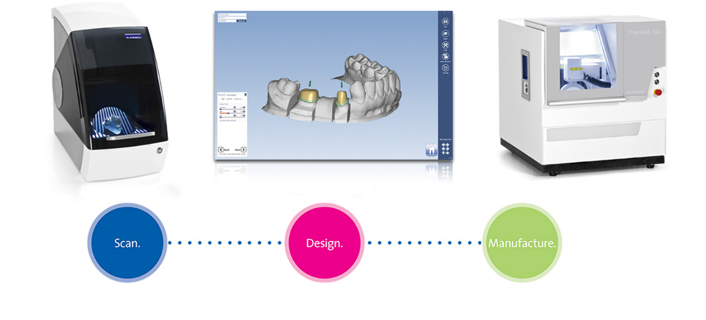
Modern prosthetic inlays, crowns, onlays, and bridges are revolutionizing dentistry through computer-aided design (CAD) and computer-aided manufacturing (CAM) processes. Utilizing 3D scanners or intra-oral scanning, impression models are digitized for precise restoration modeling by dental technicians on computer software. These digital designs are then transmitted to centralized production centers for fabrication using biocompatible, durable, and aesthetically pleasing materials like high-performance ceramics.
Since its inception in 1971, CAD/CAM technology has rapidly evolved, offering solutions ranging from single tooth inlays to extensive 14-unit full-arch restorations. Notably, the CEREC® 3D system by Sirona Dental Systems stands as the only in-office chair-side CAD/CAM system currently available. Its simplicity, versatility, and precision enable the design and milling of all-ceramic restorations chair-side, eliminating the need for traditional impressions and temporaries. Patients can now receive a final restoration within an hour of their appointment.
The optical impression technique, integral to dental CAD/CAM, encompasses various analytical methods from diagnosis to treatment. It emphasizes optical means of measurement for accuracy and efficiency in dental restoration processes. Overall, CAD/CAM systems are transforming dental practices by streamlining workflows and delivering superior outcomes in terms of fit, durability, and esthetics.
General Principles of CAD/CAM
- All true CAD/CAM systems exhibit their computer-linked functional components, although the degree of sophistication may differ.
- They typically consist of three main components:
-
- A means of data acquisition
- Restoration design
- Restorative production
- The first stage involves digital data collection from the patient's mouth, which is equivalent to traditional impression taking.
Uses of CAD/CAM in prosthodontics areas follows
- Inlays and onlays
- Crowns and bridges
- Fabrication of post and cores
- Complete denture fabrication
- Removable Partial Denture Prosthesis Fabrication
- Designing the placement of the Implant
- Designing the maxillo facial prosthesis
CLINICAL STEPS IN THE FABRICATION OF CAD/CAM CROWN
- Crown Preparation
- Optical Impression
All Ceramic Crown Preparation
- Functional cusps should be reduced by 2.0 mm, nonfunctional cusps by 1.5 mm, and the central fissure area by at least 1.5 mm to ensure adequate strength to the crown.
- Axial reduction of the tooth should be a minimum of 1.2 mm.
- The facial and lingual walls of the crown preparation should be done in two planes to avoid thin areas in the milled restoration.
- Rounded shoulder or chamfer margins, at least 1.0 mm thick, ensure a strong edge of material to prevent fracture of the restoration.
- A sloped shoulder (90°-120°) ensures a strong edge of material while providing a good transition of translucency and shade between the tooth and crown at the margin area.
- Thin areas at the margins, such as light chamfers or bevels, are predisposed to fracture and are not recommended.
Optical Impression
- POWDERING
- ACQUIRING SINGLE OPTICAL IMPRESSION
- ACQUIRING MULTIPLE OPTICAL IMPRESSION POWDERING
Background Information on Powdering
To make an optical impression it is necessary to powder (or opaque) the tooth surface to be imaged because of two reasons:
- the surface of the tooth must be covered with a non-reflective coating
- make it easier for the infrared camera to see the details.
ACQUIRING SINGLE & MULTIPLE OPTICAL IMPRESSION
- Direct systems of data acquisition are globally utilized but lack an ideal solution.
- Significant development has occurred in recent years, necessitating dentist involvement for proper selection of virtual molding equipment.
- Computer-aided design/computer-aided manufacturing (CAD/CAM) images can be obtained through direct or indirect imaging methods.
- Indirect systems involve digitalization from impression materials or casts, while direct systems capture images directly from the mouth using intraoral scanners.
- Direct acquisition systems are preferred for being less invasive, quicker, and more precise than conventional methods. They also allow for easy storage of digital images.
- Main direct image acquisition systems available on the market include:
- CEREC Bluecam (Sirona)
- Lava C.O.S. System (3M ESPE)
- iTero System (Cadent/Straumann)
- E4D System (D4D Technologies)
CEREC Bluecam (Sirona)
Introduction of CEREC: The CEREC system was introduced in 1985, pioneering the use of CAD/CAM technology for chair-side dental procedures.
High-Resolution Imaging: The CEREC AC Bluecam offers high-resolution image acquisition, similar in quality to laboratory-scanned impressions.
Contrast Powder Requirement: To create optical impressions, a thin layer of contrast powder is applied to the tooth's surface before scanning.
Blue Diode Technology: The CEREC Bluecam employs a blue diode with powerful LED for image acquisition.
Speed and Efficiency: The system provides quick and efficient results with high CAD/CAM quality, precision, and confidence. It emits a short wavelength blue light for imaging.
Fast Acquisition: In automatic mode, it can capture optical impressions of one quadrant in less than a minute, with opposite-arch impressions taking only a few seconds.
Versatile Imaging: The blue light allows imaging from difficult-to-reach areas, and the camera can be positioned close to or slightly touching the tooth surface.
Automatic Exposure: The camera automatically detects the right moment to trigger exposure, ensuring stability during image capture.
Quality Control: The CEREC 3D software eliminates substandard images automatically.
Applications: CEREC Bluecam can be used for various dental procedures, including single crowns, fixed partial dentures (FPDs), and implant-supported FPDs. It can scan prepared abutments or implant surfaces.
Surface Requirements: The system requires a nonreflective and thin, homogeneous contrast layer on the tooth surface for precise imaging.
CEREC Optispray: The system offers CEREC Optispray, a material that enhances imaging capabilities by providing a homogenous coating on the tooth surface. It's water-soluble and easy to remove.
Output Format: The CEREC Bluecam generates STL format documents that can be modified using compatible software. The resulting restoration can be milled using different equipment, making it an open scanner.
These points summarize the key features and capabilities of the CEREC Bluecam system in dentistry.
Lava C.O.S. System (3M ESPE)
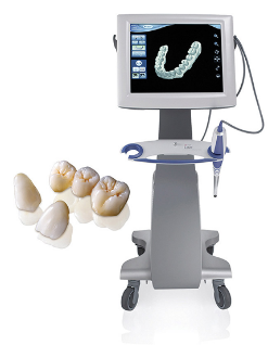
- Origin: Developed at Brontes Technologies, Lexington. Acquired by 3M ESPE in October 2006.
- Launch: Officially launched in Chicago in February 2008.
- 3D Impression Method: Uses active wavefront sampling, '3D in Motion' concept for real-time capture.
- Speed: Capable of capturing about 20 3D data sets per second.
- Components: Includes CPU, touch-screen display, scanning wand with 13.2-mm-wide tip.
- Powdering Requirement: Needs powdering for reference point location, applied thinly to avoid distortions.
- Scanning Process: Initiated by button press, software recognizes and groups images, allowing rescanning if needed.
- Digital Articulation: Maxillary and mandibular scans digitally articulated on-screen.
- Restoration Design Software: Specific software for designing restorations based on optical impressions or scanned images.
- Compatibility: Initially not compatible with implant platforms, partnerships established for compatibility with Biomet 3i, data transfer to Dental Wings software (DWOS). Considered semi-open, compatible with limited software options.
iTero System (Cadent/Straumann)

Introduction and Technology:
- iTero System introduced in 2007, using parallel confocal imaging for digital impressions of teeth and gums.
Capture Process:
- Captures 100,000 points of laser light, providing perfect focus images at over 300 depths without powders.
Components:
- System includes computer, monitor, scanner device, and peripherals.
Disinfection:
- Easily disinfected by replacing disposable scanner cover.
Applications :
- Initially limited, partnership with Straumann expanded use. Compatible with software accepting STL images.
Optical Impression Process:
- Camera guided by voice control for data collection, with automatic rescan if needed.
Data Processing:
- Data transferred to software for restoration drawing, customization.
Implant Positioning:
- Specific transfer ensures proper positioning for implant impressions.
Abutment Drawing:
- Software used for drawing structures over implant images obtained through optical impressions.
Milling Process:
- Prototypes fabricated at milling centers.
iTero System offers advanced technology for efficient and versatile dental impressions of both teeth and implants.
E4D System (D4D Technologies)
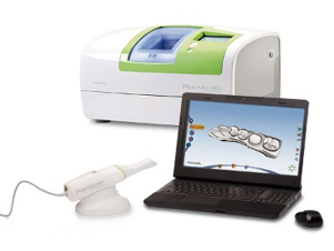
Overview of the System:
- Developed by D4D Technologies in Richardson, TX, designed for dental applications.
- Enables optical impression of various dental components without requiring powder for scanning.
Digital Casting:
- Automatically converts dental impressions into digital casts for further manipulation and various applications.
Technology Used:
- Utilizes red laser technology and micromirrors vibrating at 20,000 cycles per second to capture a sequence of images and generate detailed 3D models.
Image Acquisition Options:
- Provides manual and automatic image acquisition options, allowing flexibility for operators.
Versatility:
- Capable of scanning conventional dental impressions and casts, suitable for various dental applications.
Single-Appointment Restorations:
- Offers chairside milling unit, eliminating separate laboratory steps for creating dental restorations, resulting in shorter appointment times for patients.
Data Processing:
- Optical impression data sent to DentaLogic software for creating virtual casts and restoration design.
- Adjustments to occlusion made by scanning opposite dental arch, and precise temporary crowns can be fabricated.
In summary, the D4D Technologies dental scanning system provides advanced capabilities for efficient and precise digital impressions, restorations, and occlusion adjustments, benefiting both dental practitioners and patients.
CONE BEAM COMPUTED TOMOGRAPHY (CBCT)
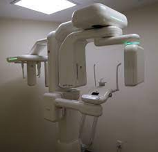
- Developed by Sir Godfrey Hounsfield in 1967, CT technology has evolved through five generations.
Generations:
- First generation: Single radiation source and detector, obtaining information slice by slice.
- Second generation: Introduced multiple detectors within the scan plane.
- Third generation: Advancements in detector and data acquisition technology.
- Fourth generation: Utilized moving radiation source and fixed detector ring, addressing scattered radiation issues.
- Fifth (or sixth) generation: Reduced motion or scatter artifacts, introduced Cone Beam CTs (CBCTs) for dental, oral, and maxillofacial applications.
CBCT Technology:
- CBCT uses a low-radiation X-ray tube and projects the beam onto a flat panel detector (FPD) or charge-coupled device (CCD) with an image intensifier.
- CBCT produces a more focused beam with less radiation scatter compared to conventional CT devices, improving X-ray utilization and reducing tube capacity.
- The tube and detector rotate around the selected region, converting primary data into slice data.
Voxel Representation:
- CT volume consists of a 3D array of voxels, each characterized by height, width, and depth.
- CBCT images have isotropic voxels, offering uniform resolution in all directions, advantageous for precise measurements.
- Voxel sizes can vary (e.g., 0.076 mm, 0.2 mm, 0.3 mm, 0.4 mm) based on imaging unit capabilities and imaging objectives.
- Smaller voxel sizes allow visualization of finer details, while larger voxels provide lower resolution.
- Voxel size should be smaller than the desired anatomical structure for adequate representation.
- Soft tissue structures may be limited in evaluation due to voxel size constraints.

Illustration of CBCT and MDCT voxel size.
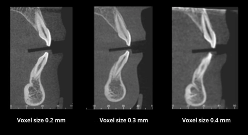
Voxel size affects details of the objects imaged.
CBCT as compared with conventional CT
Summary of CBCT Advantages Over Conventional CT:
- Lower Radiation Dose:
- CBCT uses lower effective tube current, resulting in reduced radiation dose compared to multislice CT.
- Voltage remains similar (90–120 kV), but current is lower (1-8 mA for CBCT versus 80-200 mA for multislice CT).
- Different Detection Systems:
- CBCT employs different detection systems such as Flat Panel Detectors (FPD) or Charge-Coupled Devices (CCD) with image intensifiers.
- Higher Resolution:
- CBCT offers higher resolution due to smaller isotropic voxel size.
- Reduced Metallic Artifact:
- CBCT shows fewer artifacts caused by metallic structures compared to conventional CT.
- Classifications and Subcategories:
- CBCT devices classified into four subcategories based on clinician's needs and field of view (FOV):
- Dentoalveolar (FOV < 8 cm)
- Maxillo-mandibular (FOV 8-15 cm)
- Skeletal (FOV 15-21 cm)
- Head and neck (FOV > 21 cm)
USES OF CBCT TECHNOLOGY
- Diagnosis.
- Clinical application.
- Clinical evaluation of treatment outcomes
Clinical Applications of the CBCT
- CBCT provides information for 3D models made by rapid prototyping.
- The obtained 3D models can serve as a matrix that enables precise planning of operations such as for mini-implant positions in anatomically complex sites.
- CBCT is also a tool for the evaluation of surgical and orthodontic treatment.
Use of CBCT for Diagnostics and Treatment Planning Prior to SFE
Diagnostic Capabilities: CBCT accurately measures bone dimensions, evaluates bone quality and quantity, detects bony changes, and aids in volume quantification for bone remodeling and sinus disease monitoring.
Surgical Planning: Recommended by organizations like the American Academy of Oral and Maxillofacial Radiology (AAOMR) and the European Association for Osseointegration (E.A.O.) for sinus floor elevation (SFE) planning, CBCT helps in detailed surgical planning, reducing patient morbidity.
ALADAIP Principle: CBCT imaging adheres to the "as low as diagnostically achievable and patient-indicated" (ALADAIP) principle, optimizing acquisition parameters to minimize radiation dose while ensuring diagnostic quality.
Diagnostic Evaluation: CBCT provides crucial information on sinus anatomy, root proximity, membrane thickness, sinus septum, ostium patency, and more, aiding in SFE planning and reducing complications.
Graft Suitability: CBCT assesses bone suitability for grafting, evaluates adjacent teeth for pathology, determines Schneiderian membrane thickness, identifies maxillary sinus septa, and estimates graft volume, facilitating surgical planning and cost estimation.
Surgical Approach: CBCT helps in planning the surgical approach by evaluating sinus floor width, lateral wall thickness, and the location of the alveolar antral artery.
Incidental Findings: CBCT may reveal incidental findings in the maxillary sinus, impacting treatment planning and potential complications, emphasizing the importance of appropriate field of view (FOV) selection.
In summary, CBCT plays a critical role in dental procedures, particularly in sinus floor elevation, providing comprehensive assessment and aiding clinicians in effective planning and treatment.
OCCLUSAL RECORDING SYSTEM -THE T-SCAN II
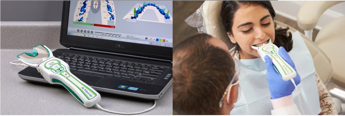
The T-Scan system revolutionizes the assessment of occlusal forces, providing unparalleled accuracy in measuring and balancing a patient's occlusion. By using an ultra-thin sensor, the T-Scan eliminates guesswork associated with traditional articulation paperwork, making evaluation as simple as a patient biting down. Offering both 2D and 3D views, it enables precise measurement of individual contact forces and visualization of occlusal relationships during movements.
In implant prosthodontics, where excessive occlusal loading can lead to material failures and implant complications, the T-Scan II proves invaluable. Prior techniques relied on subjective patient feedback, lacking neurological feedback for accurate assessment. However, the T-Scan II enhances precision by balancing forces on full arch prostheses, removing interferences, and aiding in delayed implant loading. It accurately identifies interference locations and duration of contact during excursive movements, essential for maintaining occlusal balance and longevity of prostheses. Ultimately, the T-Scan II significantly improves the quality and longevity of implant-supported restorations.
COMPUTERIZED JAW RELATION RECORDING SYSTEM
Dental software significantly enhances dental practice capabilities by providing extensive options for data analysis and presentation. It offers features such as overlaying curves for progressive treatment monitoring, zooming for detailed examination of curve segments, and dynamic motion replay for real-time or slow-motion viewing of recordings.
The software facilitates diverse case presentations with 3D movement visualization of the hinge axis and time curves. Additionally, it enables more detailed calculations of condylar data, which can be repeated across various articulators for comprehensive analysis.
Moreover, the software fosters improved communication with dental laboratories by enabling online transmission of data to technicians without requiring special hardware. It also supports the creation of patient databases and the storage of additional documents like text files and images, enhancing record-keeping and documentation capabilities.
COMPUTERIZED REMOVABLE PARTIAL DENTURE DESIGN -STELLIGRAPHE
The software patented by Doctor Gaillard and Jourda, developed by Jacques Orliac and Philippe Beaugrand, streamlines dental procedures with just a few mouse clicks, facilitating the indication of missing teeth for crown and bridge placement. Stelligraph, an integral feature, enables instantaneous realization upon validation.
Stelligraph offers characteristics extraction for various dental elements, including crowns, bridges, and the establishment of hooks, brackets, and supports. Notably, the Total Porcelain System, incorporating the dental colorimeter, Shade Eye System (SES), and Vintage Halo Porcelain, introduces a novel color concept for natural teeth, reducing ambiguity in shade taking.
This innovation significantly decreases the time required for color reconstruction and simplifies the selection of appropriate porcelain powders, even in challenging cases. Moreover, it enhances patient safety by promoting infection control measures and instilling confidence in dentists' reliability.
DIGITAL IMPRESSIONING
Digital dental impressioning devices are classified into two categories: CAD/CAM devices and dedicated intraoral digital scanners. CAD/CAM devices and dedicated intraoral digital scanners are utilized in dental practices for efficient and precise digital impressions.
The Finite Element Method (FEM) is a numerical method employed in stress analysis to solve complex structural problems by dividing structures into smaller segments. Technological advancements in computers and finite element software have enhanced the accuracy and speed of stress analysis, revealing stress and strain distribution throughout the structure under specific loads.
The FE method offers several advantages over other stress analysis methods:
- Non-invasiveness: The technique does not require invasive procedures.
- Simulation of oral structures: Structures such as teeth, alveolar bone, and the periodontal ligament can be accurately simulated, providing a close representation of the oral environment in vitro.
- Measurement of actual stress: The method allows for the measurement of actual stress experienced at any point within the structure.
- Visualization of tooth displacement: Actual tooth displacement can be visually represented graphically.
- Reproducibility: The physical properties of the material involved are not affected by reproducibility, enabling repeated studies without compromising accuracy.
- Accurate modeling: FEM has the potential to accurately model real objects with complex shapes and varying material properties.
These advantages make the Finite Element Method a valuable tool in dental research and analysis, facilitating comprehensive understanding and assessment of structural behavior in dental applications.
CONCLUSION
- The contemporary dental practice offers numerous options for preserving oral health and achieving natural aesthetics.
- Enhanced approaches in dentistry result in reduced treatment time, minimized error potential, and improved quality assurance.
- These advancements contribute to what is often referred to as the golden age of dentistry.
- Digitization has begun to influence the dental fraternity through audiovisual aids in teaching and patient education.
- Digital techniques are increasingly becoming integral to contemporary prosthodontics, with the likelihood of most procedures being based on digital methods in the near future.
- Recent literature on dental techniques helps practitioners assimilate the latest trends for the benefit of patients and the expansion of the dental profession.
- The transition from traditional to digital techniques is driven by the goal of improving patients' lives and advancing the field of dentistry as a whole.
REFERANCES
- Anusavice, Shen, Rawls; Phillip’s Science of Dental
- Materials 2013, 12th edition, Elsevier.
- Ronald L Sakguchi, John M Powers; Craig’s Restorative
- Dental Materials 2013, 13th edition, Elsevier.
- Marco Anntonio Bottino; Perception: Esthetics in Metal Free Prosthesis of Natural Teeth and Implants 2009, Artis Medicas Dentistry.
- Bernard Touati, Paul Miara, Dan Nathanson: Esthetic Dentistry and Ceramic Restorations 1999, informa healthcare
- AD Bona, AD Noguiera, OE Pecho; Optical properties of CAD/CAM ceramic sysems; J Dent 2014, 42: 1202-09
- GD Quin, AA Guiseppetti, KH Hoffman; Chipping fracture resistance of dental CAD/CAM restorative materials; Dent Mater 2014, 30(5): e112-e123
- Z Zhang, Y Tamaki, Y Hotta, T Miyasaki; Novel method for titanium crown casting using a combination of wax patterns fabricated by a CAD/CAM system and a non expanded investment; Dent Mater 2006, 22: 681-87.
- Moldovan, RG Luthardt, N Corcodel, H Rudolph; Three dimensional fit of CAD/CAM made zirconia copings; Dent Mater 2011, 27: 1273-78.
- S Giaunetopoulos, RV Noort, E Triston; Evaluation of the marginal integrity of ceramic copings with different marginal angles using two different CA/CAM systems; J Dent 2010, 38: 980-86.
- ER Batson, LF Cooper, I Duqum, G Mendonca; Clinical outcomes of three different crown systems with CAD/CAM technology; J Prosthet Dent 2014, 28(5): 234-40
- Raul Euan, OF A lvarez, JC Termes, RO Parra; Marginal adaptation of zirconium dioxide copings: Influence of the CAD/CAM system and the finish line design; J Prosthet Dent 2014, 112: 155-62.
- C Chen, FZ Trindade, N de Jager, CJ Kleverlaan, AJ Feilzer; The fracture resistance of a CAD/CAM resin nano-ceramic (RNC) and a CAD ceramic at different thickness; Dent Mater 2014, 30: 954-62.
- Jeramies Hey, F Beur, T Bensel, AF Boeckler; Single crowns with CAD/CAM fabricated copings from titanium: 6 year clinical results; J Prosthet dent 2014, 112: 150-154.
Disclaimer from Guident
The views and opinions expressed in articles published in Guident are solely those of the authors and do not necessarily reflect the views of the editors, the editorial board or the publisher, Ivory India. Guident does not endorse any product, technique or treatment described. Clinical content is intended for qualified dental and medical professionals and is not a substitute for individual clinical judgement. Guident, its editors and the publisher accept no liability for any consequence arising from the use of information published in the journal.
How to cite this article
Dr. Pooja Biswas, Dr. Sanskriti, Dr. Manesh Lahori. Digital Dentistry in Prosthodontics: Transforming Precision and Efficiency in Restorative Dentistry. Guident. 2024 May;17(6). Available from: https://guident.in/articles/prosthodontics/digital-dentistry-in-prosthodontics-transforming-precision-and-efficiency-in-restorative-dentistry.html