Managing Apert Syndrome a Multidisciplinary Way
Department of Pedodontics and Preventive Dentistry, K. D. Dental College, Mathura, U.P. India
INTRODUCTION
Apert syndrome is characterized by triad of symptoms such as midfacial hypoplasia, craniosynostosis and syndactyly of both the limbs. It is considered to be one of the most severe forms of craniosynostosis syndrome and amounts up to 4.5% of total number of cases. Craniosynostosis, in most severe form, can be described as Kleeblattschadel. Apert syndrome has an incidence of 15 cases/million live births. Apert syndrome is inherited in an autosomal dominant fashion with a prevalence of about 1 in 70,000 births. The incidence of Apert syndrome increases with advanced paternal age and is believed to result from mutations arising in the male germ cells that confer a selective survival advantage on sperm cells.
SUTURAL DEFECT : A HALLMARK FINDING
A hallmark finding of all the Apert cases in wide, patent midline defect extending from the glabella up to the posterior fontanelle. This defect can be described as sutural agenesis. However, this defect gets obliterated by the coalescence of bony islands that forms in the defect by the age of 2–4 years. This, therefore, eliminates the formation of proper suture in sagittal and metopic regions. However, suture formation can be formed in coronal, squamous, sphenotemporal and lambdoid. Coronal sutures are commonly fused at births while other sutures and synchondroses of the cranial base are patent (Fig 1)
Systemic abnormalities such as congenital heart defects, respiratory defects, gastrointestinal anomalies, central nervous system-associated anomalies such as corpus callosum anomalies, congenital defects and dermatological manifestations such as Choanal stenosis, cervical spine
CLINICAL MANIFESTATIONS
- Apert syndrome is characterized by premature fusion of cranial sutures, craniofacial anomalies and syndactyly, i.e. cutaneous and bony fusion, bilateral coronal stenosis, widened metopic and sagittal suture.
- Craniofacial features include ocular hypertelorism, down-standing palpebral fissures, midfacial hypoplasia, proptosis and flat nasal bridge. (Fig 2)
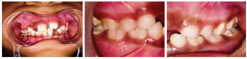
- Oral features include trapezoid shape mouth, cleft lip and palate bifid uvula, malocclusions, byzantine arch palate and shovel-shaped incisors. (Fig 2)
- Syndactyly of all the four limbs that involves 2nd, 3rd, and 4th digits in the form of osseous and cutaneous syndactyly but in severe cases all digits might appear fused along with deeply concave palm. Associated synochia of hands, simple syndactyly of fifth digit and foreshortened thumb with radial clinodactyly. (Fig 3)
- Limitations of shoulder movements, short humerus.
- Systemic abnormalities such as congenital heart defects, respiratory defects, gastrointestinal anomalies, central nervous system-associated anomalies such as corpus callosum anomalies, congenital defects and dermatological manifestations such as Choanal stenosis, cervical spine fusion (C5 and C6).
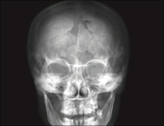
Figure 1: Radiological picture showing sutural defect

Fig 2 clinical picture shows midface hypoplasia and classical trapezoid shape mouth.
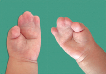
Fig 3 clinical picture shows syndactyly that involves 2nd, 3rd, and 4th digits of upper limbs.

Fig 4 clinical picture shows pointed palate and associated cleft palate.

Fig 5 clinical picture shows thickening of the alveolar process along with Frontal and temporal bulge present bilaterally with a visible construction bond
CRANIOFACIAL MANIFESTATIONS
- Middle third of the face is hypoplastic, underdeveloped and appears flat,
- The nose is small and parrot shaped
- Relative mandibular prognathism due to underdeveloped maxilla
- Apparent hypertelorism and strabismus
- Flattened orbit
- Proptosed eyes
- Horizontal groove appearance over the forehead
- High-arched palate with a median follow.
- Associated cleft lip and palate
- Bifid uvula
- Malocclusion and crowding of teeth causes thickening of the alveolar process (Fig 4)
- Delayed tooth eruption, enamel hypoplasia, ectopic tooth position, teeth agenesis, fusion long canine cusps and supernumerary teeth
- Pointed palate gives rise to protruded middle portion of the upper lip (Fig 4)
- Exophthalmos is generally from mild to moderate in severe cases; it might be associated respiratory difficulty.
- Cranium has oxycephalic appearance. Ovoid and brachycephalic shape gives forehead a high steep and prominent shape
- Frontal and temporal bulge present bilaterally with a visible construction band, isolating temporal region from the cranium (Fig 5)
- The lambdoid sutures are prematurely fused
- Exophthalmos is generally from mild to moderate in severe cases; it might be associated respiratory difficulty.
PRENATAL DIAGNOSIS
- Fetal ultrasound or anomaly scan would show
- Clenched hands.
- Clever leaf skull
- Low set ears
- Syndactyly of hands and limbs
- Hypoplastic left heart
- Choroid plexus cyst
2..PER is a noninvasive prenatal diagnosis
3.Amniocentesis will indicate the mutation analysis of
4.FGFR2 gene and will show a substitution of 5252W zzFetoscopy to visualize fetal abnormalities.
POSTNATAL DIAGNOSIS
- Clinical examination of cranium hands
- Compound tomography can reveal the pathological anatomy radiographs of skull, hand, spine, chest, elbow and feet.
- Molecular analysis of Apert syndrome as more than 98% of cases are due to substitution. Rest are thought to be due to Alu-element insertion mutation.
- Management of Apert syndrome is through multidisciplinary approach .
MANAGEMENT
1. Medical
Airway obstruction immediately after birth:
- Removal of excessive secretions
- Avoiding respiratory infection
- Humidification of the airway
- Use of nasal decongestants.
Sleep Apnea
- Treating recurrent middle ear effusion with the help of microbial therapy
- Polysomnography.
Ophthalmic
- Application of lubricants in the eye for corneal protection
- Artificial tear drops
- Genetic counselling
- Social and emotional development.
2. Surgical Management
The main aim of surgical management is the correction of facial features, fused fingers and obstructive sleep apnea.
Cranial Surgery
- Craniosynostosis requires multistage intervention starting from as early as 3 months. Craniofacial dysjunction is a new technique post-Ilizarov procedure and reported to give complete correction of enophthalmos and correction of middle third without the need of bone graft.
- Removing premature fusions
- Reshaping of cranium to allow normal development of relieving intracranial pressure.
Facial Surgery
- From as early as 3 months:
- Correction of midface hypoplasia by Le Fort III surgery
- Correction of severe sleep obstructive apnea
- Expansion of nasal and nasopharyngeal airway
- Establishing a normal dent oskeletal relationship.
Finger Surgery
Correction of cutaneous and osseous syndactyly of all the digits of hands and nose in 2 stages:
- 9–12 months of age
- Approximately 3 months later to first surgery.
Orbital Surgery
- Correction of orbital proptosis
- Correction of hypertelorism
- Correction of strabismus
- Nasal surgery focuses on:
- Nasal reconstruction due to flat nasal dorsum and obtuse nasofrontal angle—for infants and children
- Reduction of nasal tip bulk in case of adults and teenagers surgeries.
Mandibular Surgery
- Osteotomies are performed to improve the harmony of dental skeletal units and promote masticatory and esthetic functions.
3. Dental management

Disclaimer from Guident
The views and opinions expressed in articles published in Guident are solely those of the authors and do not necessarily reflect the views of the editors, the editorial board or the publisher, Ivory India. Guident does not endorse any product, technique or treatment described. Clinical content is intended for qualified dental and medical professionals and is not a substitute for individual clinical judgement. Guident, its editors and the publisher accept no liability for any consequence arising from the use of information published in the journal.
How to cite this article
Dr. Priya Verma. Managing Apert Syndrome a Multidisciplinary Way. Guident. 2019 Feb;12(3). Available from: https://guident.in/articles/pedodontics/managing-apert-syndrome-a-multidisciplinary-way.html