Management of Peri Radicular Lesions with Calcium Hydroxide
Department of Conservative Dentistry and Endodontics, Seema Dental College And Hospital, Rishikesh
Calcium hydroxide is used extensively as an intracanal medicament in various clinical situations such as to promote apexification, to repair perforation, to enhance healing of periapical lesions, to control root resorption, and to control exudation in teeth with persistent periapical inflammation. This paper presents case reports in which Ca(OH)2 was used as an intracanal medicament for treatment of periradicular lesions.
INTRODUCTION
The main goal of endodontic therapy is to eliminate or at least achieve a significant reduction of microorganisms present in the root canal system. It is well recognized that chemomechanical instrumentation alone is unable to completely disinfect the root canal system 1. The bacteria surviving root canal instrumentation proliferate between appointments. The use of intracanal medication with antimicrobial activity between appointments has been recommended to eliminate possible persistent microorganisms, particularly in case of pulp necrosis with periradicular bone loss [2]. Calcium hydroxide with its antimicrobial property has been used in endodontics for interappointment intracanal dressing. It has been demonstrated that treatment with calcium hydroxide as an interim dressing in the presence of large and chronic periapical lesions can create an environment more favorable to healing and encourage osseous repair 3.
Several bacterial species commonly found in infected root canals are eliminated after a short period when in direct contact with this substance 4.
Antimicrobial activity of calcium hydroxide is related to release of hydroxyl ions in an aqueous environment. Hydroxyl ions are highly oxidant-free radicals that show extreme reactivity resulting in the damage to bacterial cytoplasmic membrane, protein denaturation, and damage to bacterial DNA 1, 4–7.
It has been asserted that all biological actions of calcium hydroxide progress by the ionic dissociation in calcium ion and hydroxyl ion (Estrela 1995) 8. In general three types of vehicles are used: aqueous, viscous, or oily. The first group is represented by water soluble substances including water, saline, dental anesthetics, Ringer’s solution, aqueous suspension of methyl cellulose or carboxymethyl cellulose, and anionic detergent solution. When calcium hydroxide is mixed with one of these substances, Ca2+ and OH− ions are rapidly released 10. This type of vehicle promotes a high degree of solubility. The root canal may become empty in a short period, delaying the healing process. From a clinical standpoint, this means that root canal must be redressed several times until the desired effect is achieved, thereby increasing the number of appointments.
Some viscous vehicles are water soluble substances that release Ca++ and OH− ions more slowly for extended periods. They promote lower solubility of the paste when compared with aqueous vehicles, for example, glycerine, poly ethylene glycol, and propylene glycol 9.
Oily vehicle are nonwater soluble substances that promote the lowest solubility and diffusion of paste within the tissues. Paste containing this type of vehicle may remain within the root canal for longer than the paste containing aqueous or viscous vehicle, for example, olive oil, silicone oil, camphor, metacresylacetate, and some fatty acids 9.
CASE 1
A 17-year-old patient came to the Department of Conservative Dentistry and Endodontics, Seema Dental College and Hospital, Rishikesh, with complaint of pain in right side in relation to the posterior teeth of lower jaw. On oral examination, it was found that and 46 was cariously exposed. On radiographic examination, a large radiolucency in enamel and dentin approaching pulp was seen. The periapical area showed radiolucency in the mesial and distal roots of 46. Root canal treatment was decided for the tooth. Access opening was done in 46. Following the working length estimation in 46, a thorough chemomechanical preparation was performed. The root canals were irrigated with combination of sodium hypochlorite and sterile saline solution. The canals were dried with sterile paper points. After the canals were dried with paper points, Calcium Hydroxide syringe was inserted in the canals and the material was injected into the canal. The calcium hydroxide was further applied using appropriate size lentulospiral until the canal was completely filled with it. The procedure was repeated with the other three canals. The completed root canal filling with calcium hydroxide was verified with the help of radiograph. The access cavity was sealed with intermediary restorative material. Radiographs were taken at 3-month interval. Tooth showed satisfactory radiographic healing after 6 months After 6 months, removal of Ca(OH)2 paste from 46 was done using copious amount of sodium hypochlorite and saline and final rinse with EDTA. Obturation was done followed by post endodontic restoration.
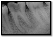
PRE OPERATIVE
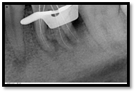
WORKING LENGTH
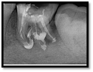
CALCIUM HYDROXIDE PLACED

3 MONTHS FOLLOW UP
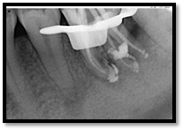
MASTER CONE

OBTURATION & POSTENDODONTIC COMPOSITE RESTORATION

3 MONTH FOLLOW UP CASE REPORT 2
A 23-year-old patient came to the Department of Conservative Dentistry and Endodontics, Seema Dental College and Hospital, Rishikesh, with complaint of pain in left side in relation to the posterior teeth of lower jaw. On oral examination, it was found that and 36 was cariously exposed. On radiographic examination, a large radiolucency in enamel and dentin approaching pulp was seen. The periapical area showed radiolucency in the mesial and distal roots of 36. Root canal treatment was decided for the tooth. Access opening was done in 36. Following the working length estimation in 36, a thorough chemomechanical preparation was performed. The root canals were irrigated with combination of sodium hypochlorite and sterile saline solution. The canals were dried with sterile paper points. After the canals were dried with paper points, Calcium Hydroxide syringe was inserted in the canals and the material was injected into the canal. The calcium hydroxide was further applied using appropriate size lentulospiral until the canal was completely filled with it. The procedure was repeated with the other three canals. The completed root canal filling with calcium hydroxide was verified with the help of radiograph. The access cavity was sealed with intermediary restorative material. Radiographs were taken at 3-month interval. Tooth showed satisfactory radiographic healing after 6 months After 6 months, removal of Ca(OH2 paste from 36 was done using copious amount of sodium hypochlorite and saline and final rinse with EDTA. Obturation was done followed by post endodontic restoration.
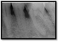
Pre operative radiograph
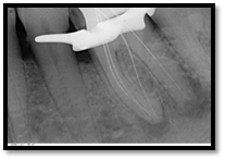
Working length
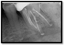
CALCIUM HYDROXIDE PLACED
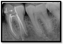
Follow up at 3 months interval
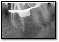
Master cone

Obturation with post endodontic composite restoration
DISCUSSION
Microorganisms are the cause of apical periodontitis and their elimination from the root canal space during root canal treatment results in predictable healing of apical pathosis. Unfortunately, the complete elimination of bacteria by instrumentation alone is unlikely to occur. In addition, pulp tissue remnants may prevent microorganisms from being entombed as well as have a negative impact on the root filling in terms of its physical properties and adaptation to the canal walls. Thus, some form of irrigation and disinfection is necessary to kill and remove microorganisms, their by-products, and residual tissue as well as remove the smear layer and other debris from the canal system. Such chemical (therapeutic) treatments of the root canal can be arbitrarily divided into irrigants, canal rinses, and interappointment medicaments. Calcium hydroxide is included in the intracanal medicaments group.
Since its introduction to dentistry calcium hydroxide (Hermann 1920) has been indicated to promote healing in many clinical situations 11, 12. Although the overall mechanism of action of calcium hydroxide is not fully understood, its antimicrobial activity is influenced by its speed of dissociation into calcium and hydroxyl ions in a high pH environment which inhibits enzymatic activities that are essential to microbial life, that is, metabolism, growth, and cellular division. Ca(OH)2 is often used to effect periapical healing by combination of its antimicrobial activity and its ability to promote hard tissue formation and periodontal healing 1, 4–7.
When calcium hydroxide is mixed with suitable vehicle, a paste is formed for use in endodontics.
The type of vehicle has a direct relationship with the concentration and the velocity of ionic liberation as well as with the antibacterial action when the pasted is carried into the contaminated area 10, 13.
Different vehicles have been added to calcium hydroxide in an attempt to enhance to its antimicrobial activity, biocompatibility, ionic dissociation, and diffusion.
When Ca(OH)2 is mixed with saline solution, Ca++ and OH− are rapidly released when the paste comes in direct contact with the tissue and tissue fluids; it is rapidly solubilized and resorbed by macrophages; the root canal becomes empty in a short period. Some studies consider 4-week period to be a reasonable time interval to expect effective therapeutic benefits from Ca(OH)2 saline dressings 2, 14–16.
CONCLUSION
Calcium hydroxide has a great value in endodontics, being indicated for several clinical conditions. In the present case, calcium hydroxide was used to treat periapical lesions. Calcium hydroxide proved to be an effective intracanal medicament and resulted in successful resolution of the periapical lesions.
REFERENCES
- C. A. S. de Souza, R. P. Teles, R. Souto, M. A. E. Chaves, and A. P. Vieira Colombo, “Endodontic therapy associated with calcium hydroxide as an intracanal dressing: Microbiologic evaluation by the checkerboard DNA-DNA hybridization technique,” Journal of Endodontics, vol. 31, no. 2, pp. 79–83, 2005. View at Publisher · View at Google Scholar · View at Scopus
- M. Trope, “Treatment of immature teeth with non-vital pulps and apical preriodontitis,” Endodontic Topics, vol. 14, pp. 51–59, 2006. View at Google Scholar
- A. Milosevic, “Calcium hydroxide in restorative dentistry,” Journal of Dentistry, vol. 19, no. 1, pp. 3–13, 1991. View at Publisher · View at Google Scholar · View at Scopus
- R. M. Simcock and M. L. Hicks, “Delivery of calcium hydroxide: comparison of four filling techniques,” Journal of Endodontics, vol. 32, no. 7, pp. 680–682, 2006. View at Publisher · View at Google Scholar ·View at Scopus
- C. Estrela, L. L. Bammann, F. C. Pimenta, and J. D. Pécora, “Control of microorganisms in vitro by calcium hydroxide pastes,” International Endodontic Journal, vol. 34, no. 5, pp. 341–345, 2001. View at Publisher · View at Google Scholar · View at Scopus
- C. Estrela, C. R. A. Esterla, and J. D. Pecora, “A study of the time necessary for calcium hydroxide to eliminate microorganisms in infected canals,” Journal of Applied Oral Science, vol. 11, no. 2, pp. 133–137, 2003. View at Google Scholar
- C. Estrela, “Two methods to evaluate the antimicrobial action of calcium hydroxide paste,” Journal of Endodontics, vol. 27, no. 12, pp. 720–723, 2001. View at Publisher · View at Google Scholar · View at Scopus
- C. Estrela, G. B. Sydney, H. F. Pesce, and O. Felippe Júnior, “Dentinal diffusion of hydroxyl ions of various calcium hydroxide pastes,” Brazilian Dental Journal, vol. 6, no. 1, pp. 5–9, 1995. View at Google Scholar · View at Scopus
- L. R. G. Fava and W. P. Saunders, “Calcium hydroxide pastes: classification and clinical indications,” International Endodontic Journal, vol. 32, no. 4, pp. 257–282, 1999. View at Publisher · View at Google Scholar · View at Scopus
- G. H. Robert, F. R. Liewehr, T. B. Buxton, and J. C. McPherson III, “Apical diffusion of calcium hydroxide in an in vitro model,” Journal of Endodontics, vol. 31, no. 1, pp. 57–60, 2005. View at Publisher · View at Google Scholar · View at Scopus
- R. J. G. Moor and A. M. J. C. de Witte, “Periapical lesions accidentally filled with calcium hydroxide,” International Endodontic Journal, vol. 35, no. 11, pp. 946–958, 2002. View at Google Scholar
- J. F. Siqueira Jr. and H. P. Lopes, “Mechanisms of antimicrobial activity of calcium hydroxide: a critical review,” International Endodontic Journal, vol. 32, no. 5, pp. 361–369, 1999. View at Publisher · View at Google Scholar · View at Scopus
- I. C. Gomes Camões, M. R. Salles, and O. Chevitarese, “Ca2+ diffusion through dentin of Ca(OH)2associated with seven different vehicles,” Journal of Endodontics, vol. 29, no. 12, pp. 822–825, 2003. View at Publisher · View at Google Scholar · View at Scopus
- M. G. Pacios, M. L. de la Casa, M. de los Ángeles Bulacio, and M. E. López, “Influence of different vehicles on the pH of calcium hydroxide pastes,” Journal of Oral Science, vol. 46, no. 2, pp. 107–111, 2004.View at Publisher · View at Google Scholar · View at Scopus
- R. Weiger, R. Rosendahl, and C. Löst, “Influence of calcium hydroxide intracanal dressings on the prognosis of teeth with endodontically induced periapical lesions,” International Endodontic Journal, vol. 33, no. 3, pp. 219–226, 2000. View at Publisher · View at Google Scholar · View at Scopus
- O. Zmener, C. H. Pameijer, and G. Banegas, “An in vitro study of the pH of three calcium hydroxide dressing materials,” Dental Traumatology, vol. 23, no. 1, pp. 21–25, 2007. View at Publisher · View at Google Scholar · View at Scopus
- Z. Mohammadi and P. M. H. Dummer, “Properties and applications of calcium hydroxide in endodontics and dental traumatology,” International Endodontic Journal, vol. 44, no. 8, pp. 697–730, 2011. View at Publisher · View at Google Scholar · View at Scopus
Disclaimer from Guident
The views and opinions expressed in articles published in Guident are solely those of the authors and do not necessarily reflect the views of the editors, the editorial board or the publisher, Ivory India. Guident does not endorse any product, technique or treatment described. Clinical content is intended for qualified dental and medical professionals and is not a substitute for individual clinical judgement. Guident, its editors and the publisher accept no liability for any consequence arising from the use of information published in the journal.
How to cite this article
Dr. Annil Dhingra, Dr. Nayanika Singh Rathore, Dr. Sumit Sabharwal. Management of Peri Radicular Lesions with Calcium Hydroxide. Guident. 2019 Feb;12(3). Available from: https://guident.in/articles/endodontics/management-of-peri-radicular-lesions-with-calcium-hydroxide.html