Management of Cystic Lesions in Endodontics
Department of Conservative Dentistry and Endodontics Seema Dental College And Hospital, Rishikesh
INTRODUCTION
Successful endodontic therapy depends on complete periapical repair and regeneration. In cases where conventional root canal therapy fails to eliminate the lesion, surgery is the last alternative.1 Periapical surgery aims to remove periapical pathology followed by achieving complete wound healing and regeneration of the bone and periodontal tissue.2,3 In modern endodontic practice, the number of indications for endodontic periapical surgery is decreasing, but still periapical surgery accounts for 3%–10% of typical endodontic practice.4 The most commonly used root end filling material is MTA because it provides superior sealing ability when compared with Amalgam, IRM and Super EBA. MTA is the best root-end filling material. This material is almost non-resorbable and non-toxic, having bacteriostatic properties to heal, and offers a good apical seal.
Histological examination showed the stimulation of cementum and hard tissue formation with minimal inflammatory response after using this hydrophilic material MTA.5Hence, in this study we have used MTA in the apical third of root to form a good apical seal in the fractured root.
CASE REPORT
A 18-year female patient came to the Department of Conservative Dentistry and Endodontics, Seema dental college and hospital with the complaint of pain in relation to the maxillary left anterior region. On diagnosis patient gave history pain i.r.t 22 and 23 since last 3 months.
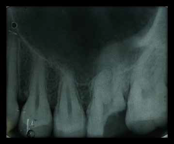
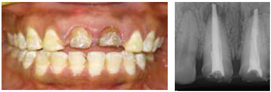
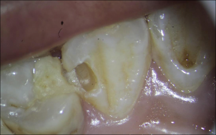
On clinical examination, there was grade I mobility . On radiographic examination revealed a well-defined periapical radiolucency of about 1.1 × 1.0 cm around the apices of maxillaryleft lateral incisor and canine. Both the teeth tested responsive to thermal and electric pulp testing. There was only mild tenderness to percussion and palpation. The diagnosis was irreversible puilpitis with chronic apical abscess with respect to maxillary lateral incisor and canine.
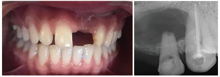
Pre-Op

Flap Raised
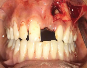
Cavity Prepared
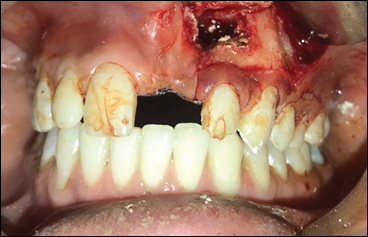
Mta Placed
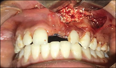
Bone Graft Placed
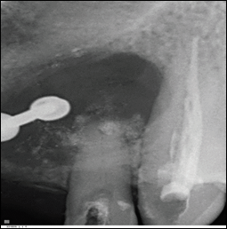
Mta Placed

Bone Graft Placed
MANAGEMENT
The treatment plan was decided as cystic anucleation in the maxillary left lateral incisor and canine followed by root end resection.
After effective local anesthesia with 2% lignocaine, bleeding points were marked after that a full-thickness Luebke Ochsenbein flap was reflected. A large defect was visible in the cortical plate with the Lateral incisor, which was enlarged to aid in complete curettage of the granulation tissue (which was sent for biopsy). This was followed by irrigation with betadine (Win Medicare; Win Medicare Dental Products, New Delhi, India) and sterile saline solution. Root end resection done. After that MTA was placed into the defect and completely used as root end filling material in teeth. The flap was repositioned and closed with 4-0 silk suture.After suture placement removal applicance placed in oral cavity for support and for proper healing.
REFERENCES
- Mazumbar P, Bhunia S. Treatment of periapical lesion with platelet rich fibrin. Indian Med Gazette 2013:28‑33.
- Singh S, Singh A, Singh S, Singh R. Application of PRF in surgical management of periapical lesions. Natl J Maxillofac Surg 2013;4:94-9.
- Mazumdar S, Joshi S, Ansari S. Experiences with the use of PRF (Plasma Rich Fibrin) in enucleated cystic cavity. J Indian Dent Assoc 2014;8:19-26.
- Hiremath H, Motiwala T, Jain P, Kulkarni S. Use of second‑generation platelet concentrate (platelet‑rich fibrin) and hydroxyapatite in the management of large periapical inflammatory lesion: A computed tomography scan analysis. Indian J Dent Res 2014;25:517-20.
- Shivashankar VY, Johns DA, Vidyanath S, Sam G. Combination of platelet rich fibrin, hydroxyapatite and PRF membrane in the management of large inflammatory periapical lesion. J Conserv Dent 2013;16:2614.
- Shivashankar VY, Johns DA, Vidyanath S, Kumar MR. Platelet Rich Fibrin in the revitalization of tooth with necrotic pulp and open apex. J Conserv Dent 2012;15:395-8.
- 7Sunitha Raja V, Munirathnam Naidu E. Platelet‑rich fibrin: Evolution of a second generation platelet concentrate. Indian J Dent Res 2008;19:42-6.
- Kiran NK, Mukunda KS, Tilak Raj TN. Platelet concentrates: A promising innovation in dentistry. J Dent Sci Res 2011;2:50-61. 9. Bhandari S, Ashwini TS, Naik R, Bandiwadekar T, Makandar S. Regenerative periapical surgery: A case report. Dent Hypotheses 2013;4:61-6.
- Boyapati L, Wang HL. The role of stress in periodontal disease and wound healing. Periodontol 2000 2007;44:195-210.
- Gassling V, Douglas T, Warnke PH, Açil Y, Wiltfang J, Becker ST. Platelet‑rich fibrin membranes as scaffolds for periosteal tissue engineering. Clin Oral Implants Res 2010;21:543-9.
- Corso MD, Toffler M, Ehrenfest DM. Use of autologous leucocyte and platelet rich fibrin (L‑PRF) in post‑avulsion sites: An overview of Choukran’s PRF. J Implant Adv Clin Dent 2010;1:27-35.
- Deenadayalan E, Kumar A, Tewari RK, Mishra SK, Iftekhar H. Management of large periapical lesion with the combination of second generation platelet extract and hydroxyapatite bone graft: A report of three cases. J Clin Diagn Res 2015;9:D24-7.
- Nagaveni NB, Kumari KN, Poornima P, Reddy V. Management of an endo-perio lesion in an immature tooth using autologous plateletrich fibrin: A case report. J Indian Soc Pedod Prev Dent 2015;33:69-73.
- Simonpieri A, Del Corso M, Sammartino G, Dohan Ehrenfest DM. The relevance of Choukroun’s platelet-rich fibrin and metronidazole during complex maxillary rehabilitations using bone allograft. Part II: Implant surgery, prosthodontics, and survival. Implant Dent 2009;18:220-9.
- Naik B, Karunakar P, Jayadev M, Marshal VR. Role of Platelet rich fibrin in wound healing: A critical review. J Conserv Dent 2013;16:284-93.
Disclaimer from Guident
The views and opinions expressed in articles published in Guident are solely those of the authors and do not necessarily reflect the views of the editors, the editorial board or the publisher, Ivory India. Guident does not endorse any product, technique or treatment described. Clinical content is intended for qualified dental and medical professionals and is not a substitute for individual clinical judgement. Guident, its editors and the publisher accept no liability for any consequence arising from the use of information published in the journal.
How to cite this article
Dr. Annil Dhingra, Dr. Shalini Sharma, Dr. Mayuri Kumari. Management of Cystic Lesions in Endodontics. Guident. 2019 Mar;12(4). Available from: https://guident.in/articles/endodontics/management-of-cystic-lesions-in-endodontics.html