Cleft Lip and Palate: A Review on Multidisciplinary Treatment Approach
Department of Pedodontics and Preventive Dentistry K. D. Dental College, Mathura, U.P. India
Cleft lip and cleft palate are the most common congenital malformations occurring in humans. These structural defects of the facial-oral complex may vary from slight notching of the lip or a small cleft in uvula to a complete separation of the lip and absence of the partition between the oral and nasal cavities. Clefts are usually associated with numerous problems such as malformed teeth, malocclusion and impairment of speech; middle ear infection and a high susceptibility to upper respiratory infections. The children affected with this anomaly are handicapped right from birth for breastfeeding, deglutition, improper growth and development of maxilla and overall face development affecting the total personality of the individual.
In India, though no regular epidemiological surveys have been conducted. From reports, the pooled incidence of major malformations in total births at 26 large hospitals, from big cities and towns from different parts of the country, is estimated to be 14.82 per 1,000 births; in other parts of the world, the incidence of this anomaly is reported to be the highest in Afghans. Its prevalence ranges from 1:500 in China to 1:800 in Asia and europe and up to 1:2,000 in Africa.
MULTIDISCIPLINARY MANAGEMENT IN CLP
For convenience, treatment is divided into four stages, which generally correspond to stages in the child’s dental development. Feeding problems are often associated with cleft infants, making it difficult to maintain adequate nutrition. These problems include insufficient suction to pull milk from the nipple, excessive air intake during feeding (requiring several burpings), choking, nasal discharge, and excessive time required for nourishment.
Some believe that such appliances are not effective in facilitating feeding. Pashayan and McNab recommend using a standard nipple that has been crosscut. This enlarged cut provides improved ejection of the milk into the infant’s mouth with a minimum of effort. Although this recommendation is appropriate for infants born with isolated clefts of the palate; obturator construction is generally indicated for those born with complete clefts of the lip and palate. This is especially important in consideration of the maxillary orthopedic molding of the alveolar segments after surgical closure of the lip.
Impression Technique and Obturator Construction
An irreversible hydrocolloid impression of the infant’s maxillary arch is made with a stock tray. The infant is held upright during the impression to prevent aspiration of excess material. Appropriate emergency equipment including forced oxygen, suction and standard airway management equipment should be available at chairside. The impression is then poured in dental stone, and a custom tray is fabricated from the model. Again, with the infant held upright, an irreversible hydrocolloid impression is made using the custom tray. The impression should exhibit good anatomic detail with coverage of the entire maxillary arch. A stone model is then produced (Figs 1A and B).
Steps in Obturator Construction
The steps in obturator construction follow: Block out excessive undercuts with modeling dough or wax. Modeling dough is preferred because it is easy to remove from the finished prosthesis. Apply a tinfoil substitute over the entire surface of the maxillary model and let it dry.
If necessary, place a dam of modeling dough on the back of the model to hold the resin in the palatal defect while curing. Pour a mixture of soft autopolymerizing acrylic resin into the cleft to the level of the palate. This will provide retention for the prosthesis by gently contouring into the available undercuts. Place the model in a warm, moist environment to cure for 20 minutes. Add autopolymerizing acrylic resin to the plate in a “salt and pepper” method, making sure the acrylic resin extends well into the mucobuccal fold area. Remove the appliance from the model and rinse the wax and modeling dough off with hot water. The appliance is then trimmed and polished.

Fig 1A and B: Impression recording and feeding plate for the newborn
Clinical Management of Initial Obturator Therapy (Birth to 3 Months)
The appliance is positioned in the infant’s mouth. Areas of excessive pressure on any intraoral tissues by the acrylic resin are identified by observation and reduced. Care is taken to keep the acrylic resin from impinging on any muscle attachments or extending to the depth of the buccal vestibule. Parents are instructed on placement and removal of the appliance and its daily cleaning. The infants are usually seen for adjustments 2 days after initial appliance delivery. Monthly observations are then scheduled. In most cases, this appliance will serve until the time of initial lip closure at approximately 3 months of age. The major advantage of the obturator during this stage of treatment is to enhance the child’s ability to obtain nourishment.
Premaxillary Orthopedics (Birth to 4 or 5 Months)
In some cases of bilateral CLP, the infant has a premaxillary segment that is either positioned severely anterior to the maxillary arch segments or deviated laterally to one side of the cleft defect. This presents a difficult clinical challenge for the surgeon before surgical closure of the lip. If lip surgery is undertaken with the premaxilla in such an abnormal position, the chances of lip dehiscence (lip separation caused by increased pressure at the suture lines) are increased. In 1686, Hofman described the use of a head cap and premaxillary strap to reposition the premaxilla. This type of apparatus is useful in anteroposterior and vertical repositioning. After delivery of the obturator, the infant is allowed to become accustomed to the appliance for a 1 week period. At the second appointment, the infant is fitted with a premaxillary retraction appliance. Acrylic “bulb” prosthesis is fitted over the protruding and laterally displaced premaxilla and anchored to the infant’s head with a Bonnet appliance. By the application of sequentially increasing differential forces to the premaxilla with elastic straps attached to the bulb prosthesis, the maxilla is brought into the facial midline. Laterally displaced premaxilla can be repositioned to the facial midline in 3–4 weeks. After the premaxilla is in the midline, the bulb appliance is replaced by a single elastic strap. Over the next 1–2 months, equal pressure is applied on the still protruding premaxilla to retroposition it into a more normal position between the lateral segments.
The rationale for use of bulb prosthesis before elastic strap retraction includes the following considerations: The bulb prosthesis affords greater control over the differential forces applied to the premaxilla. Movement of the premaxilla into the facial midline before retropositioning (rather than laterally deviated position) decreases the risk of distorting a vomer stalk. The consideration of a surgical premaxillary sit back (a procedure known for possible growth attenuation as well as other complications) is eliminated. Optimal premaxillary positioning may eliminate the need for a staged lip closure (adhesions before definitive lip repair), thereby decreasing total hospitalization time and cost. The appearance of the nose and lip is improved by the fact that the lip incisions can be surgically closed under less tension and by an underlying symmetric alignment of the alveolar segments. Airway Obstruction Infants with airway obstruction secondary to the Pierre Robin anomalad (glossoptosis, macroglossia, micrognathia) and cleft palate may require some form of surgery immediately after birth. If a nonsurgical approach (positioning oral airways or using an orogastric tube) is unsuccessful, lip-to-tongue adhesion (thus anterior positioning of the tongue and opening of the oral airway) or tracheostomy may be necessary.
Cheiloplasty (Surgical Lip Closure)
Lip surgery will significantly improve the infant’s appearance and may thereby relieve parental apprehensions and enhance acceptance of the child. Surgical closure of the cleft lip may be accomplished shortly after birth. A general “rule of tens” (10 weeks of age, 10 pounds of body weight, and 10 g of hemoglobin) is frequently used in determining optimal timing for lip closure. At the time of lip closure, when the infant is under the general anesthetic, an impression is made for new obturator to accommodate craniofacial growth during the first few months of life (Figs 2A to D).
Maxillary Orthopedics (3–9 Months)
After lip closure at about 3 months of age, maxillary arch collapse in unilateral or bilateral complete cleft is common. It is attributable to the increased tension placed on the segments by the repaired lip. To prevent this collapse, the obturator is used to provide crossarch stability and support. As pressure is exerted on the anterior segments of the maxilla by the repaired lip, orthopedic molding of the segments can be achieved. In unilateral cases, the force applied to the greater segment by the intact lip molds that segment around to approximate the lesser segment and also resists any tendency for the greater
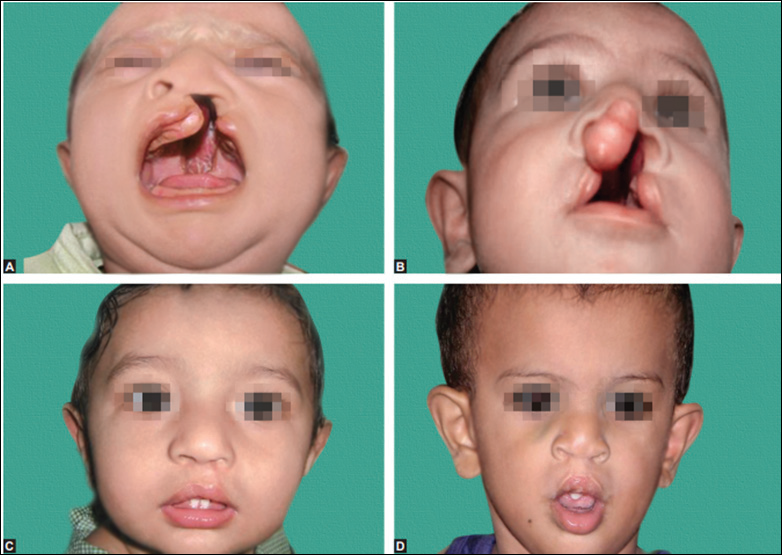
Fig 2A to D: Preoperative and postoperative pictures of cheiloplasty
and lesser segments to collapse toward the midline. In bilateral cleft cases, the repaired lip provides further retraction at the premaxilla, positioning it between the two lateral maxillary segments. When the maxillary segments are in good alignment and abutted across the cleft sites, the patient is ready for the primary cleft bone graft. This generally occurs by 6–9 months of age.
Bone Grafting of Alveolar Cleft Defects
Bone grafting of alveolar cleft defects has been a confusing issue to many patients and practitioners. According to Boyne and Sands primary bone grafting refers to bone grafting procedures to alveolar cleft defects in children less than 2 years of age. Early secondary bone grafting refers to bone grafting procedures done between 2 years and 4 years of age. Secondary bone grafting refers to reconstruction of residual alveolar cleft defects in the adult.
Palatoplasty (Surgical Closure of Palate)
Closure of the palate is accomplished between 12 months and 2 years of age. The primary purpose of palate closure by 2 years of age is to facilitate the acquisition of normal speech, because this correlates with the age at which most children begin to talk. It may also improve hearing and swallowing by aligning the cleft palatal musculature. After primary closure of the cleft palate, approximately 25% of patients demonstrate some velopharyngeal insufficiency. A persistent inability to close the nasopharynx may result in unsatisfactory speech (nasality and articulation problems), regurgitation of fluids from the nose and facial grimacing. Of the various surgical approaches to correct velopharyngeal insufficiency, the pharyngeal flap is most commonly used. The procedure is usually done when velopharyngeal insufficiency is documented in an attempt to normalize the speech of the child before he or she begins school (Figs 3A and B).
Stage II (Primary Dentition Stage—18 Months To 5 Years Of Age)
Treatment during this phase of dental development is initially focused on oral health to reduce the possibility of developing dental decay. Ectopic eruption of the primary maxillary anterior dentition is common around the cleft defect. Special care should be taken to keep these teeth free from decay because food often becomes lodged in and around the cleft defect. An increase in periodic recall examinations, possibly at 3–4 month intervals, enables the dentist to intercept areas of decay. This preventive regimen continues throughout all subsequent stages in the management of the cleft patient. In unilateral and bilateral complete clefts of the lip and palate, surgical closure of the palatal defect is postponed beyond the usual 18–24 months of age. In these cases, because of the development of speech at this age, maxillary prosthetic appliances are constructed to provide normal maxillary arch integrity. As the child
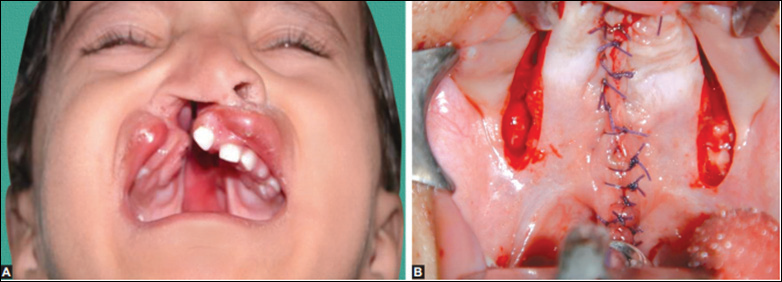
Fig 3A and B: Preoperative and postoperative pictures of palatoplasty
grows, more tissue will become available to close the palate when doing so is surgically appropriate.
Stage III (Late Primary or Mixed Dentition Stage—6 to 10 or 11 Years of Age)
Many problems encountered during this stage of dental development arise from ectopically erupting permanent central and lateral incisors or cross-bites of the posterior dental segments. Interceptive correction of a traumatic occlusion is essential to prevent destruction of enamel in the involved dentition. Maxillary expansion, to correct posterior segmental collapse, is accomplished by routine palatal expansion in patients who have not undergone primary alveolar cleft bone grafting. Once corrected, retention can be maintained by passive holding appliances.
Secondary Alveolar Cleft Bone Graft
A successful alveolar cleft bone graft satisfies several objectives. In addition to giving bony support for the teeth adjacent to the cleft and providing bone through which teeth can erupt, it offers maxillary arch continuity and aids in closure of the oronasal fistula. It also supports the alar base of the nose.
The soft tissues in and adjacent to the cleft side are incised and elevated, so that labial and palatal mucosal leaflets are everted to obtain labial and palatal closure. The tissues lining the cleft are elevated and inverted into the nose for nasal floor closure. Particulate marrow and cancellous bone harvested from the iliac crest is placed into the cleft defect, filling it from the pyriform rim to the alveolar crest before closure of the labial tissues. Providing bony support to teeth adjacent to the cleft site is of paramount importance. In most cases, one should graft bone into the cleft before beginning orthodontic tooth alignment. When the cleft is filled with normal, viable bone, the orthodontist can proceed with tooth alignment without fear of exposing a root surface into the cleft site. In fact, after a 2-month healing period, a tooth can be moved into the newly grafted bone with the expectation that the bone will respond to the tooth movement as any normal bone would.
Secondary alveolar cleft bone grafting has met with widespread acceptance. Success rates are generally in the 90% range. Morbidity has included pain in the donor site, dehiscence of mucosal flaps, and partial or complete loss of grafted bone. Infection in either the donor or recipient sites has been rare. Secondary alveolar cleft bone grafting is an important procedure that greatly facilitates total rehabilitation. Of special importance in the comprehensive dental of CLP is the dentist’s ability to provide the young patient with interim prostheses to improve facial appearance. These can be periodically adjusted to allow eruption of the developing dentition. Such treatment, when possible, should begin before the child starts formal education.
The majority of persons with CLP require some degree of orthodontic management and can be treated in a conventional manner. Treatment during this stage of dental development concentrates on comprehensive orthodontic or surgical management of the permanent dentition. The orthodontist, before initiating the indicated therapy, completes a full diagnostic examination determines the status of the patient’s current craniofacial development. In some instance, children with a cleft with a severely retrusive maxilla cannot undergo orthodontic correction with conventional therapy. In these cases, the surgical procedure most often utilized is the LeFort I maxillary advancement. This procedure is not technically feasible until the patient has a full complement of permanent dentition. The horizontal cuts to free the maxilla must necessarily be made above the apices of permanent dentition. Unerupted cuspids, or bicuspids, would make this procedure impractical. Therefore, the surgery must be deferred until the patient’s permanent dentition has erupted. Another available option is distraction osteogenesis
CONCLUSION
The congenital cleft lip and/or cleft palate anomaly has a very important place in Health Sciences because of epidemiology, racial ethnic variation, etiology, surgery, orthodontics, speech pathology and prosthetic rehabilitation. The children afflicted with this anomaly are handicapped right from birth for breastfeeding, deglutition, recurrent nasobronchial infection, defective speech along with improper growth and development affecting the total personality of the individual. The present-day society demands the total personality development of such an individual so that the individual is not a stigma as well as a burden on the society. Ever-expanding participation of dentist is essential in the rehabilitation of individuals. Hence, it is mandatory for the dentist to be familiar with complete knowledge of this complex problem to make these children esthetically, psychologically and functionally capable.
Disclaimer from Guident
The views and opinions expressed in articles published in Guident are solely those of the authors and do not necessarily reflect the views of the editors, the editorial board or the publisher, Ivory India. Guident does not endorse any product, technique or treatment described. Clinical content is intended for qualified dental and medical professionals and is not a substitute for individual clinical judgement. Guident, its editors and the publisher accept no liability for any consequence arising from the use of information published in the journal.
How to cite this article
Dr. Priya Verma. Cleft Lip and Palate: A Review on Multidisciplinary Treatment Approach. Guident. 2019 Jan;12(2). Available from: https://guident.in/articles/pedodontics/cleft-lip-and-palate-a-review-on-multidisciplinary-treatment-approach.html