Surgical Management of Failed Root Canal Treatment
Department Of Conservative Dentistry And Endodontics Seema Dental College And Hospital, Rishikesh
INTRODUCTION
Periapical lesion is a local response of bone around the apex of tooth that develops after the necrosis of the pulp tissue or extensive periodontal disease. The successful treatment of periapical inflammatory lesion depends on the reduction and elimination of the offending organism. Root canal therapy, periapical surgery, or extraction of the tooth might be the treatment alternatives.1
Periapical surgeryor Root end resectioninvolves the surgical management of a tooth with a periapical lesion which cannot be resolved by conventional endodontic treatment (root canal therapy or endodontic retreatment).It includes the curettage of all periapical soft tissues and sometimes application of different biomaterials to enhance the new bone formation in the defect site.
PRP, PRF and Bone graftshave been used for optimal healing of the periapical defect area after degranulation of the lesion. All these approaches are known as regenerative therapies.Platelet-rich plasma (PRP), introduced by Whitman et al. in 1997, is a natural source of growth factors (GF) used to enhance bone regeneration. It is an autologous concentrate of platelets suspended in plasma. It is well known that platelets have many functions beyond that of simple hemostasis. Platelets contain important GF that, when secreted, are responsible for increasing collagen production, recruiting other cells to the site of injury, initiating vascular ingrowth, and inducing cell differentiation. These are all crucial steps in early wound healing.
On the other hand, platelet-rich fibrin (PRF), a second-generation platelet concentrate, has shown to be superior to PRP. It was first developed in France by Choukroun et al. in 2001. PRF allows one to obtain fibrin membranes enriched with platelets and GF after starting from an anticoagulant-free blood harvest. Recently, studies have demonstrated that the PRF membrane has a very significant slow sustained release of many key GF for at least 1week and up to 28 days, which means that PRF could release GF with its own biological scaffold for wound healing process.
The present case report describe the management of the periapical inflammatory lesion by root end resectionusing PRF which acts as a source of GF at the healing site, thereby accelerating soft and hard tissue healing.2
CASE REPORT
A 23-year-old male patient presented to the Department of Conservative Dentistry and Endodontics, Seema dental college and hospital with the complaint of pain in relation to the maxillary left anterior region. On diagnosis patient gave history of root canal treatment i.r.t 21 and 22 twice in last six month. On clinical examination, there was grade I mobility in maxillary left centraland lateral incisor.On radiographic examination revealed a well-defined periapical radiolucency of about 1.2 × 1.0 cm around the apices of maxillary left central and lateral incisors.Both theteeth tested responsive to thermal and electric pulp testing. There was only mild tenderness to percussion and palpation. The diagnosis was irreversible pulpitis with chronic apical abscess with respect to maxillary left central and lateral incisors.
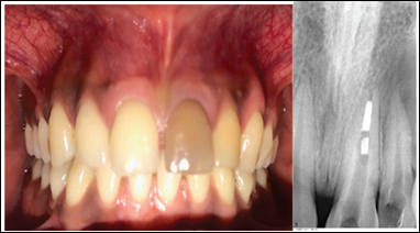
Figure 1A-Pre-Operative,Figure 1B-Pre-Operative Xray
MANAGEMENT
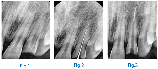
The treatment plan was decidedasrootcanal therapy in the maxillary left central and lateral incisors followed by root end resection.The canals were obturated withlateral condensation using gutta-perchapoints of 20 0.6% taper(DentsplyMaillefer, Ballaigues, Switzerland)(fig-11) and Apexit plus (ivoclarvivadent) under rubber dam isolation, 1 day prior to the surgery. Guttaperchawas extended to locate the extent of lesion.After effective local anesthesia with 2% lignocaine, a full-thickness LuebkeOchsenbein flap was reflected(fig-4). A small defect was visible in the cortical plate with the lateral incisor, which was enlarged to aid in complete curettage of the granulation tissue (which was sent for biopsy)(fig-5). This was followed by irrigation with betadine (Win Medicare; Win Medicare Dental Products, New Delhi, India) and sterile saline solution. Then theextendedguttapercha wascut.PRF was processed from 20 ml of the patient’s blood and packed into the defect to completely fill the bony defect(fig-7). The flap was repositioned and closed with 4-0 silk suture followed by COE-PAK(Gc America)(fig-8).3
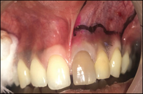
Figure 2-MarkThe Bleeding Points
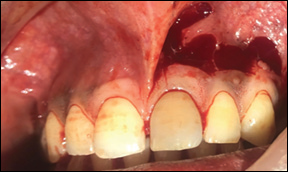
Figure 3-Incision Placed

Figure 4-Flap Raised
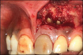
Figure 5-Granulation Tissue Removed
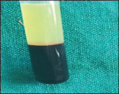
Figure 6-PRF
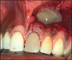
Figure 7-PRF Placed
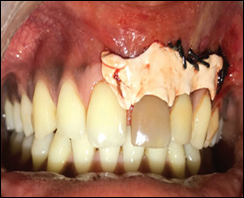
Figure 8-Suture with Coe -PAK
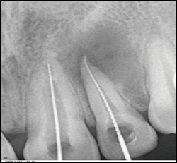
Figure 9-Working Length
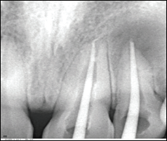
Figure 10-Master Cone Selected
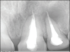
Figure 11-Obturation

Figure 12-After Root End Resection
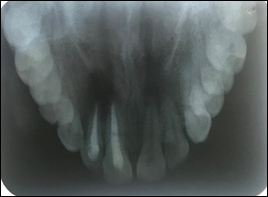
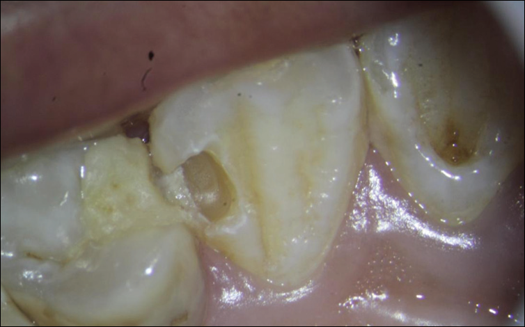
Figure13-Occlusal View After Root End Resection
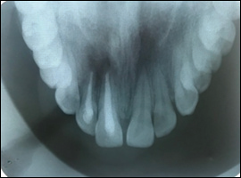
Figure 14-After 6 Month Follow-Up
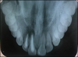
Figure 15-After 12 Month Follow Up
Discussion
Root end resection involves the surgical management of a tooth with a periapical lesion which cannot be resolved by conventional endodontic treatment (root canal therapy or endodontic retreatment). Because the term “Root end resection” consists of only one aspect (removal of root apex) of a complex series of surgical procedures, the terms “periapical surgery” or “periradicular surgery” are more appropriate. The expressions “periapical endodontic surgery” and “apical microsurgery” are also found in the literature. The objective of periapical surgery is to obtain tissue regeneration.4This is usually achieved by the removal of periapical pathologic tissue and by exclusion of any irritants within the physical confines of the affected root.A root end resection is done only after a tooth has had at least one root canal procedure and retreatment has not been successful or is not possible. For example, retreatment is often not a good option when a tooth has a crown or is part of a bridge. Retreatment of the root canal would require cutting through the crown or bridge. That might destroy or weaken the crown or bridge. A root end resection is often considered in a situation like this.An root end resection is not the same as a root resection. In a root resection, an entire root is removed, rather than just the tip.
The majority of periapical lesions are associated with endodontic pathology, except in cases of rare developmental cysts or tumors, the primary goal of treatment is orthograde occlusal approach for root canal instrumentation and obturation.
“The richest of resources in nature lie in the nature itself.” The world seems to be going back to natural substitutes for all its queries and dilemmas. Modern day medicine and surgery are certainly no exception to this rule.PRP is a natural source of GF that is used to enhance bone regeneration. But PRF, a second-generation platelet concentrate, has shown to be superior to PRP. Advantages of PRF over PRP include:
- Ease of preparation (blood centrifuged only once)
- Ease of application
- Minimal expense (sophisticated equipment not required)
- Lack of biomechanical modification (anticoagulant or thrombin or calcium chloride not required unlike PRP)3
The major role of fibrin in wound repair is haemostasis, but fibrin also provides a matrix for themigration of fibroblasts and endothelial cells that are involved in angiogenesis and are responsible for remodelling of new tissue.Platelet activation in response to tissue damage and vascular exposure results in the formation of a platelet plug and blood clot, as well as the secretion of biologically active proteins. Platelet alpha (α) granules form an intracellular storage pool of GF including platelet-derived growth factor (PDGF), transforming growth factor β (TGF-β, including β-1 and β-2-isomers), vascular endothelial growth factor (VEGF), and epidermal growth factor (EGF). Insulin-like growth factor-1 (IGF-1), which is present in plasma, can exert chemotactic effects toward human osteoblasts. After platelet activation, the α granules fuse with the platelet cell membrane, transforming some of the secretory proteins to a bioactive state. Active proteins are secreted and bind to transmembrane receptors of the target cells to activate intracellular signaling proteins.This results in the expression of a gene sequence that directs cellular proliferation, collagen synthesis, and osteoid production.
The clinical experience confirms that PRF (fig-6)can be considered as a healing biomaterial and it features all the necessary parameters permitting optimal healing, but numerous perspectives of PRF have still to be clinically tested.5
The 6 and 12 months follow up of the patients radiograph shows adequate healing periapically concluding the use of PRF as a healing biomaterial andits success in promoting regeneration of the soft andhard tissues. Long term follow up is essential to evaluate the outcome of the treatment(fig-14 and 15).
References
- Platelet-rich fibrin (PRF) as an autologous biomaterial after an endodontic surgery: Case reports.KavithaAnantula, AliveniAnnareddy.Journal of Dr. NTR University of Health Sciences 2016.
- ApicoectomybyChalmerS,J. LYONS, D.D.Sc., Ann Arbor, MichiganJour. A. D. A., January,1915.
- Role of Platelet rich fibrin in wound healing: A critical review(BalaramNaik, P Karunakar1, M Jayadev1, V Rahul Marshal)Journal of Conservative Dentistry | Jul-Aug 2013 | Vol 16 | Issue 4
- Application of platelet-rich fibrin in endodontic surgery: a pilot study.Daniele Angerame a, Matteo De Biasi b, Iva Kastriotia,Vittorio Franco c, Attilio Castaldo a, Michele Maglione.GiornaleItaliano di Endodonzia(2015).
- Apical surgery: A review of current techniques and outcome,Thomas von Arx,The Saudi Dental Journal (2011)23,9–15.
Disclaimer from Guident
The views and opinions expressed in articles published in Guident are solely those of the authors and do not necessarily reflect the views of the editors, the editorial board or the publisher, Ivory India. Guident does not endorse any product, technique or treatment described. Clinical content is intended for qualified dental and medical professionals and is not a substitute for individual clinical judgement. Guident, its editors and the publisher accept no liability for any consequence arising from the use of information published in the journal.
How to cite this article
Dr. Annil Dhingra, Dr. Anshdeep Singh, Dr. Mayuri Kumari. Surgical Management of Failed Root Canal Treatment. Guident. 2019 Jan;12(2). Available from: https://guident.in/articles/endodontics/surgical-management-of-failed-root-canal-treatment.html