Techniques for Intrusion in Deep Bite Cases-a Review Article
A B Shetty Memorial Institute of Dental Science, Mangalore, India, SMBT Dental College Ahmadnagar, India, Department of Orthodontics and Dentofacial Orthopaedics, A B Shetty Memorial Institute of Dental Science, Mangalore, India
ABSTRACT
Orthodontic anterior intrusion for correcting deep bites often constitutes an integral part of orthodontic treatment in order to improve sagittal and vertical incisor relationships, in cases of pseudo-deep overbite, where the anteriors are supraerrupted true intrusion of the incisors is indicated and also in patients with gummy smiles and periodontal compromised teeth to correct the gingival line and restore the esthetics of smile. Intrusion is mainely achived by use of verious loop mechnices or implant mechanics or differiantial force concept given by beggs.In this artical different mechanics for intrusion in beggs technique and PEA technique are mentioned.
KEYWORDS: Intrusion arch, Mechanics, Force, Mini implant.
INTRODUCTION
Deepbites can affect a person’s esthetic appearance and smile.1Anterior deepbites caused by overeruption of the maxillary incisors can be determined by using lateral cephalometric radiographs. If the lower lip covers more than 4 mm of the maxillary central incisors on a patient’s lateral cephalometric radiographs, it is the result of maxillary incisor overeruption.2Depending on the diagnosis and treatment objectives,a deep overbite can be corrected by intruding the incisors,extruding the buccal segments, or combining these treatments.1,3
Extrusion of posterior teeth drops the mandible downward and backward, and the condyle assumes a new position in the temporomandibular joint articulation. If equilibrium is achieved between function, muscles, and the temporomandibular joint after orthodontic treatment by remodeling and readaptation, the extrusion of the posterior teeth and the successful treatment of the deep overbite remain stable.
Dr.Charles j. Burston5 given forces values for deep bite correction
|
Intrusion |
Per-Side |
Total in Midline |
|
Upper central inciosr |
15-20(gm) |
30-40(gm) |
|
Upper central and lateral incisor |
30-40(gm) |
60-80(gm) |
|
Upper central lateral and canine |
60(gm) |
120(gm) |
|
Lower central incisor |
12.5(gm) |
25(gm) |
|
Lower central and lateral incisor |
25(gm) |
50(gm) |
|
Lower central lateral and canine |
50(gm) |
100(gm) |
In adults,however, the mastication muscles and altered occlusion might move the extruded posterior teeth back to the original positions until equilibrium between the softand hard tissues is obtained again and relapse occurs.4Maxillary incisor intrusion should be the preferred treatment in nongrowing patients with anterior deepbites caused by overeruption of the maxillary incisors.4
VERIOUS TYPE OF INTRUSION MECHNIQUES
Intrusion in Refined Beggs6
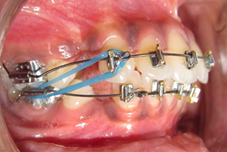
Anchor bends produced 45 grams of force and class II elastics produced 30 grams of force. Therefore the net resultant force was 15 grams on each side this 15 grams spread over three teeth amounting about 5 grams on each tooth which was too little for active intrusion
So, initially the intrusive force through anchor bend should be given around 45 grams and class II force should be 60 grams. In this situation there is more of retractive force than the intrusive force. Later the intrusive force is increased to 60 grams and the class II decreased to 30 grams which will result in more intrusion.
The increase in the force of intrusion is obtained by the increasing the degree of anchor bends gradually. Giving 30 degree bend initially and increasing to 50 degrees. The decrease in class II elastic force is done by placing the elastics for longer time i.e 3-5 days or by changing the elastics from yellow to road runner elastics.
INTRUSION TECHNIQUES IN PRE ADJUSTED EDGEWISE
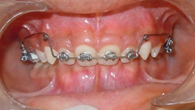
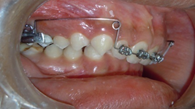
1)Ricketts Utility Arch7
Utility arch having molar section, posterior vertical step (3-4mm) in height, vestibular component, anterior vertical segment (6-8mm) in height, incisor segment.It should be fabricated in 0.019 X 0.025 TMA wire in 0.22 slot.

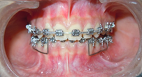

Rectangular wire is preferred to the round wire to control torque and to prevent the uncontrolled tipping In molar segment 30-40 degree tip back applied and 30-45 degree buccal root torque applied to prevent lingual tipping of molar.In incisal segment 5-10- degree lingual torque is given to prevent labial flaring of incisor. lower utility arch gives 50-75 gm of continues force.
It can be use for retraction and protraction by incorporating helix.8
2)Three piece intrusion arch9
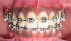
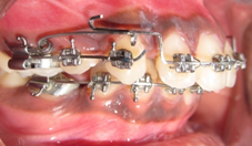
Bhavna shroff’s intrusion arch having a posterior Unit made up of 17*25 s.s. wire goes in to molar and premolar which increase the anchorage, Anterior segment made up of 17*25 s.s having an extension 2-3 mm distal to lateral incisor, which help in passing the intrusive force through center of restance of anterior segment, Intrusive spring made up of 17*25 TMA wire, which gives intrusion force on anterior segment.
A small distal force can be added by placing an elastomeric chain extending form the molars to the anterior segment of wire on each side.This force facilitate simultaneous intrusion and retraction by redirecting the force parallel to the incisor long axis.
3)K-Sir arch10
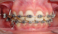
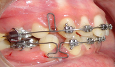

Dr Varun Kalra intrudoces K-SIR (Kalra Simultaneous Intrusion and Retraction) arch which is a modification of the segmented loop mechanics of Burstone and Nanda which can be made up of .019“x.025" TMA archwire with closed U-loops 7mm long and 2mm wide.90° bends placed in archwire at level of U-loops to obtain bodily movement and prevent tipping of the teeth into the extraction spaces.Archwire with off-center 60° V-bend placed about 2mm distal to U-loop.Off-center V-bend creates greater moment on molar, increasing molar anchorage and intrusion of anterior teeth.20° antirotation bends placed in archwire just distal to U-loops.
Trial activation performed on each loop.Archwire cinched back to activate loop about 3mm, so that mesial and distal legs are barely separated.
4)Translation arch11
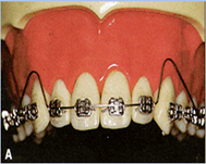
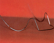
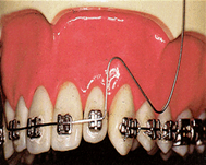
Translation Arch was given by Martina , Paduano.Translation Arch has anterior segment inserted into incisor brackets and two buccal segments inserted into gingival first molar tubes.
Vertical loops between segments extend as far as possible. Root-palatal (torque)given in the anterior segment of the arch .An arc—should be bent into each buccal segment to produce an intrusive force of 40g on the incisors. A distal activation of 2mm on each end of the arch will produce the 100g of retraction force.
5) CONNECTICUT INTRUSION ARCH12
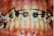

The CTA is fabricated from a nickel titanium alloy to provide the advantages of shape memory, springback, and light,continuous force distribution.Two wire sizes are available: .016" X .022" and .017" X .025".The maxillary and mandibular A tight cinch-back bend distal to the molar tube, preventing forward slippage of the wire will prevent incisor flaring during intrusion and produce some retraction of the incisors during molar tipback. Intrusion rate is1mm / 6 weeks.
It also can be use for correction of occlusal cant,flaring of incisor and close of open bite.
6) PG RETRACTION SPRING13


Pg retraction spring was given by P0UL GJESSING. 0.017 X 0.025“ or 0.016 X 0.022” SS wire is used for PG springs.
The posterior extension of the spring is always inserted in the gingival auxiliary molar tube, but the anterior extension can be attached to the lateral bracket in several ways. The most practical is to use .018"´.025" lateral incisor brackets with vertical Broussard-type slots. The anterior extension is placed in the vertical slot of the lateral incisor bracket, pulled as far occlusally as possible, and locked with a mesial bend.
The spring is activated by pulling distal to the molar tube until the two loops separate. The wire is secured with a gingival bend in the posterior extension. Reactivation to the initial spring configuration should be done every four to six weeks. Magnitude of intrusive forces produced at the anterior segment are determined by the posterior curvature of the spring , this curvature was adjusted to deliver the required force magnitude of 10-25g per side.
7) Lingual Arch for Intruding and Uprighting Lower Incisors14
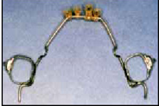
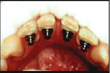

This technique was introduced by WINSTON SENIOR. An .036" lower lingual arch is soldered to first molar bands. Distal extensions form occlusal rests on the second molars to prevent distal tipping of the first molars as the incisors are intruded. Four elastic chains are attached to the anterior bridge of the lingual arch with a mosquito forceps .
After cementation of the arch, the elastics are stretched to four lingual buttons on the lower incisors . These should be bonded as far as possible from the gingival margin to facilitate intrusion.
8)Mini implant
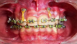
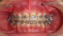
The sample patients were treated using PEA appliance with 0.022 slot MBT prescription. After the initial alignment of the incisors with 0.016 Niti wire (approximately 2 months); mini-implants were placed between the maxillary lateral incisor and canines bilaterally. Patients were treated with mini-implants of the self drilling variety (8mm in length and 1.3mm in diameter).
THE SURGICAL PROCEDURE
Under local anaesthesia micro-implant was placed apical to the free gingival margin between the maxillary lateral incisor and canines.
After two weeks, the implant was loaded with a Niti closed coil spring/E chain. A force of approximately 50gms was applied from the E chain/ spring to a 17x25 SS wire. The wire was cinched back on the molars to prevent flaring of the anterior teeth.
The mean upper incisor intrusion achieved was 2.5 mm
The movement of the maxillary molars led to the loss of sagittal and vertical anchorages during intrusion of the incisors in the Connecticut intrusion arch group, these anchorages were maintained in the implant groups.
CONCLUSION
The precise determination of the point of application of the intrusive force as well as direction are critical in intrusion of anterior teeth. This article describe the various intrusion appliance used in Refined beggs and pre-adujusted edgewise system for treatment of deep over bite cases.
Disclaimer from Guident
The views and opinions expressed in articles published in Guident are solely those of the authors and do not necessarily reflect the views of the editors, the editorial board or the publisher, Ivory India. Guident does not endorse any product, technique or treatment described. Clinical content is intended for qualified dental and medical professionals and is not a substitute for individual clinical judgement. Guident, its editors and the publisher accept no liability for any consequence arising from the use of information published in the journal.
How to cite this article
Dr. Chirag Panchasara, Dr.Muralidhar R Sastri, Dr.U.S. Krishna Nayak. Techniques for Intrusion in Deep Bite Cases-a Review Article. Guident. 2013 Jun;6(8). Available from: https://guident.in/articles/orthodontics/techniques-for-intrusion-in-deep-bite-cases-a-review-article.html