Molar Protraction
Department of Orthodontics & Dentofacial Orthopedics, Manipal College of Dental Sciences, Manipal, India, Department of Orthodontics & Dentofacial Orthopedics, Manipal College of Dental Sciences, Manipal, India
ABSTRACT
Molar protraction is always considered a tough job. This article reviews the clinical indications, associated difficulty, biomechanics, various modalities and potential problems with molar protraction.
Keywords: Corticotomy, Microimplants, Molar Protraction.
INTRODUCTION
Orthodontists are often confronted with adult patients who have large edentulous spaces in the molar areas or need molar extractions.The extraction of first permanent molar teeth in the British National Health Service accounted for nearly 12% of all extraction cases.1
Extraction of all first permanent molar has been a controversy. Those who favor claim it to prevent malocclusion, dental decay, pyorrhea, while others suggest that it causes significant occlusal disturbance, an increase in caries, and detrimental change in facial contour and appearance.
Daugaard-Jensen2 suggested that first molar extraction cases are no more time consuming than 4 premolar extraction cases and offer distinct advantages in terms of anchorage management.Houston et al3 suggested deprived social background and reduced interest in dental care as main contributory factors in children undergoing first permanent molar extractions.
CLINICAL INDICATIONS FOR EXTRACTION OF FIRST PERMANENT MOLARS:
- Extensively carious first molars.
- Hypoplastic first molars.
- Heavily filled first molars where premolars are perfectly healthy.
- Apical pathoses or root treated first molars.
- Crowding at the distal part of the arches and wisdom teeth reasonably positioned.
- High maxillary/mandibular planes angle.
- Anterior open bite cases.
Other factors include the patient’s attitude to fixed appliance therapy, the standard of oral hygiene, the amount and site of crowding, and the presence or absence of other permanent teeth. The patients suitability for this invariably lengthy course of fixed appliance therapy must be considered to ensure that the benefits of treatment far outweigh the potential risks.
Satisfactory closure of spaces is, therefore, best achieved in children and young adults.4 Adults showed less bone apposition when moving second molar teeth into the narrowed space, and poor maintenance of the closed space and, in some cases, resorption of the second molar roots was also noted.
Other authors5 concluded that complete closure could be achieved with the roots of the second molar teeth moving almost twice as far as the crowns. They agreed that most cases showed a crestal bone loss mesial to the second molars post treatment but suggested that root resorption of the second molars was only minimal.
Kessler6 contraindicated the mesial movement of second molar if the buccolingual width of the second molar alveolus is wider than the adjacent edentulous ridge. In contrast, Brown7 showed that uprighting molars produced significant reductions in the depth of existing periodontal defects; the gingival margin relocating from a coronal position to a more inferior location adjacent to the cementum of the anatomic root. He also found that alveolar bone level decreased between 0.5 and 1.0 mm, along with reduction of total pocket depth. However, he considered this amount to be therapeutically insignificant.
Ingber8 showed that forced eruption can correct isolated 1- or 2-wall infrabony osseous defects. Extrusion of a tooth from its alveolus causes a stretching of the gingival and periodontal ligament fibers. This results in a coronal shift of the bone at the base of the defect as the tooth moves occlusally. These vertical osseous defects frequently occur with an inclined alveolar crest on the mesial side of the second molar.
THEN WHY IS MOLAR PROTRACTION DIFFICULT?
Anchorage loss is more challenging in the mandible than in the maxilla, because of the structural differences between the two jaws. In the molar region, the maxilla has an average buccal cortical thickness of 1.5mm, compared with 2mm in the mandible. The rate of molar protraction is inversely related to the radiographic density or cortical thickness of the resisting alveolar bone. Because of the increased thickness of mandibular cortical bone, the rate of mandibular molar translation with skeletal anchorage is nearly half that of maxillary molar translation—approximately .34-.60mm per month.
It has been proposed by Roberts et al9 that distal root subsequently engages the more dense bone and rate of tooth movement slows down. Thus, the rate of tooth movement appears to be related to the ability of the body to remodel the relatively dense, immature bone formed by the leading root.
BIOMECHANICAL PRINCIPLE
Differential Torque- According to Thomas F. Mulligan when the bend is placed off center, the tooth (bracket or tube) located closest to the bend contains the largest moment and, therefore, indicates the anchor side.10
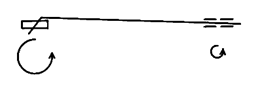
Fig 1
The wire used for molar protection is usually an 0.018 followed by an 0.020 to reduce the tipping tendency. If mesiolingual molar rotation is desired no bend is placed, as a mesial force acting at the molar tube during protraction produces the rotation as a result of the "Cue Ball" effect.
VARIOUS STRATEGIES
- Interarch elastics.
- Bimetric arch.
- Class II correctors for mandible.
- Running loop archwire
- Temporary anchorage device.
Class II ELASTICS
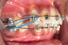
Class II elastics are quite effective as an anchorage source when it is desirable to advance or develop the lower arch forward. The retruded lower arch is quite common in strong facial patterns and can be utilized quite effectively to help achieve overall facial balance.
When employing class II elastics at a greater force level and with increased angulation, relatively greater force loss and increased side effects, such as extrusion and mesial tipping of the first molar, occur.11


Fig 2 A, B, C : showing lower molar protraction with class II elastics
BIMETRIC ARCH
It consists of 0.016×0.022 inch arch wire. The wire is twisted such that 0.016 inch segment lies in the molar region and 0.022 inch segment lies in the anterior region reinforcing anchorage.
FIXED FUNCTIONAL APPLIANCES
A. Mandibular protraction appliance (MPA)
After using part of the mandibular first molar spaces to diminish the arch length discrepancy and/or reduce the mandibular incisor protrusion, the anterior teeth are anchored with a MPA, and a closing loop archwire with activated bull loops is employed to bring the mandibular second molars forward. The third molars ordinarily follow the second molars in their forward movement.12
B.Twin Force Bite Corrector
Twin Force Bite Corrector can be used to simultaneously correct the Class II malocclusion and serve as anchorage for protraction of a mandibular molar into the edentulous space. It consists of two parallel 15mm cylinders housing nickel titanium coil springs, with a plunger incorporated into each cylinder at opposite ends. At full compression, it produces 210g of force at each end and postures the mandible forward into an edge-to-edge position.13
Similarly other fixed functional appliances FORSUS, Eureka spring, Sabbagh universal spring can also be used for molar protraction as it locks the mandible in a forward position which serves as anchorage and prevent anterior retraction.
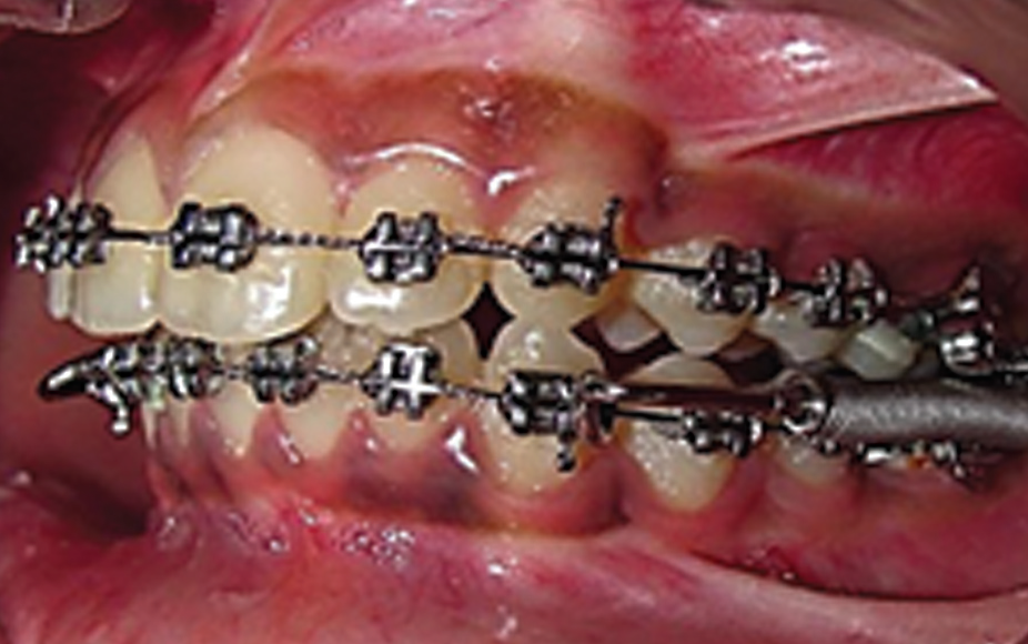
Fig 3 : showing FORSUS
RUNNING LOOP ARCHWIRE
A running loop archwire can be used to close an edentulous space in the molar area. It is usually made of 0.018 × 0.025-in stainless steel wire. It’s helical running loop is wound to the outside with an external diameter of 3 mm. The distance between the mesial end of the buccal tube and the running loop should be maintained at 5 mm, an effective tipback of 20° to 30° should be given according to the amount of mesial tipping of molars, and a slight toe-in is necessary to prevent mesiolingual rotation of the molars. Forces of 150 to 250 g can be applied from the hook of the buccal tube to the helical loop, the canine, and the micro-implant with an elastic chain.14

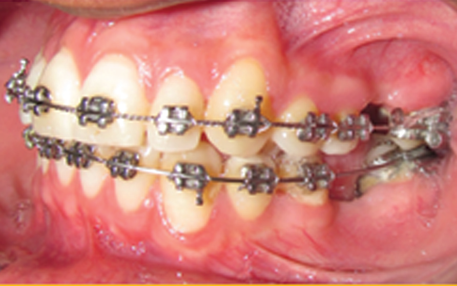
Fig 4 A, B : showing running loop
RETROMOLARIMPLANT
The anchorage implant 3.75mm × 7mm standard Branemark fixture can be used. Following local anesthesia, the implant can be placed in the retromolar area about 5mm distal to the mandibular third molar.15
PALATAL MICROIMPLANT
Two palatal microimplants 1.3x8 mm can be placed on the either side or a single palatal implant can be placed to apply force on transpalatal arch or spurs soldered on the palatal side of upper molar bands.16,17
LINGUAL ELASTIC TIED TO THE ARCHWIRE 18
This involves protraction with a balancing lingual force, such as an elastic thread tied from the lingual cleat of the molar to the archwire to avoid posterior crossbite and open bite. Other method to avoid the roll in of molars is to incorporate the second molars into the archwire to minimize arch expansion with a rectangular archwire.
An occlusal gable bend (upward V-bend) in the archwire mesial to the edentulous space to counteract molar intrusion can be placed. Alternatively, if an auxiliary slot is used, a buccal hook can be fabricated from a wire segment to protract the tooth at its center of resistance.
SLIDING BAND ON LINGUAL ARCH 18
A lingual arch with a sliding band may provide greater support than lingual elastic thread. The lingual arch consists of an0.040" wire soldered to the molar band on the side opposite the lone molar. A sliding band with a headgear tube soldered to its lingual surface is cemented to the lone molar at the same appointment. The lingual arch extends through this tube, acting as a guide rail during protraction. After protraction is complete, the clinician can cut the lingual arch from the soldered band.
THE “PUSH-PULL” TECHNIQUE18
The clinician may insert the Temporary Anchorage Device (TAD) within the edentulous space and protract from the second tooth back, using an open-coil spring to push the tooth in front of it. Advantages include simple miniscrew insertion with minimum risk of root perforation.
SIMULTANEOUS UPPER MOLAR DISTALIZATION AND LOWER MOLAR PROTRACTION
This can accomplished with the help of jig in the upper arch aided with microimplant (TAD) to stabilize the premolar and use of class II elastics.
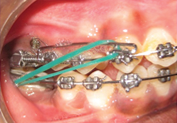
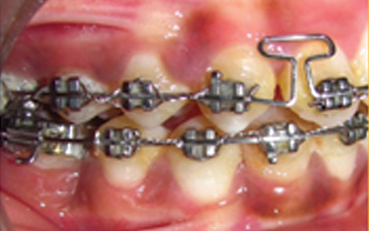
Fig 5 A, B : Simultaneous upper molar distalization and lower molar protraction
CORTICOTOMY ASSISTED ORTHODONTICS
Corticotomy surgery initiates and potentiates normal healing process by way of an accelerated transient burst of hard and soft tissue remodeling by means of a process called REGIONAL ACCELERATORY PHENOMENON (RAP). It was described by an Orthopedist Harold frost. Wilckos thought that patient could benefit from alveolar augmentation in conjunction with a decorticating procedure. Thus, corticotomy can be used to facilitate protraction.19

Fig 6 : showing corticotomy and bone graft placement to facilitate 3rd molar protraction
POTENTIAL PROBLEMS 20
- Lower Arch Space Closure
- Upper Arch Space Closure
- Timing of Extractions
- Ridge resorption
- Root Paralleling
- Class II elastics
- Arch Wire Problems
- Microimplant failure
There is always a tendency for the molars to both tilt mesially and roll lingually when elastics are applied. This tendency can be reduced by the use of full size (19/25) stainless steel arch wires. Active space closure should rarely be attempted before the patient has full alignment of all the lower teeth and is in this full-sized working arch wire.
Closure of the upper first molar extraction spaces is rarely time consuming. Indeed, because space closure occurs so readily, if a reasonable amount of the extraction space is required, consideration should be given for the use of a palatal arch with Nance button on the second molars or a headgear must be provided to the upper second molars to prevent them from moving mesially.
Effective distalization of upper premolars with a removable appliance can be achieved if the first molars are extracted early.However, extraction of the first molars must be delayed until the second molars have erupted sufficiently. If space requirement in the upper arch is minimal, earlier extraction will allow “nature” to assist with much of the space closure. The second molars are often quite high and only need to alter their eruption pathway slightly more mesially to allow them to erupt almost into the first molar sockets.
However, the lower second molars cannot completely replace the lower first molars after their extraction because of their more vertical path of eruption.
If little or no space is required in the lower arch, it is often advisable to extract lower first molars early to maximize spontaneous space closure in the lower arch. On the contrary, a lingual arch may be necessary if space is required to relieve crowding.
Missing teeth for years in adult patients show alveolar ridge resorption. The rate of resorption is greatest during the first several months to two years after extraction, but decreases thereafter.
Post-extraction resorption is significantly greater on the buccal than on the lingual side. During the first year after tooth extraction, the amount of resorption in the mandible is twice that in the maxilla—a ratio that increases to 4:1 after seven years.
Potential risks of molar protraction through an atrophic ridge include loss of attachment (particularly in the presence of plaque), dehiscence, mobility, ankylosis, root resorption, devitalization, and tooth morbidity.
If 8 to 9 mm of space has been closed between the second premolar and second molar, there is always a slight tendency for divergence between the two roots and gentle tip back bends may be placed if necessary in the final rectangular wires to fully correct the root positions. Once the crowns are correctly positioned, a dead ligature should be placed across the extraction spaces to hold them closed for a few months to allow reorganization of gingival fibres.
Use of full-sized (19/25) stainless steel arch wire and, if necessary, buccal crown torque can help prevent the lingual roll in of posteriors. The Class II elastics can also be placed from a lingual cleat on the lower molar bands, thus providing some lift to the lingual surface of the lower molars.
Long span of arch wire can cause trauma to the soft tissues as well as deflection of the arch wire during mastication. This can be avoided by placing 0.9 mm internal diameter stainless steel tubing over the arch wire in the extraction space. The tube should be only 1 to 2 mm short of the interbracket span for maximum rigidity.
This can be attributed to root proximity and greater buccal tissue mobility.20
CONCLUSIONS
Molar protraction if controlled can yield great results. In the near future, mandibular molar protraction with orthodontic Temporary Anchorage Device (TAD) may become the standard of care.Opening the mandibular molar spaces and closing them with a fixed prosthesis does not appeal to many patients because of partial destruction of the abutment teeth.
When patients have longstanding edentulous spaces, the vertical dimension of the alveolar ridges at the sites seriously diminish, which may limit implant placement. However, the decision on whether to proceed with orthodontic tooth movement through an atrophic ridge must be made on a case-by-case basis.
REFERENCES
- Bradbury AJ. A current view on patterns of extraction therapy in British health service orthodontics. Br Dent J 1985;159:47-50.
- Daugaard-Jensen I. Extraction of first molars in discrepancy cases. Am J Orthod 1973;64:115-36.
- Houston WJB, Stephens CD, Tulley WJ. A textbook of orthodontics, 2nd edition, Oxford: John Wright; 1992. p. 196-9.
- Stepovich ML. A clinical study on closing edentulous spaces in the mandible. Angle Orthod 1979;49:227-33.
- Hom BM, Turley PK. The effects of space closure of the mandibular first molar area in adults. Am J Orthod 1984;4:457-69.
- Kessler M. Interrelationships between orthodontics and periodontics. Am J Orthod 1976;70:154-72.
- Brown IS. The effect of orthodontic therapy on certain type of orthodontic defects. J Periodontol 1973;44:742-56.
- Ingber JS. Forced eruption – a method of treating isolated one or two wall infrabony osseous defects- rationale and case report. J Periodontol 1974;45:199-206.
- Roberts, Arbuckle, Analoui. Rate of mesial translation of mandibular molars using implant-anchored mechanics, Angle Orthod 1996;66:331-8.
- Mulligan T. Common sense mechanics-part 10. J ClinOrthod 1980;14(6):412-16.
- Schumacher HA, Bourauel C, Drescher D. Analysis of forces and moments in arch guided molar protraction using Class I and Class II elastics-An in-vitro study. J OrofacOrthop. 1996;57(1):4-14.
- Filho, Coelho, White. Closing mandibular first molar spaces in adults. World J Orthod 2006;7:45-58.
- Amirparviz R. Davoody, Jonathan Feldman, Flavio A. Uribe, Ravindra Nanda. Mandibular Molar Protraction with the Twin Force Bite Corrector in a Class II Patient. J ClinOrthod 2011;4:223-8.
- JongmoonChae, Sangcheol Kim. Running loop in unusual molar extraction treatment. Am J OrthodDentofacialOrthop 2007;132:528-39.
- Roberts, Marshall, Mozsary. Rigid endosseous implant utilized as anchorage to protract molars and close an atrophic extraction site. Angle Orthod 1990;60:135-52.
- Proffit WR. Contemporary Orthodontics. 4th ed. St Louis, Mosby Year Book 2009.
- Nanda R. Temporary anchorage devices in orthodontics. Mosby 2008. Page 130,131,135.
- Sandler PJ, Atkinson R, Murray AM. For four sixes. Am J OrthodDentofacialOrthop 2000;117:418-34.
- Wilcko MT, Wilcko WM, Pulver JJ, Bissada NF, Bouquot JE. Accelerated Osteogenic Orthodontics Technique: A 1-Stage Surgically Facilitated Rapid Orthodontic Technique With Alveolar Augmentation. J Oral MaxillofacSurg 2009;67:2149-59..
- Kravitz ND, Jolley T. Mandibular Molar Protraction with Temporary Anchorage Devices. J ClinOrthod 2008;6:351-5.
Disclaimer from Guident
The views and opinions expressed in articles published in Guident are solely those of the authors and do not necessarily reflect the views of the editors, the editorial board or the publisher, Ivory India. Guident does not endorse any product, technique or treatment described. Clinical content is intended for qualified dental and medical professionals and is not a substitute for individual clinical judgement. Guident, its editors and the publisher accept no liability for any consequence arising from the use of information published in the journal.
How to cite this article
Dr. Siddharth Mehta, Dr. Surendra Lodha, Dr. Anjali Mehta. Molar Protraction. Guident. 2013 Aug;6(10). Available from: https://guident.in/articles/orthodontics/molar-protraction.html