Management of Deep Overbite
Department of Orthodontics, Manipal College of Dental Sciences, Manipal, India, Department of Orthodontics, Manipal College of Dental Sciences, Manipal, India, Department of Public Health Dentistry, Manipal College of Dental Sciences, Manipal, India
Abstract
Deep overbite and flared incisors are challenging to treat orthodontically. This case report demonstrates correction of a maxillary anterior spacing in a Class I, deep bite dentition by using an intrusion and retraction utility arch wire along with the classification, morphologic characteristics, treatment considerations and treatment options for deep bite. With proper case selection, this approach permits a conservative resolution to a common esthetic complaint.
Key words: Intrusion, Deep bite, Utility archwire.
INTRODUCTION
Deep bite has been considered as one of the most common malocclusions and the most difficult to treat successfully. GRABER1 has defined ‘Deep bite’ as a condition of excessive over bite, where the vertical measurement between the maxillary and mandibular incisal margins is excessive when the mandible is brought into habitual centric occlusion. This case report describes the use of utility arch to correct deep overbite while simultaneously closing spaces.
DO DEEP OVERBITES REQUIRE CORRECTION?
If not treated, deep bites can result in trauma to the palatal mucosa behind the upper incisors or to the labial gingiva of the lower incisors.2 This can result in painful soft tissue and periodontal defects.
CLASSIFICATION

MORPHOLOGIC CHARACTERISTICS OF DEEP-BITE:
Dentoalveolar Deep-Bite caused by infra occlusion of molars has following symptoms:
- Molars partially erupted
- Large inter-occlusal space
- Lateral tongue posture or thrust
- Distances between maxillary and mandibular basal planes and occlusal plane are short
Dentoalveolar Deep-Bite caused by supra-eruption of incisors has following symptoms
:- incisal margins of incisors extend beyond the functional occlusal plane
- molars are fully erupted
- Excessive curve of spee
- Small inter-occlusal space
Skeletal Deep-Bite has following symptoms:
- Horizontal growth pattern
- Short Lower anterior facial height
- Long posterior facial height
- Normal ratio of upper to lower anterior face height 2:3, in skeletal deep-bite it is reduced to 2:2.5 or 2:2.8
- The horizontal cephalometric planes are approximately parallel to each other
- Small inter-occlusal clearance
TREATMENT CONSIDERATIONS 3,4
1. Soft tissue considerations
a) Interlabial gap: 2 to 3 mm is normal. If interlabial gap is excessive, molar extrusion should be avoided.
b) Smile line: In case of gummy smile, intrusion of maxillary incisors should be done.
c) Lip length: In cases of short upper lip, intrusion should be carried out.
2. Dental considerations
Incisor intrusion is ideal to treat deep bite in cases of supraeruption and gummy smile. It maintains the vertical dimension. Upto 4 mm of incisor intrusion can be achieved.
3. Skeletal considerations
In case of decreased lower anterior face height, extrusion of molars is acceptable but it should be attempted only in growing children. If the same is attempted in adults, the stability of the result will be questionable. In patients with increased face height, intrusion of anteriors should be considered.
Treatment Options:
Deep overbites can be corrected by four types of tooth movements:
1. Extrusion of posterior teeth: Extrusion of molars during orthodontic treatment results in opening of the bite in the incisor region. On average, a 1-mm molar extrusion results in 2 to 2.5 mm of bite opening in the incisor region. This is probably the most common and easiest, although not always the best, method to correct deep overbites.
2. Flaring of anterior teeth: An increase in the proclination of upper or lower incisors causes a decrease in overbite. This method to correct deep overbites has limited use. Only patients with lingually tipped incisors can withstand some degree of flaring.
3. Intrusion of incisors: Intrusion of upper or lower incisors reduces deep overbite. Unless pure intrusive mechanics are included in the appliance system, however, intrusion of incisors is almost impossible. This method is probably the best to correct overbites in children as well as adults.
4. Surgical5: In adult patients, orthognathic surgery in combination with orthodontics is often the treatment of choice either because of severity of the problem or reluctance of the patient to under lengthy treatment.
With these four options as well as any combination of them available to correct deep bites, it is important to understand under what circumstances which option is optimal. A well-devised treatment plan is necessary to select the most optimal option.
CASE REPORT
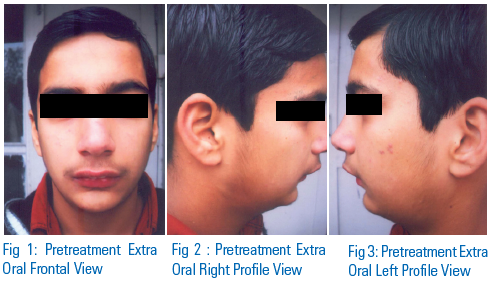
A 18years-old post-pubertal female patient came to the department with the chief complaint of spacing between upper front teeth. She was physically healthy and had no history of medical or dental trauma. The extraoral examination [Figure 1] of the patient showed good facial symmetry and a convex profile. Her upper and lower lips were significantly procumbent with respect to the soft tissue line Sn-Pg (Subnasale-Pogonion), and her interlabial gap was 6 mm at rest. She presented with an acute nasolabial angle and a deep labiomental fold. The intraoral examination [Figure 2] showed proclined upper incisors, spacing in both arches, Angle Class I malocclusion with an overjet of 5.5mm, and overbite of 6mm (75%). Upper midline was coinciding and lower midline was shifted to the left by 2mm. The curve of spee is 2.5mm. Cephalometric analysis showed a Class-I skeletal relationship with a horizontal growth pattern [Figure 3]. The panoramic radiograph [Figure 4] showed all permanent teeth including developing third molar buds.
TREATMENT OBJECTIVES
1. The treatment plan was to retract and intrude the upper and lower anteriors.
2. Maximum anchorage would be required during retraction and intrusion of the anterior teeth.
3. The lower midline had to be corrected.
4. The final objective was to achieve a stable functional occlusion with normal overjet and overbite.
5. Attaining a pleasing profile.
TREATMENT PROGRESS
Both arches were fully banded and bonded with .022" preadjusted edgewise appliances. A continuous maxillary and mandibular archwire were inserted. A transpalatal arch, custom-made with 0.036” stainless steel is placed between the right and left


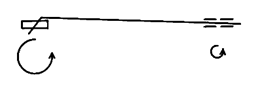
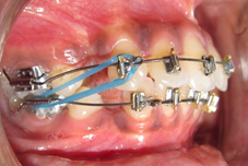
permanent first molars to control molar width as well as axial inclination in the frontal plane. After initial leveling and aligning [Figure 5], an intrusion and retraction utility arch is fabricated with 0.019 × 0.025 TMA wire. The anterior segment of the utility arch is placed directly in to the brackets of the anterior teeth and the posterior segment is placed in to auxiliary tube on the first molar. Buccal teeth are joined together by means of a 0.019 × 0.025 SS wire so as to form a buccal stabilizing unit The duration of utility arch was 6 months to achieve the desired intrusion and retraction of upper incisors [Figure 6]. Anterior bite plan was given to intrude the lower incisiors for 2 months. This is followed by individual canine intrusion with the help of elastics [Figure 7]. Since the incisors have already been intruded, it is necessary to join them to the posterior segments by an anterior wire inserted in the buccal teeth stepped occlusally around the canine. This wire holds the incisors in place and adds further anchorage for the intrusion of the canines. The duration of canine intrusion was 2 months. At the end of treatment, the anterior teeth were positioned into proper alignment, resulting in a pleasing smile [Figure 8].
DISCUSSION
A literature review indicated different opinions regarding the amount of force for intrusion of incisors and usually light, continuous forces were stated to be more suitable.6,7 The utility arch wire was fabricated with TMA wire because of the advantage of low-load deflection rate or deliver relatively constant force.
Two basic side effects could be anticipated from intrusive mechanics.8,9 Looking from the lateral view, a moment is created which tends to alter the plane of occlusion of the buccal segment; in the upper arch the plane is steepened. To minimize these changes, a number of principles are used in the intrusive mechanisms that have been described. The forces are kept as low as possible. The largest numbers of teeth are present in the buccal segment; these teeth are relatively rigidly connected by a buccal stabilizing segment and across the arch with a lingual arch.
The second major side effect produced by an intrusive arch can be seen from the frontal view. With an intrusive force on the incisors, there is an equal and opposite extrusive force on the molars. Since the extrusive force is operating buccally at a tube, it can be seen that a moment is created that tends to tip the crowns lingually and the roots buccally. One of the functions of the lingual arch is to prevent any undesirable change in axial inclination of the molars or change in width. The lingual arch also has the advantage that one does not have to be that precise in forming the widths of the intrusive arch since this is adequately controlled by the lingual arch. Lingual arches are not only helpful during the stage of intrusion, but they also help resist side effects at almost any stage of treatment
It is usually not possible to intrude all six anterior teeth at one time without producing undesirable axial inclination change in the posterior segment. Using the suggested force values, typically 100 gm. of force on a side is required to intrude the incisors and the canines. This force would produce a larger moment resulting in the tipping of the posterior teeth more rapidly than the anterior intrusion. Since this tipping is not required, intrusion mechanics will not be successful.
CONCLUSION
Not all patients with deep overbite should be treated with the same mechanics. Some patients require intrusion of the anterior teeth, while others require primarily extrusion. Orthodontic treatment of deep bite can be very rewarding if we are ready to accept the challenge of anticipating the changes, on the basis of a sound problem list and treatment goals.
REFERENCES
- Graber TM; Orthodontics: Principles and Practice. 3rd ed. Philadelphia; W.B. Saunders Company,1972. pp. 740-789
- Proffit WR, Fields HW. Contemporary Orthodontics. 3 rd ed. St Louis, Mosby;2000.
- Nanda R. Biomechanics and Esthetic Strategies in Clinical Orthodontics.
- Nanda R. The differential diagnosis and treatment of excessive overbite. Dent Clin North Am. 1981;25:69-84.
- Bell WH, et al. Simultaneous repositioning of the maxilla, mandible and chin. In: ed 2. Surgical correction of dentofacial deformities. vol 3:Philadelphia: W. B. Saunders Co.,; 1984.
- Ricketts RM, Bench RW, Gugino CF et al. Bioprogressive Therapy.Book 1. Denver, Colo: Rocky Mountain Orthodontics; 1978.
- Steigman S, Michaeli Y. Experimental intrusion of rat incisors with continuous loads of varying magnitude. Am J Orthod 1981;80:429-36.
- Charles R. Burstone. Deep overbite correction by intrusion. Am J Orthod 1977;72:1-22.
- Bhavna Shroff, Won M. Yoon, Steven J. Lindauer, Charles J. Burstone. Simultaneous intrusion and retraction using a three-piece base arch. The Angle Orthodontist 1997;67:455-461.
Disclaimer from Guident
The views and opinions expressed in articles published in Guident are solely those of the authors and do not necessarily reflect the views of the editors, the editorial board or the publisher, Ivory India. Guident does not endorse any product, technique or treatment described. Clinical content is intended for qualified dental and medical professionals and is not a substitute for individual clinical judgement. Guident, its editors and the publisher accept no liability for any consequence arising from the use of information published in the journal.
How to cite this article
Dr. Ritesh Singla, Dr. Madhumitha N., Dr. Nishu Singla. Management of Deep Overbite. Guident. 2013 Aug;6(10). Available from: https://guident.in/articles/orthodontics/management-of-deep-overbite.html