Mandibular Canine with Two Roots and Two Canals
Department of Conservative Dentistry and Endodontics Subharti Dental College, Meerut, India
Abstract
Human mandibular canines do not present an internal anatomy as simple as could be expected; there are such canines with a single root and two canals, two roots or fused roots. The existence of mandibular canines with more than one root canal is a fact that clinicians ought to keep in mind, in order to avoid failure during endodontic treatment. In spite of the low incidence of lower canines with one root and two canals, this possibility cannot be forgotten, in as much as the presence of a second canal in these teeth leads to difficulties in endodontic treatment. The precise knowledge of the dental endocanalicular system’s anatomy is essential in the success of the root canal therapy. This paper describes two clinical cases of mandibular canines with two roots and two canals.
INTRODUCTION
The vast majority of the problems that occur during the canal treatment are due to the insufficient knowledge of the anatomy of the endodontic space1.The main culprit in the failure of the endodontic treatment is the omission to obturate one of radicular canal because of ignoring the morphological variations of the dental anatomy. In order to perform a high quality endodontic treatment, which will ensure the tooth’s long lasting durability, it is imperative that the dentist take into account the morphology of the radicular canals and the variations of the entire canalicular system before beginning the endodontic treatment2,3.
Generally, mandibular canines contain a single root and root canal. Regarding anatomical variations associated with mandibular canines, approximately 15% contain two canals with one or two foramina4,5. A study of the internal anatomy, direction, number of roots and size of 830 extracted human mandibular canines showed that 98.3% of these teeth contain a single root, 92.2% consist of one canal and one foramen, 4.9% have two canals and one foramen, and 1.2% contain two canals and two foramina. The occurrence of two canals and even more two roots is rare, ranging from 1%to 5% 6. Although the prevalence is low, but the clinicians should consider all the possible variations in the number of roots and pulp canals of mandibular canines. This paper reports cases of patients with mandibular canines having two roots and two canals (incidence 1.2%).
CASE 1
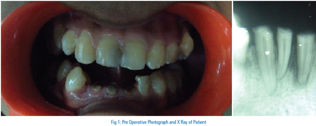
A 40 year old female patient reported to department of conservative dentistry and endodontics with the chief complaint of pain in lower left anterior region for last six months. Pain was continuous in nature and radiographic examination revealed carious canine with an indication of a separate root). Subsequently, a radiograph with a mesial angulation was shot which revealed two roots and two canals-buccal and lingual (fig 1) . The access cavity was prepared using Endo access bur and Endo Z bur (Dentsply Maillefer, Switzerland). After reaching the pulp chamber, the roof and overhanging dentin from lateral walls were removed. The orifice of labial canal was located however the access cavity had to be modified for location of lingual canal. Lingual extension of the access cavity was done with Endo Z bur (fig 2). For the straight line access, GG drills were used with crown down method to enlarge the orifices. Irrigation was done using 5.25% sodium hypochlorite and saline simultaneously. Working length was estimated with an apex locator (Propex II, Dentsply,Maillefer) using ISO #15 k- file and confirmed radiographically(fig 3). Canal anatomy corresponded to Vertucci type I canal configuration. Both the canals were instrumented till ISO #20 size k- file. Canals were then prepared with crown down technique using Protaper files till size F2 protaper. Subsequently, the canals were obturated with F2 protaper gutta percha points using AH plus sealer (Dentsply, Maillefer) (fig 4). Post endo restoration was done with composite.
CASE 2
A 46-year-old female complained of pain in the right lower front tooth region. Clinical examination revealed that tooth 43 had deep occlusal caries. A thermal test with ethyl chloride produced severe, long-lasting pain. A diagnostic radiograph did not reveal periapical pathology but did reveal occlusal caries reaching pulp and also indicating an extra canal. A mesially angulated radiograph was taken which confirmed our findings of two canals(fig 5). After local anesthesia, rubber dam (Hygenic,Coltene Whaledent Inc.,USA) was applied. Endodontic access preparation was performed with Endo Access bur and an Endo Z bur (Dentsply/ Maillefer, Ballaigues, Switzerland). The canal anatomy represented. Further lingual modification and careful exploration of the access cavity with the aid of operating loupes and DG-16 explorer demonstrated the presence of an extra canal lingual to the main canal. Exploration and negotiation of two root canals were done with a ISO # 15 K-file (Dentsply Maillifer, Ballaigues, Switzerland). Root canal length estimation was done using apex locator (Propex II, Dentsply Maillefer, Switzerland), which was confirmed radiographically (fig 6).
The root canal configuration consisted of two canals leaving the floor of the pulp chamber, converging into one canal and exiting at a single apical foramen (Vertucci type 2 canal configuration). Canals were then prepared with crown down technique using Protaper files till size F1 protaper. The root canals were irrigated with 2.5% NaOCl during the preparation procedure and 17% EDTA was used as a final irrigation to remove the smear layer after the preparation procedure. The canals were filled by Protaper F1 gutta percha points and AH Plus sealer (fig 7). After obturation, the access cavity was sealed with composite.
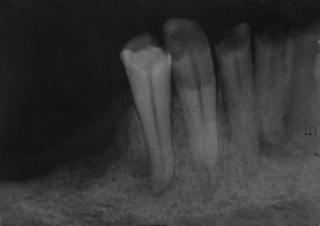
Fig 1 : (case1) Preoperative radiograph
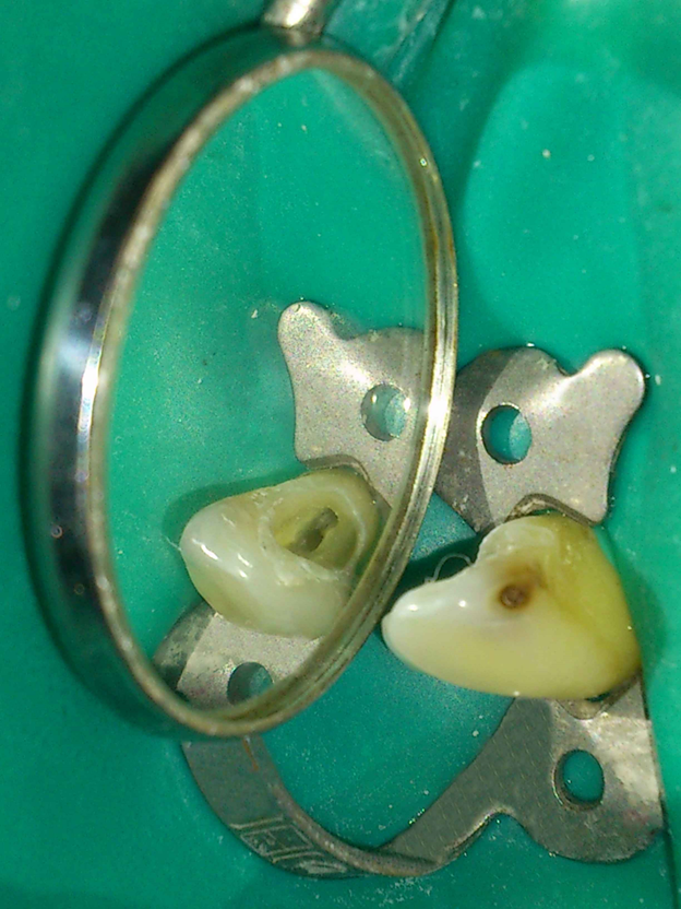
Fig 2 : 2 orifices
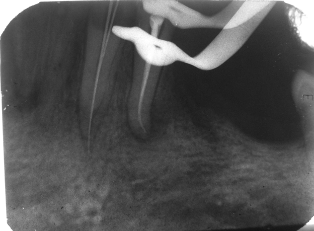
fig 3-working length
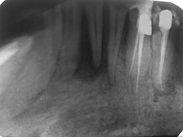
Fig 4 : obturation radiograph
DISCUSSION
It is important to recognise the possible permutations of canal morphology in endodontic therapy. Some root canals are often left untreated because the clinicians fail to identify additional root canals or anatomical variations. Treating extra canals may be challenging; however the inability to find and properly treat the root canals may cause failure7. If the clinician is alert to the clues that suggest the extra canal’s presence, most of them can be found. It is essential that clinicians know the clinical and radio- graphic signs that suggest the presence of extra canals. Endodontic exploration and a thorough and careful radio- graphic evaluation could lead to identification or suspicion of additional canals.
Radiographs taken from different angles, along with a file in place, might be helpful in finding and locating extra canals. The relationship of the two canal orifices is also significant, as the closer the orifices are to each other, the greater the chance that the two canals join at some point within the body of the root.8
Laurichesse et al. described the second root of mandibular canines in only 1% of cases. A previous study that investigated the internal anatomy, direction and number of roots and size of 830 extracted human mandibular canines found only 1.7% of the teeth with two roots and two seperate canals9,10. D Arcangelo et al. reported two cases of endodontic treatment of mandibular canines with two roots10.A case report of two roots in mandibular canine was also presented by Heling et al. yet the tooth exhibited three root canals11.
CONCLUSION
Successful and predictable endodontic treatment requires knowledge of root canal morphology and its variation. Radiographs and magnification devices are important tools in diagnosing and treating such complicated cases.
REFERENCES
- Al-Qudah AA, Awawdeh LA, Root canal morphology of mandibular incisors in a Jordanian population, Int Endod J, 2006, 39(11):873–877.
- .Vertucci FJ, Root canal anatomy of the human permanent teeth, Oral Surg Oral Med Oral Pathol, 1984, 58(5):589–599.
- Caliskan MK, Pehlivan Y, Sepetçioglu F, Türkün M, Tuncer SS, Root canal morphology of human permanent Teeth in a Turkish population, J Endod, 1995, 21(4):200–204.
- Pineda F, Kuttler Y. Mesiodistal and buccolingual roentgenographic investigation of 7,275 root canals. Oral Surg Oral Med Oral Pathol 1972;33:101-110.
- Vertucci FJ. Root canal anatomy of the mandibular anterior teeth. J Am Dent Assoc 1974;89:369-371
- Pecora JD, Sousa Neto MD, Saquy PC. Internal anatomy, direction and number of roots and size of human mandibular canines. Braz Dent J 1993;4:53-57
- Gupta S, Jaiswal S, Arora R. Endodontic management of permanent mandibular left first molar with six root canals. Contemporary Clinical Dentistry 2012,Vol 3(5):130-133
- Çaliskan MK, Pehilvan Y, Sepetçioglu F, Türkün M, Tuncer S. Root canal morphology of human permanent teeth in a Turkish population. J Endod 1995; 21:200-204
- .Green D. Morphology of the pulp cavity of the permanent teeth. Oral surgery, oral medicine, and oral pathology. 1955;8(7):743-59.
- D'Arcangelo C, Varvara G, De Fazio P. Root canal treatment in mandibular canines with two roots: a report of two cases. International Endodontic Journal. 2001;34(4):331-4.
- Heling I, Gottlieb Dadon I, Chandler N. Mandibular canine with two roots and three root canals. Dental Traumatology. 1995;11(6):301-2
Disclaimer from Guident
The views and opinions expressed in articles published in Guident are solely those of the authors and do not necessarily reflect the views of the editors, the editorial board or the publisher, Ivory India. Guident does not endorse any product, technique or treatment described. Clinical content is intended for qualified dental and medical professionals and is not a substitute for individual clinical judgement. Guident, its editors and the publisher accept no liability for any consequence arising from the use of information published in the journal.
How to cite this article
Dr. Shikha Jaiswal, Dr. Namrata Sharma, Dr. Sachin Gupta. Mandibular Canine with Two Roots and Two Canals. Guident. 2013 Aug;6(10). Available from: https://guident.in/articles/endodontics/mandibular-canine-with-two-roots-and-two-canals.html