Conventional Treatment of -a Tooth Within a Tooth
Costa (P.G. Student), Department of Conservative Dentistry and Endodontics SDM College of Dental Sciences, Dharwad
Abstract
Dens invaginatus is a developmental disturbance of teeth. The clinical importance of dens invaginatus results from the risk of pulpal disease. It can be detected clinically as well as radiographically. A case of unilateral dens in dente not extending up to the apical third in maxillary right lateral incisor is reported. Non-surgical endodontic treatment was performed.
Keywords:-Case report, dens invaginatus, maxillary lateral incisor, unilateral
Dens invaginatus is a developmental anomaly affecting permanent dentition and, less commonly, primary dentition. It is characterized by the inversion or infolding of the enamel and dentin towards the pulp chamber, usually appearing as an opening on the surface of the crown.
Salter first described anatomical anomaly in 1855 as “a tooth within a tooth”. Ploquet first described it in 1974, as he found this malformation in a whale’s tooth.
Dens invaginatus is also celled as Dens in dente, invaginated odontome, dilated gestant odontome, dilated composite odontome, tooth inclusion, and dentoid in dente.1
This defect is commonly diagnosed incidentally on radiographs unless the patient presents with pain and/or swelling associated with the involved tooth. Clinically, unusual crown morphology or a deep foramen coaceum may lead the dentist to suspect a tooth with dens invaginatus.
The commonly involved teeth are permanent maxillary lateral incisors, central incisors, premolars, canines and then molars. A maxillary predominance is seen.
Coronal invaginations may be due to increased localised external pressure, focal growth retardation or growth simulation in certain areas of the tooth bud, and an inadequate nutrition in certain areas of a single tooth germ.1,2
Radiographically, it shows a radiopaque invagination, equal in density to enamel, extending from the cingulum into the root canal. It can be identified easily because of the enamel lining is more radiopaque than the surrounding tooth structure.3
Oehlers describes dens invagination as occurring in three forms:
Type 1 – An enamel-lined minor form occurring within the confines of the crown of the tooth and not extending beyond the cementoenamel junction.
Type 2 – An enamel-lined form (like Type 1) which invades the root but remains within its confines and may not communicate with the dental pulp.
Type 3 – A form which penetrates through the root perforating at the foramen with no immediate communication with the pulp.
Teeth with dens in dente have been successfully treated endodontically from a non-surgical approach.4,5,6,7,8
Deep invaginations are difficult to access and clean. As a consequence, the early development of caries and subsequent necrosis of the dental pulp are clinical implications associated with dens invaginatus. Early diagnosis is desirable because of the fact that the teeth with dens invaginatus are predisposed for caries, pulp infections, and eventually periapical infections.
CASE REPORT
A 26 year old male patient visited the Department of Conservative dentistry and Endodontics with a chief complaint of pain in the upper front region. Apparently about 3 years before, the tooth was associated with pain and swelling, and was treated by his local dentist for the same with root canal treatment (emergency access opening). But the patient did not maintain his recall visits with his previous dentist.
On clinical examination, left maxillary lateral incisor was similar in shape to its contralateral tooth. Food lodgement was seen within the tooth as the temporary restoration placed must have come off in the due course. The tooth was tender on percussion.
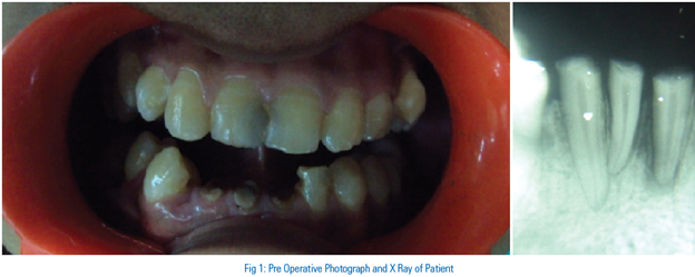
The intra-oral periapical radiograph revealed incomplete root canal treatment (Fig 1) and the presence of dens invaginatus with the left maxillary lateral incisor (22). The radiograph also revealed an internal anatomy consistent with a type II coronal dens invaginatus. Radiographic examination of the contralateral tooth confirmed that the condition was unilateral.
Rubber dam was placed and stabilized using wedgets on 22. Access was regained especially with regards to the dens invaginatus. An ISO #25 K-file and #20 K-file were placed in the main canal and dens invaginatus canal respectively. Working length was then determined using radiographs (Fig 2) followed by proper cleaning and shaping procedures. Standardized technique of biomechanical preparation was followed within the canals along with copious irrigation with sodium hypochlorite and saline. The canals were dried with paper points and access was closed with CavitTM. After 2 days, the temporary restoration was removed and on inspection the canals were dry. The root canal system was then obturated (Fig 3,4) with gutta-percha and AH-PLUS sealer using lateral condensation technique followed by warm vertical compaction. The access cavity was then restored using type II glass ionomer cement.


DISCUSSION
Dens invaginatus constitutes a challenge to endodontic treatment, due to its complicated root canal system.
Tagger (1977) and Hovland & Block (1977) the first to present dens in dente cases treated with conventional root canal therapy.
Dens invaginatus commonly occurs bilaterally4,5, 9,10,11,12, 13,14but in this case, radiographic examination proved that only one lateral incisor in the maxilla was affected.
Dens in dente could be diagnosed clinically as well as radiographically. In some cases, the affected tooth would present with a clinical crown larger than the contralateral side.7,8,15,16,17,18,19,20 In this case the affected tooth was similar in size to its contralateral one.
Duckmanton et al reported a case wherein the maxillary central incisor showed Dens in dente which remained vital for five years. The patient here didn’t approach a dentist due to pain or swelling but the left central incisor was ‘bigger’ than the right central incisor, therefore, aesthetics being his major concern.17 And so is with this case, as the patient reported with a discoloured tooth.
Dens invaginatus is clinically significant due to the possibility of the pulp being affected. Due to the tortuous lingual anatomy, it is possible for caries to develop inside the dens invaginatus without any clinically detectable lesion. Since the enamel lining is thin and in close proximity to the pulp chamber, a carious lesion could easily perforate the pulp chamber. Therefore, pulpitis and necrotic pulps are often associated with this anomaly.
The invagination presented in this case penetrated only to the level of the crown and did not communicate with the pulp although communication usually exists between the root canal and the invagination; this is in agreement with a case done by Jimenez-Rubio et al11.
In an Oehlers type II invagination, the anomaly does not extend to the apex of the tooth and hence access to the apical third of the root canal is less problematic. The canal must be debrided and shaped well, as we know the apical third has a high percentage of lateral canals.
With the most severe cases, periapical surgery21,22,23, is required to seal the root canals. Lindner et al proposed intentional replantation with retrograde surgery in otherwise hopeless cases.24 In the case presented, only non-surgical treatment was needed.
CONCLUSION
Early diagnosis and intervention can prevent pulp necrosis and potential loss of tooth. The accurate knowledge of variations in the morphology of pulp cavity will greatly assist the dentist in performing successful endodontic therapy.
REFERENCES
- Hulsmann M. Dens invaginatus: aetiology, classification, prevalence, diagnosis, and treatment considerations. Int Endod J 1997; 30: 79 - 90
- de Sousa SM, Bramante CM. Dens invaginatus: treatment choices. Endod Dent Traumatol 1998; 14: 152 – 8
- White SC, Pharoah MJ. Oral radiology principles and interpretation 4th ed. St Louis, Mosby, 2000, pp 314 – 315
- Banner H. Bilateral dens id dente in mandibular premolars. Oral Surg 1978; 45: 827 – 828
- Roland NM. Periapical lesions associated with dens id dente. Oral Surg Oral Med Oral Pathol 1979; 48: 190
- Vajrabhaya L. Nonsurgical endodontic treatment of a tooth with double dens in dente. J Endod 1989; 15: 323 – 325
- Bramante CM, de Sousa SMG, Tavano SMR. Dens invaginatus in mandibular first premolar. Oral Surg Oral Med Oral Pathol 1993; 76: 389
- Szajkis S, Kaufman AY. Root invagination treatment: A conservative approach in endodontics. J Endod 1993; 19: 576 – 578
- Bottomley WK, Johnson MR. Multiple dens in dente in a dentition. Oral Surg Oral Med Oral Pathol 1982; 54: 478
- Hosey MT, Bedi R. Multiple dens invaginatus in two brothers. Endod Dent Traumatol 1996; 12: 44 – 47
- Jimenez-Rubio A, Segura JJ, Jimenez-Planas A, Llamas R. Multiple dens invaginatus affecting maxillary lateral incisors and a supernumerary tooth. Endod Dent Traumatol 1997; 13: 196 – 198
- Mupparapu M, Singer SR. A rare presentation of dens invaginatus in a mandibular lateral incisor occurring concurrently with bilateral maxillary dens invaginatus: case report and review of literature. Aust Dent J 2004; 49: 90 – 93
- Canger EM, Kayipmaz S, Celenk P. Bilateral dens invaginatus in the mandibular premolar region. Indian J Dent Res 2009; 20: 238 – 240
- Vardhan TH, Shanmugam S. Dens evaginatus and dens invaginatus in all maxillary incisors: report of a case. Quintessence Int 2010; 41: 105 – 107
- Tavano SMR, Sousa SMG, Bramante CM. Dens invaginatus in first mandibular premolar. Endod Dent Traumatol 1994; 10: 27 – 29
- Khabbaz MG, Konstantaki MN, Sykaras SN. Dens invaginatus in a mandibular lateral incisor. Int Endod J 1995; 28: 303 – 305
- Duckmanton PM. Maxillary permanent central incisor with abnormal crown size and dens invaginatus: case report. Endod Dent Traumatol 1995; 11: 150 – 152
- Jung M. Endodontic treatment of dens invaginatus type III with three root canals and open apical foramen. Int Endod J 2004; 37: 205 – 213
- Mupparapu M. A review of dens invaginatus (dens in dente) in permanent and primary teeth: report os a case in a microdontic maxillary lateral incisor. Quintessence Int 2006; 37: 125 – 129
- Keles A, Cakici F. Endodontic treatment of a maxillary lateral incisor with vital pulp, periradicular lesion and type III dens invaginatus: a case report. Int Endod J 2010; 43: 608 – 614
- Burton DJ, Saffos RO, Scheffer RB. Multiple bilateral dens in dente as a factor in the etiology of multiple periapical lesions. Oral Surg 1980; 49: 496 – 499
- Benenati FW. Complex treatment of a maxillary lateral incisor with dens invaginatus and associated aberrant morphology. J Endod 1994; 20: 180 – 182
- Ikeda H, Yoshioka T, Suda H. Importance of clinical examination and diagnosis: a case of dens invaginatus. Oral Surg Oral Med Oral Pathol Oral Radiol Endod 1995; 79: 88 – 91
- Lindner C, Messer HH, Tyas MJ. A complex treatment of dens invaginatus. Endod Dent Traumatol 1995; 11: 153 – 155
Disclaimer from Guident
The views and opinions expressed in articles published in Guident are solely those of the authors and do not necessarily reflect the views of the editors, the editorial board or the publisher, Ivory India. Guident does not endorse any product, technique or treatment described. Clinical content is intended for qualified dental and medical professionals and is not a substitute for individual clinical judgement. Guident, its editors and the publisher accept no liability for any consequence arising from the use of information published in the journal.
How to cite this article
Dr. Vivian Flourish D, Dr. Mahantesh Yeli, Dr. Geeta Hiremath. Conventional Treatment of -a Tooth Within a Tooth. Guident. 2013 Aug;6(10). Available from: https://guident.in/articles/endodontics/conventional-treatment-of-a-tooth-within-a-tooth.html