
Management of Endodontically Treated Teeth- an Insight
Hasram Das Dental College Ghagiadabad UP, India, K.D Dental College, Mathura, UP, India, Department of PRosthodontics
Abstract
The clinical decision as to whether an endodontically treated tooth requires a post and a crown poses a challenge to dental practitioners. The longevity of endodontically involved teeth has been greatly enhanced by continuing developments made in endodontic therapy and restorative procedures. It has been reported that a large number of endodontically treated teeth are restored to their original function with the use of intraradicular devices. These devices vary from a conventional custom cast post and core to one visit techniques, using commercially available prefabricated post systems. In the last few decades, various prefabricated posts systems have been developed. The selection of post design is important, because it may have an influence on the longevity of the tooth.
This article reviews the major pertinent literature on this topic, with an emphasis on major decision-making elements in post placement and restoration of endodontically treated teeth.
The restoration of endodontically treated teeth is a topic that is extensively studied and yet remains controversial from many perspectives. It generally is agreed that the successful treatment of a badly broken down tooth with pulpal disease depends not only on good endodontic therapy, but also on good prosthetic reconstruction of the tooth after endodontic therapy is completed. The purpose of this review is to organize this topic into its component parts and provide evidence-based principles that are sound from a restorative as well as an endodontic perspective.
HISTORICAL REVIEW

Fig 1: A 6 unit bridge, pivoted in lateral incisor, with canines cantilevered.
The concept of using the root of a tooth for retention of a crown is not new. In the 1700s Fauchard inserted wooden dowels in canals of teeth to aid in crown retention. Over time the wood would expand in the moist environment to enhance retention of the dowel until, unfortunately, the root would often fracture vertically. Additional efforts to develop crowns retained with posts or dowels in the 1800s were limited by the failure of the “endodontic” therapy of the era. Several of the 19th century versions of dowels also used wooden “pivots” but some dentists reported the use of metal posts favored by Black (1869) in which a porcelain-faced crown was secured by a screw passing into a gold-lined root canal. In the early 1800s Dubois de Chemant described Porcelain pivot crowns. Early pivot crowns used seasoned wood (white hickory) pivots. Metal pivot retention was achieved by various means such as threads, pins, surface roughening, and split designs that provided mechanical spring retention. The Richmond crown was introduced in 1878 and incorporated a threaded tube in the canal with a screw retained crown. It was later modified to eliminate the threaded tube and was redesigned as a 1-piece dowel and crown , which lost its popularity quickly because they were not practical. This was obviously evident when divergent paths of insertion of the post space and remaining tooth structure existed, especially for abutments of fixed partial dentures. One piece dowel crown restorations also presented problems when the crown or FPD required removal and replacement. These difficulties led to development of a post and core restoration as a separate entity with an artificial crown cemented over a core and remaining tooth structure. With the advent of scientific endodontic therapy in the 1950s, the challenges increased for restorative dentistry. Teeth that were extracted without hesitations were now successfully treated with predictable endodontic therapy; and a satisfactory restorative solution was necessary.
CHANGES IN ENDODONTICALLY TREATED TEETH
Classic studies proposed that the dentin in endodontically treated teeth is substantially different than dentin in teeth with “vital” pulps. It was thought that the dentin in endodontically treated teeth was more brittle because of water loss and loss of collagen cross-linking.
However, more recent studies dispute this finding. In 1991, Huang et al. compared the physical and mechanical properties of dentin specimens from teeth with and without endodontic treatment at different levels of hydration. They concluded that neither dehydration nor endodontic treatment caused degradation of the physical or mechanical properties of dentin.
Sedgley and Messer tested the biomechanical properties of dentin endodontically treated teeth and compared them to their contralateral “vital” pairs. Aside from a slight difference in hardness, the properties were comparable. The study did not support the conclusion that endodontically treated teeth are more brittle.
These and other studies support the interpretation that it is the loss of structural integrity associated with the access preparation, rather than changes in the dentin, that lead to a higher occurrence of fractures in endodontically treated teeth compared with “vital” teeth .
Access preparations result in increased cuspal deflection during function and increase the possibility of cusp fracture and microleakage at the margins of restorations. In most endodontically treated teeth, there also is missing tooth structure caused by caries or existing restorations.
Randow and Glantz reported that teeth have a protective feedback mechanism that is lost when the pulp is removed, which also may contribute to tooth fracture.
Fennis et al. reported significantly more fractures in teeth with endodontic treatment. Taken together, these studies indicate that restorations that enhance structural integrity would be expected to increase the prognosis of endodontically treated teeth exposed to heavy masticatory loading forces.
RESTORATIVE FACTORS THAT AFFECT THE PROGNOSIS OF ENDODONTIC TREATMENT
Contamination
Contamination of the root-canal system by saliva, often referred to as “coronal leakage” or “coronal microleakage,” is a potential cause of endodontic failure . In addition, recurrent caries or fractured restorations may lead to recontamination of the root canal system. Under the best of conditions, the oral environment is rich in microorganisms, and dental restorations must withstand repeated exposure to physical, chemical, and thermal stressors. It is a difficult environment in which to maintain a hermetically sealed system. Bacterial byproducts and endotoxins can penetrate to the apex in an even shorter time than bacteria. When the root canal space has been grossly contaminated, retreatment should be considered. This is especially true if there has been persistent contamination.
Protective measures
Aseptic treatment techniques should be used, including the use of a rubber dam. Once root-canal treatment is completed, immediate restoration of the tooth is recommended whenever possible. When this is not possible, the root-canal system should be protected by sealing the canals and floor of the pulp chamber with intracoronal barriers.
Bonded materials such as glass-ionomer cement or composite resin are preferred. The canal orifices are countersunk with a round bur, and the floor of the chamber is cleaned of excess gutta-percha and sealer. The chamber floor is etched and primed if a resin material is used or “conditioned” if using glass-ionomer cement or resin-modified glass ionomer. The barrier material is then placed over the floor of the chamber and light cured, and a temporary restoration is placed with or without a cotton pellet in the chamber. The intracoronal barrier protects the root-canal system from contamination during the period of temporization and while the restorative dentistry is performed.
Post spaces, in particular, should be restored immediately because of the difficulties associated with maintaining the temporary seal. In vitro studies by Fox and Gutteridge and Demarchi and Sato showed that teeth restored with temporary posts had about the same amount of contamination as the controls that had no restorations.
Preservation of tooth structure is important when restoring the coronal portion of the tooth. Coronal tooth structure should be preserved to provide resistance and retention form for the crown.
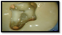
Fig-2 The orifices were countersunk with a round bur; the floor of the chamber was etched and primed and then sealed with a clear resin. The gutta-percha is visible and easily accessible.
INDICATIONS FOR A POST
The primary purpose of a post is to retain a core in a tooth with extensive loss of coronal tooth structure. Posts should only be used when other options are not available to retain a core. The need for a post varies greatly between the anterior and posterior teeth.
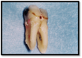
FIg 3.Post space preparation involves risk. An unnecessary post was placed in this largely intact mandibular molar. Perforation occurred, resulting in eventual loss of the tooth.
Anterior Teeth
A post is of little or no benefit in a structurally sound anterior tooth and increases the chances for a non restorable failure . In most cases, the remaining coronal tooth structure is quite thin after it has received root-canal treatment and been prepared for a crown. Anterior teeth must resist lateral and shearing types of forces, and the pulp chambers are too small to provide adequate retention and resistance without a post. Anterior teeth may require complete coronal coveragealong with post and cores. This is common when large proximal restoration are present, caries has undermined the remaing marginal ridges, or the majority of the incisal edge has been lost due to trauma.The amount of remaining coronal tooth structure and the functional requirements of the tooth determine whether an anterior tooth requires a post.
Molars
Endodontically treated, molar teeth should receive cuspal coverage, but in most cases, do not require a post. Unless the destruction of coronal tooth structure is extensive, the pulp chamber and canals provide adequate retention for a core buildup . Molars must resist primarily vertical forces. In those molars that do require a post, the post should be placed in the largest, straightest canal, which is the palatal canal in the maxillary molars and a distal canal in the mandibular molars . Rarely, if ever, is more than one post required in a molar. When a post is required because of lack of adequate remaining coronal tooth structure, if more than 60% is missing, it should generally be placed only in the largest canal; that is the palatal canal in the maxillary molar and the distal in the mandibular molar. When the molar is to be used as an abutment tooth, a post is commonly used.
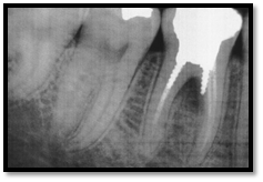
Fig 4 The mesial roots in mandibular molars tend to be thin mesiodistally and the canals often are curved. They are a poor choice for post placement.
Premolars
Premolars are usually bulkier than anterior teeth, but often are single-rooted teeth with relatively small pulp chambers. For these reasons, they require posts more often than molars. Premolars are more likely than molars to be subjected to lateral forces during mastication. When restoring an endodontically treated premolar, a decision regarding post placement is made based on the remaining coronal tooth structure, the functional requirements of the tooth, and an evaluation of the forces that acts on the tooth. If an endodontically treated premolar has increased functional stresses acting on the crown due to loss of the periodontuim and is to serve as an abutment for a removable partial denture a post may be indicated . Conversely, if a premolar has a relatively short crown and functions more like a small molar, then a post is not indicated. When a post is indicated for placement in a maxillary premolar the delicate morphologic anatomy must be considered . Post systems that require minimal enlargement and reshaping of the canal space, such as tapered posts, are best suited for maxillary premolar.

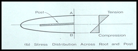
Fig 5 Experimental stress distribution in endodontically treated tooth with cemented post.when tooth is loaded the lingual surface in tension and facial surface in compression. Centrally placed cemented post lies in axis
The primary purpose of a post is to retain a core that can be used to retain the definitive prosthesis. Posts do not reinforce endodontically treatcd teeth and are not necessary when substantial tooth structure is present after teeth have been prepared . A post and core may help prevent coronal fractures when the remaining coronal tooth structure is very thin after tooth preparation.

Fig 6.No post and core was used in the mandibular second molar abutment because substantial tooth structure was present after endodontic treatment was complete.
Principles for posts
Retention and Resistance
Post retention refers to the ability of a post to resist vertical dislodging forces. Retention is influenced by the
1. Post’s length- From laboratory studics, it is apparent that a length guideline ideally would be three fourths of the root length, but this dimension is not achievable without compromising the apical seal on many teeth. Clinically, each tooth must be individually evaluated for root length and amount of remaining gutta percha before establishing the desired post length. With long-rooted teeth, achieving a length as close as possible to three fourths of the root length is desirable, whereas many teeth will have posts that are equal in length to the crown because of limited root length and the need to retain 4 to 5 mm of apical gutta percha.
2. Diameter and taper- Shillingburg et al measured 700 teeth and related the root dimensions to post diameter. They recommend a post diameter of 0.7 mm for mandibular incisors. The maximum recommended diameter was 1.7 mm for maxillary central incisors. They indicate that the post tip diameter should be at least 1.5 mm less than the root diameter at that point and the post should be 2.0 mm less than the root width at its midpoint. Differing results have been found regarding the effect of post diameter on post retcntion. Post and core diameter should be controlled to preserve root structure so that perforations are less likely to occur, and the tooth can resist root fracture.Post diameters should not exceed one third of the root diameter at any location, and post tip diameter should usually be 1 mm or less.
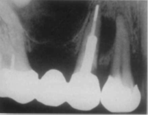
Fig 7 The post present in the maxillary second premolar has a tip diameter that exceeds the recommended 1 mm and creates a root perforation. Note the mesial bone radiolucency.
3. The luting cement – All post need to be cemented inside the root canal. The cementing medium enhances the retention, aids in stress distribution and seals the microgaps between the tooth and the post. Historically, zinc phosphate was the cement of choice. Recent studies advocated the use of low viscosity resin cement in combination with the removal of the smear layer from canal walls. This permits a movement of resin into the exposed dentinal tubules which is commonly refered to as “dentin bonding”. It increases the post retention.
4. Whether a post is active or passive -Parallel posts are more retentive than tapered posts . Active posts are more retentive than passive posts. From a retention standpoint, the laboratory evidence indicates that threaded posts are the most retentive followed by cemented, parallel-sided posts. Serrations increase post retention. Cemented, tapered posts are the least retentive, but some clinicians reported that they have successfully used this design and did not have retention problems.
Resistance refers to the ability of the post and tooth to withstand lateral and rotational forces which is influenced by the:
1. Remaining tooth structure - The amount of remaining coronal tooth structure is also a critical factor in determining the post selection. Hoag and Dwyer (1982) determined that the amount of tooth structure present is more important than the material from which the post and core is fabricated (amalgam, resin, or cast gold).The bulk of the tooth above the restorative margin should be at least 1.5mm to 2mm to achieve resistance. The use of cast post and cores in restoring endodontically treated teeth with moderate to severe coronal tooth loss has demonstrated a success rate of 90.6% after 5 years of service When ample coronal dentin remains non-metal posts such as a carbon fiber posts were deemed successful.
2. The post’s length and rigidity- post length influence the stress load along the root. Whereas enlargement of canal increases the cervical stress, post placement will decrease the stress in this region. Short wide post lead to elevated stress concentration in cervical region. It has been shown that post length is less important for fracture resistance than ferrule effect.
3. The presence of antirotation features- Intra-orally, post and core restored teeth are subjected to various types of forces. Torsional forces on the post-core-crown unit may lead to loosening and displacement of the post from the canal. Burgess et al (1992) emphasized on the importance of an antirotational feature and concluded that resistance to torsional forces is integral to the survival of the post-core-crown unit. Active post designs provide greater torsional resistance than a passive post.
4. The presence of a ferrule- A ferrule or circumferential band of metal that engages cervical tooth structure has been advocated as part of a post and core preparation to help encompass tooth structure and prevent fracture.
A restoration lacking resistance form is not likely to be a long-term success, regardless of the retentiveness of the post.

Fig-8 This cast post and core had adequate length for retention, but failed because of the lack of resistance form.
An important factor related to resistance is failure mode. All post systems have some percentage of clinical failure. However, some posts cause a higher percentage of failures that result in teeth that are non restorable. For example, teeth restored with less rigid posts, such as fiber posts, tend to have failures that are more likely to be restorable. Teeth prepared with a ferrule also tend to fail in a more favorable mode. The type of core material also can affect failure mode.
Preservation of Tooth Structure
Whenever possible, coronal and radicular tooth structure should be conserved. In most cases, preparation of a post space should require minimal removal of additional radicular dentin beyond the requirements for root-canal treatment. Further enlargement only weakens the root . It has been shown that cemented metal posts do not strengthen the root . Bonded posts are reported to strengthen the root initially , but this strengthening effect is probably lost over time as the tooth is exposed to functional stresses and the resin bond to dentin weakens . Minimal enlargement of the post space means the post must be made of a strong material that can withstand functional and parafunctional forces.
THE FERRULE EFFECT
The “ferrule effect” is important to long-term success when a post is used. A ferrule is defined as a vertical band of tooth structure at the gingival aspect of a crown preparation. It adds some retention, but primarily provides resistance form and enhances longevity. A ferrule is a metal ring or cap intended for strengthening. The word probably originates from combining the Latin for iron (ferrum) and bracelets (viriola) (Brown 1993).A dental ferrule is an encircling band of cast metal around the coronal surface of the tooth. It has been proposed that the use of a ferrule as part of the core or artificial crown may be of benefit in reinforcing root-filled teeth. A protective, or ‘ferrule effect’ could occur owing to the ferrule resisting stresses such as functional lever forces, the wedging effect of tapered posts and the lateral forces exerted during the post insertion.
Barkhordar and Radke, applying an angular lingual load to extracted maxillary central incisors, found that a significantly higher load is required to fracture the teeth with a cervical bevel. Milot and Stein used clear plastic analogues of maxillary central incisors and found that a cervical bevel significantly increases the resistance of the analogue to fracture from an angular lingual load. Hemmings et al investigated the resistance of six post and core designs to torsional forces. The cervical collar was found to be the most favorable, embracing resistance and reducing tooth fractures.
Stankiewicz and Wilson in 2002 stated that a ferrule with 1 mm of vertical height has double the resistance to fracture versus teeth restored without a ferrule. Other studies have shown maximum beneficial effects from a ferrule with 1.5 to 2 mm of vertical tooth structure . A study by al-Hazaimeh and Gutteridge reported no difference in fracture resistance with or without a 2-mm ferrule using prefabricated posts and resin cement. However, the fracture patterns were more favorable when a ferrule was present. The majority of the fractures in the teeth without a ferrule were nonrestorable.
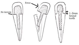
Fig 9:The ferrule effect occurs owing to the artificial crown bracing against the dentine extending coronal to the crown margin. Overall, it can be concluded that a ferrule is desirable, but should not be provided at the expense of the remaining tooth/root structure.
Retrievability
Although nonsurgical endodontic treatment enjoys a reputation as a highly successful treatment, some studies have reported lower rates of success . For this reason, it is important that posts can be retrieved if endodontic retreatment becomes necessary.
In most cases, metal posts can be removed effectively and safely.
Most fiber posts also are reported to be easy to retrieve . In contrast, ceramic and zirconium posts are considered to be very difficult and sometimes impossible to retrieve.
Retrievability should be considered when treatment planning a post. Post removal can be performed by means of conventional rotary instruments and solvents. Other commercially available systems to remove posts include the Masseran Kit, Post Removal System, Endodontic extractors, and ultrasonic unit Roto-Pro bur and a combination of tube extractors with cyanoacrylate will aid in post removal by breaking up the cement .
Abbott (2002) reported that post removal is a predictable procedure, if appropriate techniques and devices are used, and root fracture is a rare occurrence. He also concluded the following:
(1) Masseran kit and ultrasonics are effective in removing fractured cast posts and parallelsided posts.
(2) Unscrewing of the threaded screw posts.
(3) Eggler post removal (Ruddle) in retrieving cast posts.
(4) Ultrasonics in retrieving parallel-sided posts.
Ideal properties of post
Wagnild et al (2002) summarized the ideal physical properties of a post that include:
(1) Maximum protection of the root.
(2) Adequate retention within the root.
(3) Biocompatible / noncorrosive
(4) Maximum retention of the core and crown.
(5) Maximum protection of the crown margin cement seal.
(6) Pleasing esthetics
(7) Radiopaque
Classification of post
By Stephen Cohen
A.Preformed dowels and custom cast dowelsb
B.Metallic and non metallic dowels
C.Rigid and flexible dowels
D.Aesthetic and nonesthetic dowels
By Ricketts et al
1 . Active Posts
A. Pre tapped posts
B. Self threading
2. Passive Posts
A. Custom made or prefabricated
B. Parallel smooth or serrated
C. Tapered smooth or serrated
Wein's Classification
A. Tapered smooth sided
B Parallel, serrated and vented posts
C. Parallel threaded posts
D. Parallel threaded split shank posts.
According To Rosensteil
A.Prefabricated post
B.Tapered smooth sided posts
C.Tapered serrated posts
D.Tapered Threaded posts
E.Parallel smooth sided
F.Parallel serrated posts
G.Parallel Threaded posts
According to material used
A.Wooden posts
B.Type III and Type IV gold alloy
C.Stainless steel
D.Gold Platinum alloy
E.Titanium/ Titanium alloy
F.Brass
G.Nickel chromium and Cobalt chromium
H.Cobalt chromium molybdenum alloy
I.Amalgam
J.Carbon fiber
K.Glass fiber
L.Ceramic Posts, Zirconium dioxide post
Disclaimer from Guident
The views and opinions expressed in articles published in Guident are solely those of the authors and do not necessarily reflect the views of the editors, the editorial board or the publisher, Ivory India. Guident does not endorse any product, technique or treatment described. Clinical content is intended for qualified dental and medical professionals and is not a substitute for individual clinical judgement. Guident, its editors and the publisher accept no liability for any consequence arising from the use of information published in the journal.
How to cite this article
Dr.Ajal Gupta, Dr. Kamna Nagrani. Management of Endodontically Treated Teeth- an Insight. Guident. 2013 Jun;6(8). Available from: https://guident.in/articles/endodontics/management-of-endodontically-treated-teeth-an-insight.html


