Implant Placement Following Ridge Preservation Using Synthetic Bone Graft Substitute
Department Of Periodontology, Yenepoya Dental College, Mangalore, India, New Delhi, India
INTRODUCTION
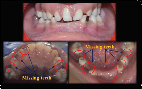
Requirements for successful implant treatment is perfect implant placement, proper hard and soft tissue form and osseointegration of implant. These need adequate alveolar bone volume and positive ridge architecture coupled with a suitable surgical technique. However, following extraction of teeth the alveolar bone resorbs, this may differ from subject to subject and location to location in the same subject which in turn causes bone volume deficient and poor ridge architecture for placement of dental implant.1
At the time of extraction or following it, alveolar ridge preservation reduces external ridge resorption and increase socket bone formation.
A consensus report recently advocates that minimal dimensional changes are observed within 6-8 weeks of tooth extraction2. If an implant to be placed after 6-8 weeks of extraction, it is advised for ridge preservation. If placement of implant before 6-8 weeks or immediately after extraction it is advantageous of performing ridge preservation at that time. Even though placement of implant is not outlined, ridge preservation should be taken into consideration at critical sites to maintain a possibility for implant in future. In prosthodontic aspect it is to be considered for esthetic purpose at site of pontic.
Grafting of extraction sockets in both clinical trials and experimental preclinical studies showed anticipated and positive way to preserve dimensions and architecture of the alveolar ridge3.Various kinds of bone grafts, barrier membranes and growth-factor preparations, and many different surgical techniques and protocols have been proposed4.Yip et al.5 proposed the ideal bone grafting material should have specific characteristics which should be osteoconductive, osteoinductive and biocompatible. It should be progressively replaced by newly formed bone, exhibiting controlled breakdown and resorption and it must be capable to continue contour of ridge in site of augementation. Furthermore it should have adequate mechanical properties and minimal risk of disease transmission.
Alloplasts represent group of synthetic and highly biocompatible bone grafting materials.6 The use of these material do not cause any risk of transmitting infections or diseases and their availability is infinite7. Bioactive osteoconductive materials include Calcium phosphate ceramics and are known to promote neovascularization due to its osteoinductive properties.8
Beta-tricalcium phosphates (b-TCP) are extensively used in orthopedics and in dentistry in the form of particulate grafts or cements. To enhance biological fulfillment and mechanical properties, other synthetic materials can be combined with b-TCP.9 Coating poly (lactic-co-glycolic) acid (PLGA) with b-TCP produces hardening, stable, porous biomaterial which serves as a bioactive scaffold for reconstruction of bone. Where in need for additional membranes for retention and stabilization of the graft material in the bone defect is minimized.10 Increased bone regenerations is achieved by stability throughout the grafted site, and the reduced micromotions of the graft particles. These micromovements between bone and any implanted material may activate differentiation of mesenchymal cells to fibroblasts instead of osteoblasts, and inhibit bone formation, which results in the development of fibrous tissue.11
CASE REPORT
This case report enlightens the use of beta tricalcium phosphate for socket preservation. A 25 year-old male with a non-contributory medical history, presented to department of periodontology with mobility of first premolar (tooth 14). Clinical examination showed grade II mobility and decay in relation to 14. As the prognosis was poor, 14 was subjected for extraction with socket preservation for future dental implant placement.
Steps:
- Extraction: Atraumatic extraction of the tooth is done by preserving the buccal plate and the interradicular septa under local anesthesia (Figure 1)
- Socket evaluation: Visual inspection and probing of the bony socket using a periodontal probe to determine the status of the buccal & lingual plates was carried out.
- Socket curettage: Ensure thorough cleaning down to the alveolar apex.
- Inspecting the marginal soft tissue and removing inflamed tissue.
- Thorough rinsing using sterile saline solution
- Application of the biomaterial. GUIDOR easy-graft CRYSTAL (biphasic calcium phosphate, a compound of 60 % hydroxyapatite and 40 % ß-tricalcium phosphate). Easy-graft CRYSTAL bone augmentation material that was placed directly from the syringe into the defect. (Figure 2). The mouldable adhesive granules can be shaped in the defect. In contact with blood, the material hardens within minutes to form a porous defect analog.
The easy-graft products are 100 % alloplastic and do not contain substances of animal or human origin. They consist of coated biphasic calcium phosphate (compound of 60 % hydroxyapatite and 40 % ß-TCP). The hydroxyapatite portion remains integrated in the newly formed bone for long-term volume stability.
Before filling the socket, remove any blood that has already coagulated. This should ensure more efficient bleeding into the socket. Freshen the wound again if necessary. Leave the liquid component (BioLinker) in contact with the granules for about 30 seconds.
Before application, excessive BioLinker was discarded. Push mixed material to the opening of the syringe so that the strand of material slightly protrudes. Apply the material in one process to achieve a continuous bone graft substitute mass. The material should be shaped for no longer than 30 seconds followed by suturing. (Figure 3)
Post operative OPG was taken after ridge preservation (Figure 5). Favourable ridge dimensions was obtained after ridge preservation for implant placement (Figure 6).After about 5 months implantation was done using indirect sinus lift technique.(Figure 6 & 7) Supracrestal incision was made and a full thickness flap was raised. The implant bed was prepared to approximately 1 mm below the floor of the maxillary sinus. The osteotomes were chosen to extend the preparation area both horizontally and vertically until elevating the Schneiderian membrane without perforation. The graft material is placed where indicated, the implant is inserted (Figure 8), and the wound is closed. (Figure 9 ) Prosthesis was loaded after 6 months. (Figure 11)
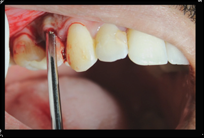
Figure 1: Atraumatic extraction of the first premolar
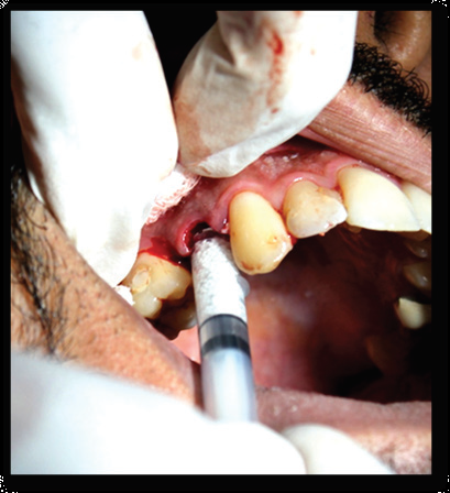
Figure 2: Application of the biomaterial. GUIDOR easy-graft CRYSTAL

Figure 3: Sutures placed
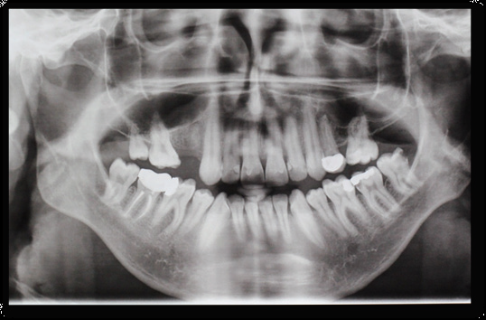
Figure 4: Post operative OPG after Ridge preservation
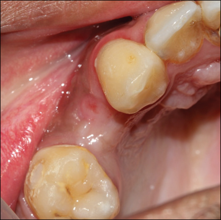
Figure 5: Healed Socket before Indirect Sinus Lift
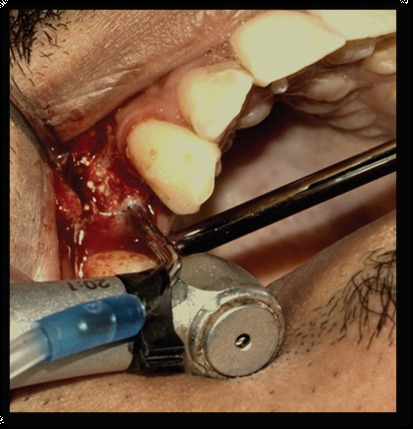
Figure 6: Implantation Procedure

Figure 7 : Indirect sinus lift
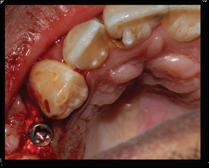
Figure 8: Implant placed
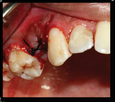
Figure 9: Sutures placed after implant placement
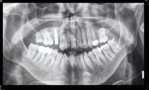
Figure 10: OPG Showing After Implant Placement

Figure 11: After Prosthesis placement
DISCUSSION
In this case a minimally invasive, flapless protocol was followed. No membrane was used to cover the grafted post-extraction site. Mechanical stability was provided as a result of the unique biomechanical properties of the bone graft substitute due to the absence/lack of use of membrane to cover the grafted post-extraction site.
The use of β-TCP as a resorbable socket grafting material can preserve space for bone formation until new bone is formed. This helps in regenerating vital bone of higher quality even without the embedment of non-resorbable bone matrix. This helps the periimplant bone to adapt in accordance to Wolff’s law through functional remodeling after the implant is loaded.
Also normal bone healing and remodeling, reduction in bone –to-implant contact, and negative effect on the overall quality and architecture of the bone surrounding the implants are attributed to the presence of residual non-resorbable or slowly resorbable grafting material.13
Ridge preservation is a procedure which involves the placement of graft material within the socket, with the use of either a membrane or an advanced or rotated flap procedure for closure.
A wide range of ridge preservation materials and techniques have been formulated but with minimal scientific knowledge. Furthermore it lacks a critical cost/benefit analysis in relation to the various treatment alternatives.14
TCP is one such material which has been widely investigated over the past few years. It is known to have two crystalline forms; alpha TCP and beta TCP, of which beta TCP exhibits a good biocompatibility and osteoconductivity. It is thereby used as a novel alternative as bone replacement material.
A study carried by Leventis et al in patients requiring single extraction, using (ð›½-TCP) coated with poly (lactic-co-glycolic acid) (PLGA) were utilized as a grafting material that does not necessitate primary wound closure. The clinical observations revealed excellent soft tissue healing without loss of attached gingiva in all cases. Histological and histomorphometrical analysis revealed pronounced bone regeneration (24.4 ± 7.9% new bone) in parallel to the resorption of the grafting material (12.9 ± 7.7% graft material) while high levels of primary implant stability were recorded.10
Another study by Kakar et al that grafting of intact post-extraction sockets using a biphasic in situ hardening bone graft substitute results in an effective preservation of the ridge contour and sufficient new bone formation in the grafted sites, which is imperative for successful implant placement.15
TCP grafts hence be studied in terms of resorption as well as bone regenerative potential. Resorption of TCP graft is a resultant of two main factors, namely
a) Dissolution of biological fluids in the absence of osteoclasts around the grafted material
b) Presence of osteoclast mediated resorption; via osteoclast like giant cells present within the defect.
Beta TCP has also shown to have regenerative capacities similar to that of autogenous bone, FDBA, DFDBA and collagen sponge. Human clinical studies have availed the use of TCP biomaterials for the repair of periapical and marginal periodontal defects as well as alveolar bone defects.16
CONCLUSION
In the presented case, a technique of ridge preservation is elicited using an in-situ hardening alloplastic bone graft substitute in a minimally invasive manner to obtain a predictable outcome. This technique resulted in the formation of bone capable of remodeling and maturing after loading and also able to support the implant placement after a 5 month healing period.
A flapless technique may be used in future that may reduce patient morbidity, which may be attributed to the in situ hardening property of the material. This thereby preserves the attached keratinized gingival. Thus, both hard- and soft-tissue preservation and regeneration have been utilized in achieving an optimal soft-tissue profile both esthetically and functionally allowing for improved long-term implant stability.
REFERENCES
- Darby I, Chen S, De Poi R. Ridge preservation: what is it and when should it be considered. Australian dental journal. 2008 ;53(:11-21.
- Chen ST, Wilson TG, Hammerle CHF. Immediate or early placement of implants following tooth extraction: review of biologic basis, clinical procedures and outcomes. Int J Oral Maxillofac Implants 2004;19(Suppl):12–25.
- Jambhekar, S.; Kernen, F.; Bidra, A.S. Clinical and histologic outcomes of socket grafting after flapless tooth extraction: A systematic review of randomized controlled clinical trials. J. Prosthet. Dent. 2015; 113: 371–382.
- Horowitz, R.; Holtzclaw, D.; Rosen, P.S. A review on alveolar ridge preservation following tooth extraction.J. Evid. Based Dent. Pract. 2012; 12 (Suppl. 3), 149–160.
- Yip, I.; Ma, L.; Mattheos, N.; Dard, M.; Lang, N.P. Defect healing with various bone substitutes. Clin. Oral Implants Res. 2015; 26:606–614.
- Gao, C.; Peng, S.; Feng, P.; Shuai, C. Bone biomaterials and interactions with stem cells. Bone Res. 2017;5: 17059
- Araújo, M.G.; Liljenberg, B.; Lindhe, J. b-tricalcium phosphate in the early phase of socket healing:An experimental study in the dog. Clin. Oral Implants Res. 2010; 21: 445–454.
- Henkel, J.; Woodruff, M.A.; Epari, D.R.; Steck, R.; Glatt, V.; Dickinson, I.C.; Choong, P.F.; Schuetz, M.A.;Hutmacher, D.W. Bone regeneration based on tissue engineering conceptions—A 21st century perspective.Bone Res. 2013;1: 216–248.
- Harel, N.; Moses, O.; Palti, A.; Ormianer, Z. Long-term results of implants immediately placed into extraction sockets grafted with b-tricalcium phosphate: A retrospective study. J. Oral Maxillofac. Surg. 2013;71: E63–E68.
- Leventis, M.D.; Fairbairn, P.; Kakar, A.; Leventis, A.D.; Margaritis, V.; Lückerath, W.; Horowitz, R.A.;Rao, B.H.; Lindner, A.; Nagursky, H. Minimally invasive alveolar ridge preservation utilizing an in situ hardening b-tricalcium phosphate bone substitute: A multicenter case series. Int. J. Dent. 2016;2016: 5406736.
- Dimitriou, R.;Mataliotakis, G.I.; Calori, G.M.; Giannoudis, P.V. The role of barrier membranes for guided bone regeneration and restoration of large bone defects: Current experimental and clinical evidence. BMCMed. 2012;10: 81.
- Leventis MD, Fairbairn P, Dontas I, et al. Biological response to β-tricalcium phosphate/ calcium sulfate synthetic graft material: An experimental study. Implant Dent. 2014;23(1):37-43
- Chan HL, Lin GH, Fu JH, Wang HL. Alterations in bone quality after socket preservation with grafting materials: a systematic review. Int J Oral Maxillofac Implants. 2013;28(3):710-720.
- Kassim B, Ivanovski S, Mattheos N. Current perspectives on the role of ridge (socket) preservation procedures in dental implant treatment in the aesthetic zone. Australian dental journal. 2014;59(1):48-56.
- Kakar A, Rao BH, Hegde S, Deshpande N, Lindner A, Nagursky H, Patney A, Mahajan H. Ridge preservation using an in situ hardening biphasic calcium phosphate (β-TCP/HA) bone graft substitute—a clinical, radiological, and histological study. International journal of implant dentistry. 2017; 3(1):25.
- Sheikh Z, Hamdan N, Ikeda Y, Grynpas M, Ganss B, Glogauer M. Natural graft tissues and synthetic biomaterials for periodontal and alveolar bone reconstructive applications: a review. Biomaterials research. 2017;21(1):9.
Disclaimer from Guident
The views and opinions expressed in articles published in Guident are solely those of the authors and do not necessarily reflect the views of the editors, the editorial board or the publisher, Ivory India. Guident does not endorse any product, technique or treatment described. Clinical content is intended for qualified dental and medical professionals and is not a substitute for individual clinical judgement. Guident, its editors and the publisher accept no liability for any consequence arising from the use of information published in the journal.
How to cite this article
Dr. Shashikanth Hegde, Dr. Vino Tito V Kurien, Dr. Ashish Kakar. Implant Placement Following Ridge Preservation Using Synthetic Bone Graft Substitute. Guident. 2019 Jan;12(2). Available from: https://guident.in/articles/implantology/implant-placement-following-ridge-preservation-using-synthetic-bone-graft-substitute.html