
A Cost Effective Indigenous Approach for Hybrid Fixed Implant Prosthesis - a Case Report Using Indident Implants
New Delhi India
ABSTRACT
This article presents the complete oral rehabilitation using Indigenous Titanium Dental Implants (INDIDENT) developed by Institute of Nuclear Medicine & Allied Sciences, DRDO. Four External hex type implants were placed in the mandibular arch. An implant supported fixed Hybrid prosthesis was designed over the implants against a conventional tissue supported maxillary denture. Hybrid prosthesis is used to describe a prosthesis, which is composed of different materials. The Hybrid denture in this study consisted of a metal framework with acrylic and denture teeth attached to it and were cemented onto the abutments over the implants. The treatment modality shown is different from the traditional hybrid prosthesis, which is generally screw retained. A cement retained fixed prosthesis is described in this article with a follow –up period of eight years.
Keywords: hybrid denture, indigenous dental implant , INDIDENT
INTRODUCTION
Conventional rehabilitation of complete tooth loss has limitations for many people. Lack of retention and stability are the most common complications related to complete removable prosthesis. Complete removable dentures are often poorly tolerated by the oral tissues. Continual ridge resorption is a problem faced by denture wearers necessitating constant adjustments, replacements of dentures and reduced ability to retain and control the prosthesis1,2. A common treatment plan for an edentulous patient uses implants to support the mandibular prosthesis and a traditional soft tissue supported maxillary denture3-5. This combination resembles a single complete maxillary denture opposing mandibular natural dentition.
Patients with Implant borne dentures are free of the injuries and problems associated with tissue borne dentures. Chewing efficiency has shown to be comparable to that of dentate patients when an implant borne prosthesis is utilized6. Thus the implant borne prosthesis provides the patient with the best substitute for the natural dentition available at present.
Due to high cost of dental implants, most of the cripples in our country could not afford the latest concepts of skeletal rehabilitation by implants being practiced in all the developed nations. With the development of indigenous technology by Defence Research & Development Organization in this field of DENTAL IMPLANTS, large section of our population will be benefited in the days to come.
This article describes the complete oral rehabilitation using cost effective indigenously developed Dental Implants (INDIDENT) by Institute of Nuclear Medicine & Allied Sciences. The case represents a HYBRID -TYPE implant supported fixed prosthesis for the mandibular arch against a conventional tissue supported maxillary denture.
Hybrid dentures are a way to replace teeth when all or most of the teeth are missing on the upper or lower jaw. They are especially useful when a lot of bone is lost and patient is having a difficult time wearing a denture. The prosthesis consists of a metal frame cemented onto the implants .The teeth and acrylic plastic are added to simulate the teeth and gums.
The inability to consistently achieve a passive fit with a multiple-implant ceramo-metal prosthesis led to the development of the fixed-detachable Hybrid prosthesis.7 The high temperatures necessary for firing porcelain onto the metal framework may result in warpage and lack of passivity8. Zarb and Symington9 and Lundquist & Carlsson10 developed the hybrid prosthesis by attaching denture teeth with heat-polymerized acrylic resin to a cast metal substructure.
The case presents a fixed Hybrid prosthesis with cement retention over four implants with a follow up period of eight years.
CASE REPORT
A 45-year-old woman, ex- nursing staff reported to the Dental Depatment OPD with complaint of difficulty in eating with her present denture.
On clinical examination it was revealed that the patient had lost all her teeth at the age of sixteen due to periodontitis. Further she had been wearing the denture for the last 25 years and complained about the loose fitting lower denture. It was further noted that the upper denture usually becomes loose while pronouncing word ‘S’. Clinically, it revealed that the patient had a flat lower alveolar ridge and the upper ridge also had marked resorption. A complete medical review revealed the patient with good general health. An extraoral examination revealed a normal class I jaw relationship, normal TM joint. Muscle tone and width of the Masseter muscle appeared normal.
Intra oral findings showed marked alveolar ridge resorption in upper and a flat lower alveolar ridge. Mucosa appeared healthy pink and devoid of flabby tissue. Fig – 1 shows resorbed mandibular alveolar ridge.
The first step in formulating a treatment plan for dental rehabilitation using implants is determination of sufficient bone quality and bone quantity. So the radiological examination was done with the help of panoramic radiographs and intraoral periapical radiographs .The Bone was found to be healthy and the bone height was assessed by the panoramic x-ray .A marker of known size was placed directly on the mucosa when the panoramic x-ray was obtained. The markers revealed that the radiograph had a 20% distortion. So the actual bone height calculated was 18.5mm in lower left premolar and canine areas and 19.5 mm in lower right premolar and canine area. The bone width was determined with the help of bone caliper, which came to be 6.5 mm at right side of mandible, and 6 mm at left side of mandible.A mandibular fixed Implant supported prosthesis was planned for the patient. The treatment outcome and the surgical procedure involved for the implant placement was explained to the patient.
Four indigenous titanium External hex, single stage, transmucosal implants of 3.5 mm in diameter and 12 mm in height were selected and planned for the implant-supported prosthesis.
A splint was designed to ascertain the implant site for the four implants. The implant placement was carried after giving inferior alveolar nerve block on both sides. The flap was reflected from the canine to canine region .A pilot drill of 2.5mm diameter was employed with consecutive bone drilling with 3.0 mm and 3.2mm diameter drill .A speed reduction handpiece was used to have a controlled torque. Continuous irrigation with chilled saline was used to lower the heat generated by the bone drilling .The parallelism was maintained by the help of paralleling pins and the depth of bone drilling ascertained by the markings on the drills starting from 8mm, 10mm, and12mm. Bone tap corresponding to 3.5 mm diameter implant was used to guide the placement of implant into the prepared site .The implants were placed into the prepared site with the help of implant hex inserters. Fig –2 – shows four external hex implants placed in the mandibular arch. The healing plugs were placed over the four implants. Sutures were placed and the patient was put on antibiotics for five days. General condition of the patient was fair and the patient was comfortable at the time of discharge. Sutures were removed after seven days.
Postoperative Panoramic x-rays were taken immediate after implant placement, after 6 weeks and after three months.
After three months of healing period, the implant-supported prosthesis was planned for the mandible opposing a traditional soft tissue supported maxillary denture.
The implants were firm having no vertical bone loss. Osseointegration and absence of radiolucency around implants were confirmed by panoramic radiograph and with the help of RVG. Fig -3 shows four implants with abutments placed on to it in the mandibular arch.

Fig 1 - Photograph showing resorbed mandibular alveolar ridge.

Fig 2 Photograph showing Four External hex implants
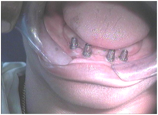
Fig-3 Photograph showing four implants with abutments in the mandibular arch
PROSTHETIC ATTACHMENT
IMPRESSION
The object of the impression is to transfer the implant positions to a master working cast. An accurate impression is to be taken for obtaining an exact replica of the implant, abutments and adjacent soft tissues.
The abutments were screwed onto the implants and impression was taken. Preliminary impression was made with irreversible hydrocolloid material and a custom tray was constructed of self-curing acrylic resin. A custom tray was used to make the final impression with Addition Type Silicone impression material, as this material is preferred for accurate location of the abutments onto the impression.11
The abutments were dried and impression material loaded with care not to incorporate air bubbles into the material. The loaded impression tray was then seated. On removal from the mouth the impression was carefully inspected. The abutments with the implant analogs were relocated into the impression .Fig - 4 shows the impression of the implants in which the abutment with implant analogs are relocated. The impression was placed on the vibrator and poured in Dental stone. The analogs were splinted together with the help of soft acrylic to prevent any movement while pouring the impression, which may lead to inaccuracy in the impression.

Fig-4 Photograph showing impression of the implants with analogs

Fig-5 Photograph showing hybrid prosthesis
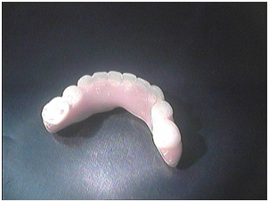
Fig-6 Photograph showing hybrid prosthesis
So, a master cast having implant analogs and abutments placed over it was prepared having a perfect replica of implants placed in the mandible.
The maxillary complete denture impression was made using an acrylic custom tray. The master casts were recovered and trimmed and record bases and occlusion rims were fabricated.
DESIGN OF THE SUPERSTRUCTURE
A fixed mandibular implant supported denture was planned. The mandibular and maxillary casts were articulated .The framework for the prosthesis was waxed over the implants with abutment .The design of the framework was similar to the conventional hybrid prosthesis.
Since the implants were placed in the anterior segment of the arch, the posterior section of the framework is cantilevered posteriorly from the distal abutment. The cantilever was extended upto first molar region. In the mandibular prosthesis, the cantilever should not exceed 12 mm when using four implants.12
A metal framework was constructed over the four implants. A split was given in the middle of the metal framework. This acted as the Stress Breaker.The Stress breaker was provided in the metal framework to avoid any deformation resulting from occlusal stress to the implants. The metal framework was tried in to evaluate and verify a passive and accurate fit intraorally.
The mandibular denture teeth were waxed to the hybrid framework and a final wax try in was performed to verify and correct maxillomandibular relations.
The maxillary complete denture was invested / flasked and processed by use of the maxillary master cast as any conventional complete denture The mandibular hybrid prosthesis was invested without the master cast. The internal aspects of the casting that fit on the abutments were blocked and prosthesis was invested. The investing, flasking and processing procedures were then completed. The prosthesis were then finished and polished, a clinical remount was performed to allow for refinement of occlusal contacts Fig- 5,6 shows the hybrid prosthesis having a metal framework with acrylic attached to it.
The final hybrid prosthesis was cemented over the implant abutments providing a mandibular Implant supported prosthesis opposing a conventional tissue supported maxillary denture. Fig - 7
To facilitate oral hygiene and avoid food impaction, a space of app 2-3 mm was provided between the residual ridge and the framework. It acted as the self-cleansing area for the prosthesis. Oral hygiene instructions were given to the patient and reviewed after every one month.
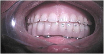
Fig 7 - Photograph showing Hybrid prosthesis cemented over four implants
FOLLOW-UP
Regular check-up for the patient was followed for eight years. Radiological evaluation consisted of measurement of vertical bone loss at regular intervals. The implants showed no mobility and no radiolucency was present in the periimplant area. Fig –8 shows the panoramic radiograph of the patient after one year.
So the case represented a successful implant supported fixed hybrid prosthesis.
DISCUSSION
Implant dentistry requires a complex blend of diagnostic, treatment planning, surgical prosthetic and maintenance skills in order to achieve maximum success rates.
As a result, implants offer a number of various treatment options and have added options to successful prosthodontic rehabilitation. The type of prosthesis depends on patient’s chief complaint. For determination of the prosthesis design the existing problems are evaluated to determine if a fixed or removable restoration is desired. The prosthesis should be discussed and determined primarily for predictable result of implant treatment.
The term Hybrid prosthesis is used to describe a prosthesis that is composed of different materials.13 In implant dentistry the term hybrid implies a combination of a metal framework with a complete denture It is also suggested that this prosthesis be called a metal-resin implant fixed complete denture.14
Hybrid prosthesis is defined as, when acrylic & Denture teeth are added to a metal superstructure. Hybrid prosthesis is fabricated over a metal framework and retained by screws threaded into the implant abutments.15 It s more commonly used in the mandible than in maxilla. The prosthesis is secured to implants placed in the anterior portion of the mandible, while the posterior part of prosthesis is cantilevered from the implants.
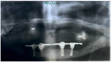
Fig 8- OPG showing hybrid prosthesis with stress breaker and cantilever
A hybrid prosthesis is generally given / planned
- When the original bone height is decreased by natural resorption at the time of implant placement and
- To place the incisal edge of teeth in proper position for esthetics, function, lip support and speech, and when the excessive vertical dimension is to be restored which requires teeth, which look longer.
In Hybrid Denture the restored gingival colour and contour give the teeth a more natural appearance in size and shape and mimic the interdental papilla region.
In the present case, hybrid implant prosthesis was planned for the patient. The prosthesis given was different than the traditional hybrid denture .It was fabricated over a metal framework in the mandibular arch and was cemented to the implant abutments rather than retained by screws to the abutments. The cement –retained fixed-detachable hybrid prosthesis provides an added advantage over screw retained hybrid prosthesis.16
SUMMARY & CONCLUSION
A cost-effective approach to oral rehabilitation using four indigenous dental implants has been presented in this article. The prosthesis provided is the hybrid prosthesis A cement retained fixed Hybrid prosthesis is presented in this study. A definite treatment planning of the prosthesis design is very helpful for the successful outcome of the implant treatment.
BIBLIOGRAPHY
- Campbell R: A comparative Study of the resorption of the alveolar ridges in denture wearers and non-denture wearers.
- Journal of American Dental Association, 60: 143-153, 1960
- Josefowicz: The Influence of wearing dentures on the residual ridge: A comparative study, Journal of Prosthetic Dentistry24: 137-144, 1970
- Branemark PI et al : Osseointegrated Implants in the treatment of edentulous jaw:Experience from a 10-year study(Scandinavian journal of Plastic and Reconstructive Surgery) I 1977 Suppl. 16
- Adell R et al: A 15 year study of Osseointegrated Implants the Treatment of Edentulous jaw,International Journal of Oral Surgery 10: 387, 1981
- Alberktsson T et al: Osseointegrated oral Implants,ASwedishMulticentric Study of 8139 consecutively inserted Noblepharma Implants Journal of Periodontology, 59:287, 1988
- Haroldson T, Carlsson GE: Chewing Efficacy in patients with Osseointegrated Oral Implants bridges.Swedish Dental Journal 3: 183-191, 1979.
- TharpG. Implant update. Miss Dent Assoc. J 1998; 54:22-3
- Misch CE. Contemporary implant dentistry.2nd ed. St Louis: Mosby; 1999 P 69, 549-93
- Zarb GN Symington JM: Osseointegrated Dental Implants Preliminary report on replication study Journal of Prosthetic Dentistry50: 271-276, 1983.
- LundquistS, Carlsson GE. Maxillary fixed prostheses on osseointegrated implants.J Prosthet Dent 1983;50:262-70
- Schelb E, Norliny BK: Impression materials and Techniques (In) Malone WPK, Koth DL (eds): Tylman’s Theory and Practice of Fixed Prosthetics, edition 8. St. Louis Ishiyaka, Euroamerica, 1989.
- Taylor TD: Fixed implant rehabilitation on the edentulous maxilla. Int J Oral Maxillofac Impl 6: 329-337, 1991.
- The Glossary of Prosthodontic terms .J Prosthet Dent 1999: 81(1) 39-110.
- Simon H, Yanase RT.Terminology for implant prostheses Int J Oral Maxillofac Implants. 2003 Jul-Aug; 18(4): 539-43.
- Michael S Block, JN Kent, LR Guerra.Implants in Dentistry: Ch 11 Hybrid Denturesp112.
- GeorgeW.Cobb,AM Metcalf, Douglas Parsell , Gary W ReevesAn alternate treatment method for a fixed-detachable hybrid prosthesis: A clinical report. The J Prosthet Dent 2003; 89: 239-43.
Disclaimer from Guident
The views and opinions expressed in articles published in Guident are solely those of the authors and do not necessarily reflect the views of the editors, the editorial board or the publisher, Ivory India. Guident does not endorse any product, technique or treatment described. Clinical content is intended for qualified dental and medical professionals and is not a substitute for individual clinical judgement. Guident, its editors and the publisher accept no liability for any consequence arising from the use of information published in the journal.
How to cite this article
Dr. Nidhi Adya Saluja Dr. Bobin Saluja Dr. Maj Gen GK Thapliyal. A Cost Effective Indigenous Approach for Hybrid Fixed Implant Prosthesis - a Case Report Using Indident Implants. Guident. 2017 Dec;11(1). Available from: https://guident.in/articles/implantology/a-cost-effective-indigenous-approach-for-hybrid-fixed-implant-prosthesis-a-case-report-using-indident-implants.html

