Orthodontic Disimpaction of Buccally Impacted Canines
Post Graduate Institute of Dental Science, Rohtak
ABSTRACT
Buccally impacted canines specially present in Sector III & IV are difficult to disimpact. This case report demonstrates successful management of bilateral buccally impacted canines one of which is present in Sector IV.
INTRODUCTION
Teeth with a delayed eruption time or that are not expected to erupt completely on the basis clinical and radiographic assessment are called as impacted teeth.1 Permanent maxillary canines are the second most frequently impacted teeth; the prevalence of their impaction is 1-2% in the general population.2,3 This is most likely due to an extended development period and the long, tortuous path of eruption before the canine emerges into full occlusion.2-4Other causes for canine impaction could be due to arch length tooth size discrepancy, early loss or prolonged retention of deciduous teeth. Methods of diagnosis that may allow for early detection and prevention should include a family history, visual and tactile clinical examinations by the age of 9-10 years and a thorough radiographic assessment.Palatally impacted maxillary canines occur twice as frequently as buccally. But buccally impacted maxillary canines are also found very frequently. This case report represents a case of bilaterally buccally impacted maxillary canine managed by closed flap technique and orthodontic traction.
DIAGNOSIS AND TREATMENT PLANNING
A 15year old female reported to the department of orthodontics and dentofacial orthopaedics with chief complaint of forwardly placed upper front teeth. On extraoral examination patient had symmetrical face, acute nasolabial angle with upward tip nose, protruded lips and convex facial profile. Patient had full incisor display and gingival display of 0.5 to 1mm on smiling. (Figure 1)Intraorally super Class I molar relationship on both sides, missing13, 23; retained63,73; ectopically erupted 33 present in crossbite relationship; overjet of 2mm and overbite of 20%. Spacing was present in maxillary arch and crowding in mandibular arch.(Figure 1)Orthopantomogram showed presence of both13, 23 and third molars, with sequential IOPAs and SLOB rule both 13 and 23 were impacted labially. Right maxillary canine 13 was present in sector IV and left maxillary canine 23 was present in sector II according to the classification of Lindauer et al.5(Figure 2)
Lateral cephalogramshow protruded maxillary and mandibular jaw bases (SNA 95°, SNB 90°), proclined upper and lower incisors (upper 1 to NA 26° & 6mm; lower 1 to NB 37° & 11mm) and a vertical growth pattern (FMA 34°)(Figure 2)
TREATMENT OBJECTIVES AND TREATMENT PLAN
Main treatment objectives were to improve convex facial profile, eruption of impacted maxillary canines, alignment of ectopically erupted lower canine and to achieve class I molar and canine relationship.
Treatment plan was to extract all four 1st premolars followed by disimpaction of canines and retraction of anterior teeth and finally finishing and detailing.
TREATMENT PROGRESS
Retained 53 and 63 were extracted along with extraction of all four first premolars. Banding and bonding was done with MBT 022 slot bracket kit with placement of 012 NiTi arch wire for initial leveling and alignment. Sequential arch wires used were as follows: U/L 012 NiTi, 016 NiTi, 017 x 025 NiTi,, 019 x 025 NiTi, 019 x 025 SS arch wire. Maxillary incisors were consolidated followed by elevation of a full thickness flap for canine exposure in both 1st and 2nd quadrant. Orthodontic attachment lingual button was bonded and a traction wire was attached for distal traction followed by occlusal eruption. Flap was closed and a closed traction was thereby done for the prevention of infection and maintain hygiene. For the high into the bone traction of 13 a mini-implant was placed between 15 and 16. Initially a distal force was applied using this mini-implant as an anchor to prevent impingement on the root of 12 followed by occlusal traction for eruption. A normal distal and occlusal for was however applied for 23 using 26 as anchor tooth. Alignment and leveling of all the teeth was followed by closure of remaining spaces and finishing and detailing. Total treatment time was of 18 months.
TREATMENT RESULTS
Significant improvement in facial profile, well aligned maxillary and mandibular arches, normal overjet and overbite, Class I molar and canine relationship were achieved by the end of treatment.(Figure 3,4)
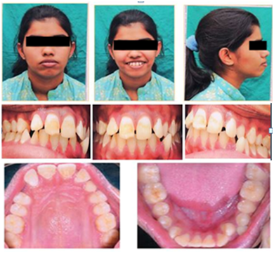
Figure 1: Pre-treatment Extraoral& Intraoral Photographs
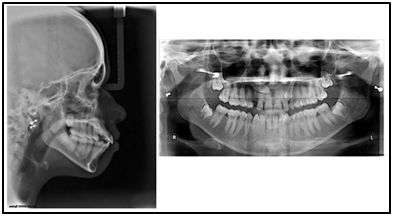
Figure 2: Pre-treatment Lateral Cephalogram& OPG

Figure 3: Post-treatment Extraoral& Intraoral Photographs

Figure 4: Pre &Post comparison of Lateral Cephalogram
Table 1
|
Cephalometric Values |
Pre Treatment |
Post Treatment |
|
SNA |
95° |
95° |
|
SNB |
90° |
90° |
|
WITS |
6mm |
4mm |
|
FMA |
34° |
32° |
|
Upper Incisor to NA |
26° & 6mm |
14.5° & 2mm |
|
Lower Incisor to NB |
37° & 11mm |
31° & 7mm |
|
Lower incisor to Mand. plane |
96° |
90° |
|
Nasolabial Angle |
98° |
103° |
|
Upper lip to E line |
2mm |
-1mm |
|
Lower lip to E line |
5mm |
2.5mm |
CONCLUSION
With thorough treatment planning and its proper execution, canines impacted in difficult positions (like Sector IV) can also be aligned to their normal position giving patients healthy and attractive smile.
REFERENCES
- Thilander B, Jakobsson SO. Local factors in impaction of maxillary canines. ActaOdontolScand 1968; 26:145-68.
- Rayne J. The unerupted maxillary canine. Dent Pract Dent Rec 1969;19:194-204.
- Bass TB. Observations on the misplaced upper canine tooth. Dent PractDent Rec 1967; 18:25-33.
- Hitchin AD. The impacted maxillary canine. Br Dent J 1956; 100:1-14.
- Lindauer SJ, Rubenstein LK, Hang WM, Andersen WC, Isaacson RJ. Canine impaction identified early with panoramic radiographs. J Am Dent Assoc1992;123:91–92 ,95–97
Disclaimer from Guident
The views and opinions expressed in articles published in Guident are solely those of the authors and do not necessarily reflect the views of the editors, the editorial board or the publisher, Ivory India. Guident does not endorse any product, technique or treatment described. Clinical content is intended for qualified dental and medical professionals and is not a substitute for individual clinical judgement. Guident, its editors and the publisher accept no liability for any consequence arising from the use of information published in the journal.
How to cite this article
Dr. Manisha Kamal Dr. Shweta Airan Dr. Manish Airan Department of Orthodontics & Dentofacial Orthopaedics. Orthodontic Disimpaction of Buccally Impacted Canines. Guident. 2017 Dec;11(1). Available from: https://guident.in/articles/orthodontics/orthodontic-disimpaction-of-buccally-impacted-canines.html