
Surgical Management of Periapical Lesion with Palatal Perforation: A Case Report
Department of Conservative Dentistry and Endodontics, Pacific Dental College and Research Centre, Udaipur
ABSTRACT
Aim of the Endodontic surgery to remove periapical pathology followed by achieving regeneration of the periodontal tissue and bone. Traumatic injuries to the anterior teeth are one of the most common of cyst-like apical periodontitis. Chronic periapical infection and microbial attacks on the periapical area results apical granulation tissue and long-standing infection resulting periradicular bone resorption and degradation of the apical periodontal ligament. Some of the apical granulomas turn into cysts. A 35-year-old male patient referred to the department with a chief complaint of pain and swelling on left palatal region since last 2 months. Patient is having history of trauma few years back. On examination, Ellis class I fracture in tooth 21 and 22 was present and the swelling was extending from 21 to 24 on palatal region. Radiographic examination of IOPA revealed large periapical radiolucency irt
21,22. CBCT has also been done for confirmation of the lesion size and location. CBCT revealed evidence of left side palatal perforation seen in maxilla. Periradicular surgery should
be considered as an extension of nonsurgical treatment, because the underlying etiology of the disease process and the objectives of treatment are the same i.e. prevention or
elimination of apical periodontitis.
INTRODUCTION
Successful endodontic treatment depends on periapical repair and regeneration. In cases where conventional root canal therapy fails to eliminate the lesion.1 As there is direct physio pathological relationship in between pulp and periapical region, bacteria–fungi and other cell components can activate inflammatory process in the periapical region causing resorption, leading to formation of abscess, granulomas, and periapical cyst. They have potential to heal nonsurgically using an interim dressing like Ca(OH)2 successfully.2 Calcium Hydroxide has a wide range of antimicrobial activity against common endodontic pathogens, but is less effective against Enterococcus faecalis and Candida albicans. Calcium hydroxide is also an effective anti-endotoxin agent.3 If the lesion is still persistent, surgery is the last alternative.
Aim of the Endodontic surgery to remove periapical pathology followed by achieving regeneration of the periodontal tissue and bone. In modern endodontic practice, due to help of recent advances in diagnosis & material, the number of indications for endodontic periapical surgery is decreasing, but still periapical surgery accounts for 3%–10% in endodontic practice.4 Traumatic injuries to the anterior teeth are one of the most common of cyst-like apical periodontitis. Chronic periapical infection and microbial attacks on the periapical area results apical granulation tissue and long-standing infection resulting periradicular bone resorption and degradation of the apical periodontal ligament. Some of the apical granulomas turn into cysts.5 Long standing infection causes periapical cyst varies from 6 to 54%.
CASE REPORT
A 35-year-old male patient referred to the department of conservative dentistry and endodontics, Pacific Dental College and Research Centre, Udaipur with a chief complaint of pain and swelling on left palatal region since last 2 months. Patient is having history of trauma few years back. On examination, Ellis class I fracture in tooth 21 and 22 was present and the swelling was extending from 21 to 24 on palatal region. Tender on percussion was present in tooth 21 and 22. Electric pulp vitality test gave a negative response in relation to maxillary left central and lateral incisor.
INVESTIGATION
Radiographic examination of IOPA revealed root canal treatment has been initiated in tooth 21 and 22 and also revealed large periapical radiolucency from distal side of right maxillary incisor to mesial side of left maxillary canine which led to a provisional diagnosis of large periapical cyst (Fig. 1). CBCT has also been done for confirmation of the lesion size and location (Fig. 2). CBCT show 3×2 cm large periapical and palatal perforation in irt 21 & 22.
Treatment options were explained to the patient and took patient’s consent for RCT in 21 and 22 followed by surgery. Multiple visit endodontic treatment was performed in tooth 21,22 and calcium hydroxide as intracanal medicament has been placed for 3 weeks. Patient recalled after 3 weeks. The lesion was as it was earlier, no changes seen. So, we planned for surgery. After that upper alginate impression was taken to make maxillary obturator.

Fig 1: Pre operative radiograph

Fig. 2: CBCT i.r.t 21,22
CYST ENUCLEATION PROCEDURE
Under all aseptic condition local anesthetic agent given (lignocaine 2%). Vertical incision given on right side of the second premolar to left side of the second premolar. A full thickness mucoperiosteal flap reflected 5 mm away from the defect bone (Fig. 3). After removal of granulation tissue and cyst obturation was done in relation to 21 and 22 (Fig. 4). Bony cavity was completely cleaned, PRF placement done and sutures were placed (Fig. 5). And obturator has been given (Fig. 6). Patient recalled after 1 week and suture removal done (Fig. 7). Patient recalled after 3 months for follow up. Radiograph showed satisfactory healing of the lesion (Fig. 8).
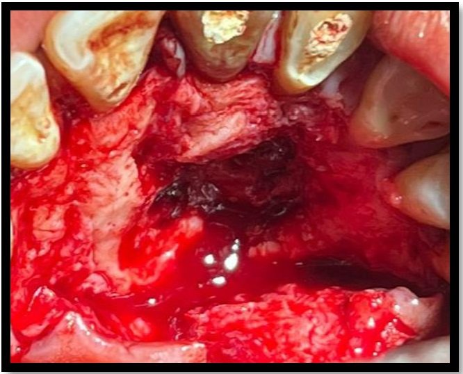
Fig. 3: Flap Raised
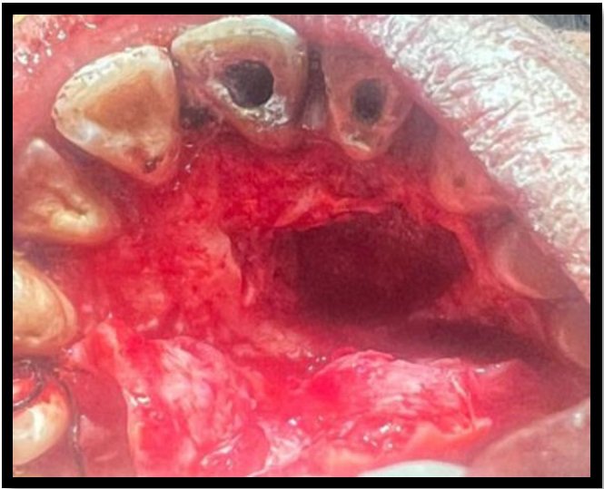
Fig. 4: Cyst Enucleated

Fig. 5: Sutures placed
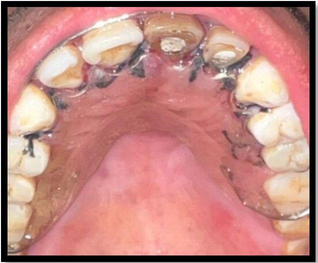
Fig. 6: Obturator

Fig. 7: after one week suture removal

Fig. 8: After 3 month follow up
DISCUSSION
In our case report periapical resulting from long-standing pulpal necrosis and microbial infection due to trauma. In the most of the case, the development of the radicular cyst at the periapical area resulting from continuous microbial infection from the pulp.6 The term, ‘cyst’ is derived from the Greek word, ‘Kystis’, meaning, ‘sac or bladder’. Cyst is defined as a pathological cavity that is usually lined by epithelium and which has a centrifugal, expansive mode of growth.7 There has been evidence of bone-resorbing factors such as receptor activator of nuclear factor kappa-B ligand and osteoprotegerin, which have a role in facilitating the cystic expansion.8
Pathogenesis of radicular cyst has comprised of three distinct phases: the phase of initiation, the phase of cyst formation and the phase of enlargement.9 Radiographically, most radicular cyst appears as round or pear-shaped radiolucent lesion in the periapical region.10 The treatment of all the periapical lesion must be primarily treated by nonsurgical root canal therapy. If, lesion persist then plan for surgical treatment like enucleation, marsupialization or decompression when lesion is large.7
Conservative approach of treatment of any lesion is always preferable over surgery as most periapical lesions are the result of an inflammatory response to bacterial infection within the root canal.11 Nonsurgical root canal treatment involves cleaning and disinfecting the root canal system, thereby reducing the bacterial load and creating an environment in which periapical healing can occur.12 To prevent tooth loss, surgical solution for the inflammatory periapical lesions was considered in the present case as it did not respond favorably to conservative treatment.11
Regeneration of the bone after surgery results from (1) Primary wound closure, (2) Angiogenesis as blood supply, (3) Source of undifferentiated mesenchymal cells, (4) Space maintenance, (5) Stability of the wound (PASS principle) and for good healing we can go with Platelet Rich Fibrin (PRF).13
In this present case we have used PRF for better healing of the lesion. This has a three-dimensional architecture and physiologically all the constituents of a blood sample favor wound healing and immunity. PRF comprises of a multitude of growth factors like Insulin Like Growth Factor (IGF), Transforming Growth Factor β1 (TGF β1), Platelet Derived Growth Factor (PDGF) etc., exhibiting varied potent local properties such as cell migration, cell attachment, cell proliferation and cell differentiation.14 Recent studies shown that, regeneration of lost tissue PRF has proved its potential to regenerate in oral and maxillofacial regions. It was developed by Choukroun et al. in 2001, PRF accumulates platelets and releases cytokines in a fibrin clot, this makes it an ideal biomaterial for wound healing of larger periapical bony defects.15
CONCLUSION
Periradicular surgery should be considered as an extension of nonsurgical treatment, because the underlying etiology of the disease process and the objectives of treatment are the same i.e. prevention or elimination of apical periodontitis.
The administration of PRF increases the number of growth factors present in the wound and aids in tissue healing and bone formation.
REFERENCES
- Prasanthi NN, Chittem J, Simpsy GS, Sajjan GS. Surgical management of a large inflammatory periapical lesion with platelet-rich fibrin. J Interdiscip Dentistry 2017;7:76-9
- Maral SA, Borkar AC, Tandale AB, Nighot NB, Mujumdar SV, Khade SS. Nonsurgical management of persistent periapical lesions in the anterior region - A systematic review. J Int Clin Dent Res Organ 2020;12:8-17
- Mustafa, Mohammed. (2012). Role of Calcium Hydroxide in Endodontics : A Review. Global Journal of Medicine and Public Health. 1. 66-70.
- Hiremath H, Motiwala T, Jain P, Kulkarni S. Use of second-generation platelet concentrate (platelet-rich fibrin) and hydroxyapatite in the management of large periapical inflammatory lesion: A computed tomography scan analysis. Indian J Dent Res 2014;25:517-20.
- Tejasvini B Prajapati et al (2021). Biomimetic Management of a Large Periapical Lesion with an Iatrogenic Defect: A Case Report. Saudi J Oral Dent Res, 6(5): 179-183.
- Nik Abdul Ghani NR, Abdul Hamid NF, Karobari MI. Tunnel’ radicular cyst and its management with root canal treatment and periapical surgery: A case report. Clin Case Rep. 2020 May 25;8(8):1387-1391. doi: 10.1002/ccr3.2942. PMID: 32884760; PMCID: PMC7455399.
- Kadam NS, Ataide Ide N, Raghava P, Fernandes M, Hede R. Management of large radicular cyst by conservative surgical approach: a case report. J Clin Diagn Res. 2014 Feb;8(2):239-41. doi: 10.7860/JCDR/2014/5763.4069. Epub 2014 Feb 3. PMID: 24701544; PMCID: PMC3972573.
- Tekkesin MS, Mutlu S, Olgac V. The role of RANK/RANK/OPG signalling pathways in osteoclastogenesis in odontogenic keratocysts, radicular cysts, and ameloblastomas. Head Neck Pathol. 2011;5(3):248-253.
- Kolari V, Rao HTA, Thomas T. Maxillary and mandibular unusually large radicular cyst: A rare case report. Natl J Maxillofac Surg. 2019 Jul-Dec;10(2):270-273. doi: 10.4103/njms.NJMS_6_18. Epub 2019 Nov 12. PMID: 31798272; PMCID: PMC6883872.
- Deshmukh J, Shrivastava R, Bharath KP, Mallikarjuna R. Giant radicular cyst of the maxilla. BMJ Case Rep. 2014 May 2;2014:bcr2014203678. doi: 10.1136/bcr-2014-203678. PMID: 24792022; PMCID: PMC4025383.
- Tolasaria S, Das UK. Surgical and nonsurgical management of bilateral periapical lesions in the maxillary anterior region. J Surg Tech Case Rep. 2011 Jan;3(1):44-8. doi: 10.4103/2006-8808.78473. PMID: 22022655; PMCID: PMC3192519.
- Morrow SG, Rubinstein RA. Endodontic Surgery. In: Ingle JI, Bakland LK, editors. Endodontics. 5th ed. Hamilton:BC Decker Inc; 2003. p. 405-570
- Prasanthi NN, Chittem J, Simpsy GS, Sajjan GS. Surgical management of a large inflammatory periapical lesion with platelet-rich fibrin. J Interdiscip Dentistry 2017;7:76-9
- Sinha N, Kamboj A, Chopra SS, Rathod A, Mohanty D, Mathur P. Management of a Large Periapical Lesion by Endodontic Surgery Using Platelet Rich Fibrin. J Dent Oral Biol. 2022; 7(4): 1200.
- Pal SS, Agrawal S, Singh M, Gupta A. PRF: An autologous biomaterial for surgical management of periapical lesion – A case report. Asian J Oral Health Allied Sci 2022;12:10.
Disclaimer from Guident
The views and opinions expressed in articles published in Guident are solely those of the authors and do not necessarily reflect the views of the editors, the editorial board or the publisher, Ivory India. Guident does not endorse any product, technique or treatment described. Clinical content is intended for qualified dental and medical professionals and is not a substitute for individual clinical judgement. Guident, its editors and the publisher accept no liability for any consequence arising from the use of information published in the journal.
How to cite this article
Dr. Nehali Thakkar, Dr. Jitendra Lohar, Dr. Krati Sahu. Surgical Management of Periapical Lesion with Palatal Perforation: A Case Report. Guident. 2023 May;16(6). Available from: https://guident.in/articles/endodontics/surgical-management-of-periapical-lesion-with-palatal-perforation-a-case-report.html


