
A Comparative Evaluation of Etidronic Acid and EDTA Irrigating Solutions in Dentinal Demineralisation and Removal of Smearlayer: A Systematic Review
ABSTRACT
AIM: The aim of this systematic review is comparative evaluation of Etidronic acid and EDTA solutions in dentinal demineralization and removal of smear layer.
METHODS: The study started with focused electronic searches of the PUBMED database and other subject-specific databases, then went through a thorough manual search and citation mining process to find all articles reporting a comparison of the effectiveness of etidronic acid and EDTA in dentinal demineralization and the removal of the smear layer in human extracted teeth that were then analysed under SEM.
RESULTS: A moderate to high risk of bias was present in seven out of the included papers. The best available scientific evidence demonstrates that EDTA and etidronic acid do not significantly differ in the elimination of the smear layer and dentinal demineralization.
CONCLUSION: Even though Etidronic acid is better at smear layer removal and less dentin demineralization still the results are unclear and further research is needed for better conclusions.
KEYWORDS: EDTA, Etidronic acid , Dentinal demineralization, smear layer removal.
INTRODUCTION
A successful root canal procedure requires the right biomechanical preparation techniques and standards, as well as irrigation, disinfection, and obturation.1 Complete removal of the damaged dentin, necrotic pulp, debris, and bacteria is required for root canal preparation. The creation of a smear layer that covers the whole root canal wall is facilitated by dentin removal. It is composed of organic and inorganic components that have bacteria, necrotic materials, and odontoblastic processes in it.2 The smear layer blocks the diffusion of intracanal medications into the dentinal tubules and irregularities in the root canal. It also prevents obturating substances from sticking to the root canal's surface.4 A thin layer (1-2 m thick) and a deeper smear plug (40 m thick) make up the smear layer.3
By allowing medication and irrigants to penetrate more deeply, smear layer removal is crucial for disinfecting the root canal system. Moreover, it aids in the penetration of root canal sealants into dentinal tubules, which enables obturating materials better adhere to root canal walls.4
Many studies have been done on how effective irrigating solutions are at removing the smear layer. Any of the currently available root canal irrigants, including NaOCl, cannot completely eliminate the smear layer since it comprises both inorganic and organic material. It is necessary to utilise sodium hypochlorite (NaOCl) and a chelating agent or acid that dissolves inorganic tissue in that order. Hence, the suggested procedure for removing the smear layer is NaOCl, followed by EDTA (ethylenediaminetetraacetic acid) or citric acid.3 Because it is impossible to employ EDTA and sodium hypochlorite solutions at the same time, this rinse calls for two different irrigation methods. NaOCl loses its ability to dissolve tissue when combined with EDTA since there is almost no free chlorine left.1
Sodium hypochlorite and etidronic acid (1-hydroxyethane-1, 1-diphosphonic acid, HEDP), which must be added to the NaOCl rinsing solution (as powder) immediately before use, have recently been combined to create a novel irrigating solution; the powder (in pre-weighed capsules) is now marketed as "Dual Rinse" (Medcem, Weinfelden, Switzerland).5
It has been found that the sodium hypochlorite solution in which the chelating agent HEDP is dissolved keeps it stable and active for one hour. It is anticipated that this irrigant will replace frequently used irrigation solutions and combine the properties of a chelating agent with those of sodium hypochlorite. The fact that both reagents are present during the cleaning and shaping process is a possible advantage of employing this new irrigant. Moreover, sodium hypochlorite's antibacterial effects may be improved if smear layer formation is avoided throughout the treatment. However, using a chelating agent during the procedure may cause the dentinal walls of the root canal to erode and sustain injury.6 In this study, we evaluated the efficiency with which EDTA and etidronic acid demineralized the dentin and eliminated the smear layer.
METHODS AND PROTOCOL
This systematic review was carried out utilising PRISMA (Preferred Reporting Items for Systematic Reviews and Meta-Analyses) standards. The main enquiry for this evaluation was "How Etidronic acid solution causes elimination of smear layer and dentinal demineralization compared to EDTA."
According to the null hypothesis, EDTA and etidronic acid have the same effects on dentinal demineralization and smear layer removal.
LITERATURE SEARCH
In-vitro experiments were exhaustively searched for irrigation solutions containing EDTA, etidronic acid, or both. The keywords "Smear layer," "Dental erosion," "EDTA," "Etidronic acid," or "in vitro" "Scanning electron microscopy" were used to search the PUBMED database and subject-specific data base, which includes the INTERNATIONAL ENDODONTIC JOURNAL (IEJ), JOURNAL OF ENDODONTICS (JOE), and AUSTRALIAN ENDODONTIC JOURNAL (AEJ). Searches were performed on the PUBMED database and the remaining journals from July 2010 to November 2022. Additionally, a manual forward search was done on the chosen articles' references list.
SELECTION OF STUDIES AND DATA EXTRACTION
The titles and abstracts of the articles in the search results were checked separately by two distinct authors. As the full texts of pertinent publications were reviewed, the eligibility standards were observed. It was made a sheet for systematic data extraction. The following factors were gathered from the chosen studies: sample size, irrigation solution, tooth kinds, assessment using SEM, other parameters investigated in the studies besides smear layer and dentinal degradation, and research outcomes.
ELIGIBILITY CRITERIA
Prior to conducting a literature search, selection criteria for studies were created. The inclusion and exclusion criteria are summarized in Table 1.

TABLE 1: Inclusion and Exclusion Criteria.
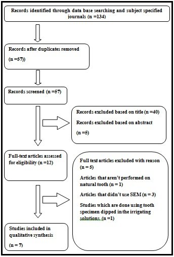
PRISMA flow chart
Risk of Bias and Quality of Evidence:
Each paper was individually examined for quality by two authors. The overview quality assessment questionnaire (OQAQ) score system was used to calculate the risk of bias in the selected studies. Nine questions with a 0–18 score range were devised in order to satisfy these quality assessment requirements. Articles in this systematic review were deemed to be of high quality if they had an OQAQ score of 12 or higher. Greaves et al. changed the overall quality assessment questionnaire that was used to measure the quality. Table 2 provides the bias potential.

TABLE 2: Bias potential
RESULTS
The first search strategy returned 134 references after removing duplicate entries from the numerous databases that were searched. A preliminary screening assessment based on the titles and abstracts of the references reduced the number of sources to 57 papers. The entire texts of the other articles were then obtained and updated by two authors. The systematic review solely incorporated in vitro studies that evaluated the removal of the smear layer and dentinal demineralization after the extraction of human teeth. Table 3 frames the final 7 articles acquired.
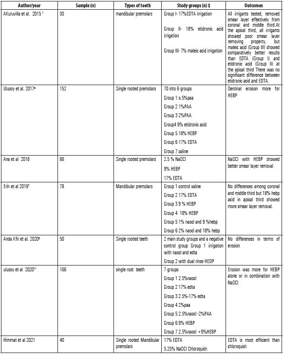
TABLE 3: Summary of the main characteristics of the included studies
DISCUSSION
The current study sought to assess the effects of EDTA and etidronic acid on the demineralization of the dentin and elimination of the smear layer. The current study demonstrated that a procedure using either EDTA treatment or etidronic acid treatment could minimise the smear layer in instrumented root canals.
Eick et al. originally described using scanning electron microscopy to find the smear layer in 1970. With the use of a scanning electron microscope, the surface modifications brought on by dental erosion can be seen. The scanning electron microscope offers unmatched imaging and analytical performance thanks to its cutting-edge electron optics and signal detection technologies. The new user-friendly graphical user interface offers thorough picture observation and presentation functions and was developed with intuitive logic. It is designed to be used for a variety of purposes, including the storage of sophisticated materials and biological samples. Compared to other methods, SEM provides a clearer picture of the dentinal tubules and smear layer.7
Although EDTA and NaOCl appear to be the most efficient combination for removing smear layers so far, they cannot be used concurrently because EDTA solution can chemically interact with NaOCl and lower the amount of free chlorine.1The intertubular and peritubular dentine can easily be penetrated by EDTA in this combination, speeding up the dentine's disintegration and causing a noticeable erosion of the canal wall.
Etidronate has the benefit of mixing with NaOCl without affecting its antibacterial properties. Since HEDP is a mild chelator, it may be less damaging to dentin than EDTA. However, the elimination of the smear layer may take longer with these treatments. In terms of periapical tissues, it is biocompatible.6
De-Deus et al. claim that because etidronic acid and NaOCl have a sluggish and weak impact on inorganic components, it took around 300 seconds to entirely remove the smear layer.8
Anda K. Fir et al. demonstrated that there was no difference in the frequency and seriousness of erosion between the two groups. Most of the cases in both groups of cases exhibited no or very little erosion, while 10–26% of the cases had significant erosion. In each of the groups, there was just one instance of severe erosion. As a result, the short presence of EDTA following the endodontic process did not induce any more erosion than the continuous presence of the HEDP chelator.9 This conclusion is consistent with Tartari et alfindings .'s in their investigation, which showed that dentine demineralization was more mildly induced by HEDP than by EDTA.10
Etidronic acid, however, induced changes in root dentin and a decrease in nanohardness, which led to more dentinal demineralization than EDTA, according to ulusoy et al.11
Erik et al. claim that while there were no differences between the two treatments' abilities to remove smear layers from the coronal and middle thirds, etidronic acid was more effective at doing so from the apical third.7
According to Ana et al., etidronic acid and NaOCl shown improved smear layer removal.
According to Ulusoy et alstudy, .'s 9% and 18% Etidronic acid were more effective than EDTA at removing the smear layer in the apical third.6
The proposed null hypothesis states that neither EDTA nor etidronic acid differs from one another in terms of removing smear layers and demineralizing dentin.
CONCLUSION
The featured research varied widely from one another. Due to these discrepancies, there is contradicting evidence.
Further research is needed before we can draw firm conclusions about the demineralization of dentin and removal of the smear layer by etidronic acid.
REFERENCES
- Kuruvilla A, Jaganath BM, Krishnegowda SC, Ramachandra PK, Johns DA, Abraham A. A comparative evaluation of smear layer removal by using edta, etidronic acid, and maleic acid as root canal irrigants: An in vitro scanning electron microscopic study. J Conserv Dent. 2015 May-Jun;18(3):247-51
- Khademi A, Feizianfard M. The effect of EDTA and citric acid on smear layer removal of mesial canals of first mandibular molars, a scanning electron microscopic study. J Res Med Sci 2004;2:80–88.
- Violich DR, Chandler NP. The smear layer in endodontics—a review. Int Endod J 2010;43(1):2–15
- Mahajan V, Kamra A, Dahiwale S. The effect of 17% EDTA and MTAD on smear layer removal and on erosion of root canal dentin when used as final rinse: an in vitro SEM study. J Int Clin Dent Res Organ 2010;2(3):113–118. DOI: 10.4103/2231-0754.95282.
- Kfir A, Goldenberg C, Metzger Z, Hülsmann M, Baxter S. Cleanliness and erosion of root canal walls after irrigation with a new HEDP-based solution vs. traditional sodium hypochlorite followed by EDTA. A scanning electron microscope study. Clinical Oral Investigations. 2020 Oct;24:3699-706.
- Ulusoy Ö?, Zeyrek S, Çelik B. Evaluation of smear layer removal and marginal adaptation of root canal sealer after final irrigation using ethylenediaminetetraacetic, peracetic, and etidronic acids with different concentrations. Microsc Res Tech. 2017 Jul;80(7):687-692.
- Emre Erik C, Onur Orhan E, Maden M. Qualitative analysis of smear layer treated with different etidronate concentrations: A scanning electron microscopy study. Microscopy Research and Technique. 2019 Sep;82(9):1535-41.
- De?Deus G, Paciornik S, Mauricio MH. Evaluation of the effect of EDTA, EDTAC and citric acid on the microhardness of root dentine. International Endodontic Journal. 2006 May;39(5):401-7.
- Kfir A, Goldenberg C, Metzger Z, Hülsmann M, Baxter S. Cleanliness and erosion of root canal walls after irrigation with a new HEDP-based solution vs. traditional sodium hypochlorite followed by EDTA. A scanning electron microscope study. Clin Oral Investig. 2020 Oct;24(10):3699-3706.
- Tartari T, Borges MMB, de Araújo LBB, Vivan RR, Bonjardim LR, Duarte MAH. Effects of heat in the properties of NaOCl alone and mixed with etidronate and alkaline tetrasodium EDTA. Int Endod J. 2021 Apr;54(4):616-627.
- Ulusoy Ö?, Mant? A?, Çelik B. Nanohardness reduction and root dentine erosion after final irrigation with ethylenediaminetetraacetic, etidronic and peracetic acids. Int Endod J. 2020 Nov;53(11):1549-1558
Disclaimer from Guident
The views and opinions expressed in articles published in Guident are solely those of the authors and do not necessarily reflect the views of the editors, the editorial board or the publisher, Ivory India. Guident does not endorse any product, technique or treatment described. Clinical content is intended for qualified dental and medical professionals and is not a substitute for individual clinical judgement. Guident, its editors and the publisher accept no liability for any consequence arising from the use of information published in the journal.
How to cite this article
Dr. Naga Mahitha, Dr. Deepika Priyadarsini, Dr. Devalla Venu Babu. A Comparative Evaluation of Etidronic Acid and EDTA Irrigating Solutions in Dentinal Demineralisation and Removal of Smearlayer: A Systematic Review. Guident. 2023 Apr;16(5). Available from: https://guident.in/articles/endodontics/a-comparative-evaluation-of-etidronic-acid-and-edta-irrigating-solutions-in-dentinal-demineralisation-and.html


