Management of External Root Resorption: A Case Report
Department of Conservative Dentistry and Endodontics, Seema Dental College And Hospital, Rishikesh
ABSTRACT
External root resorption is a complex pathological process that affects the integrity of tooth roots, leading to potential tooth loss. Although it is predominantly detected by radiographs, in some cases root resorption may be identified by clinical symptoms such as pain, swelling and mobility of the tooth. Treatment alternatives are case?dependent. This article discusses a case report on external root resorption and its management.
Keywords: Root Resorption, External Resorption, MTA
INTRODUCTION
External root resorption is a pathological condition characterized by the progressive loss of dental root structure due to various factors. It affects both primary and permanent dentition and can lead to significant functional and aesthetic consequences.1,2
External root resorption can be attributed to a range of factors. Trauma, such as dental injury or orthodontic treatment, is a common cause, as it disrupts the protective layers surrounding the tooth root. Orthodontic forces, if excessive or prolonged, can also induce resorption. Additionally, certain inflammatory conditions, such as periodontal disease, cysts, or tumors, may contribute to root resorption. Systemic factors, including genetic predisposition and certain medical conditions, can increase the susceptibility to resorption.4,10
This article aims to present a clinical case depicting external root resorption and its management with the use of MTA as material of choice.
CASE REPORT
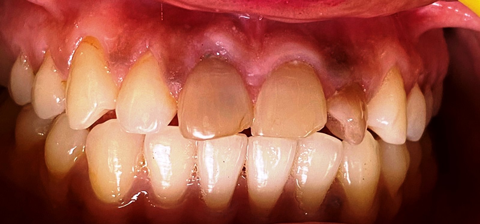
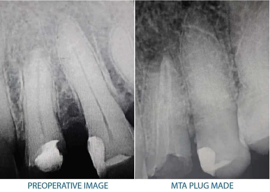
A 48 years old male patient reported to the department of Conservative dentistry and Endodontics with the chief complaint of broken tooth in the upper left tooth region since 6-7months. The patient gave a history of root canal treatment being initiated for the same tooth 1 month back. Clinical Examination revealed temporary restoration irt 24. Radiographic finding revealed previously initiated root canal treatment with external root resorption.
After complete examination, a treatment plan for non-surgical root canal treatment irt 24 followed by MTA plug and fiber post and core were made.
On the first visit, temporary restoration was removed and working length determination was done using apex locator. Following working length determination, cleaning, and shaping of root canal system was done using a crown down technique using 2.5% NaOCl and normal saline as irrigating solutions. The canal was dried were paper points and MTA was mixed as per manufacturer’s instructions and a apical plug of 3-4mm was made for both buccal and lingual canal. After the plug was made, a wet cotton gauze was placed and provisional restoration was given. The patient was recalled after 1 day and the temporary restoration was removed and the set of MTA was verified.
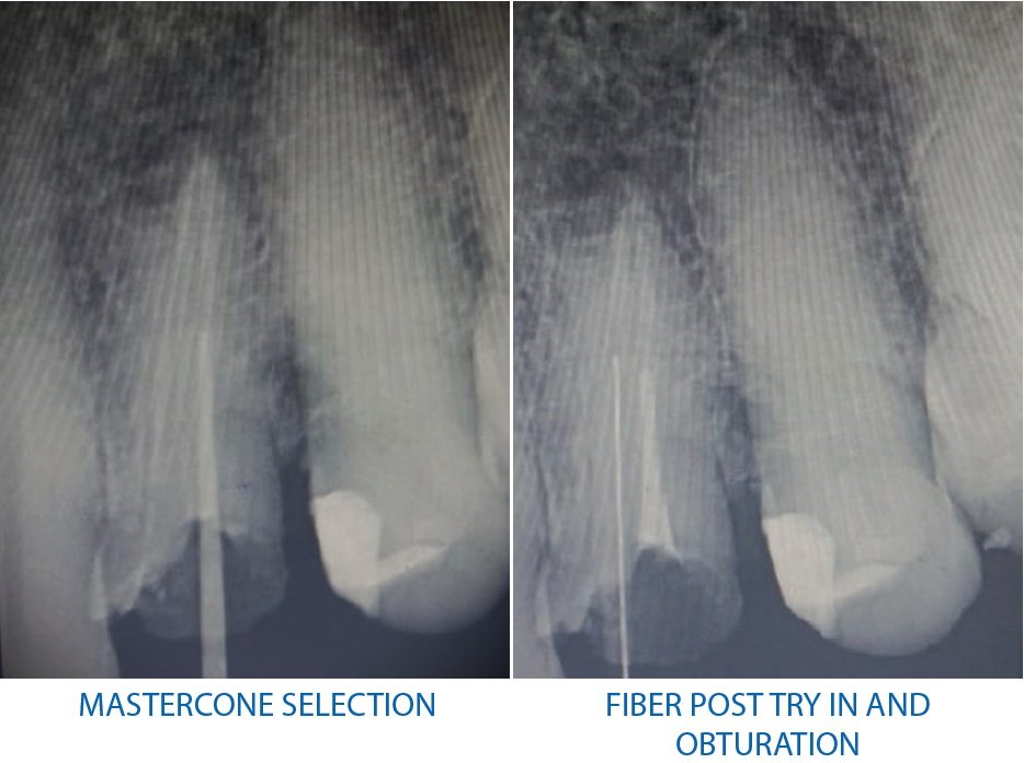
The lingual canal was then enlarged using peeso reamers to receive a fiber post and the buccal canal was obturated. Fiber post try was done for the lingual canal followed by its cementation and core build up. Tooth preparation was then done and indirect putty impression was recorded for the fabrication of prosthesis. Once the prosthesis was fabricated, it was cemented using glass ionomer cement.


DISCUSSION
External root resorption is a challenging condition that can compromise the integrity and stability of affected teeth. External root resorption may present with various clinical manifestations, depending on the severity and location of the resorptive process. In early stages, patients are often asymptomatic, and the condition may only be detected during routine dental examinations. As the resorption progresses, symptoms such as tooth sensitivity, pain, and mobility may develop.3,5,6
The management of external root resorption depends on several factors, including the extent of resorption, patient age, and overall dental health. In cases of mild to moderate resorption, regular monitoring with radiographic evaluations may be sufficient. However, active treatment is often necessary to preserve tooth function and aesthetics.7,8,11
Non-surgical approaches involve identifying and addressing the underlying cause, such as treating periodontal disease or removing a source of trauma. In some cases, placing a protective barrier, such as a biocompatible material, over the resorptive defect can help prevent further progression.9
Severe cases of external root resorption may require surgical intervention. Various techniques, including root canal therapy, root resection, or extraction, may be employed based on the individual circumstances.12
This case report described the use of MTA aa a barrier to prevent further progression of the defect.
Traditional treatment options for external root resorption have limitations in terms of sealing the resorptive defect and promoting repair. However, the use of mineral trioxide aggregate (MTA) as a plug has emerged as a promising treatment approach.13
MTA is a biocompatible material with excellent sealing properties and biocompatible characteristics. The placement of an MTA plug in the resorptive defect can effectively seal the area, preventing bacterial infiltration and promoting the regeneration of periapical tissues. MTA promotes the formation of a mineralized tissue barrier, thereby preventing further resorptive activity and facilitating the repair process. Moreover, MTA exhibits antimicrobial properties, reducing the risk of infection.16
In a study by Pace et al. (2019), a significant reduction in the size of external root resorption defects was observed after the placement of MTA plugs.14 Another study by Yu et al. (2017) reported successful healing of invasive cervical resorption lesions using MTA plugs with a high success rate.15 Additionally, a systematic review by Patel and Sayed (2016) demonstrated favorable outcomes with MTA plugs in the treatment of external root resorption.16
CONCLUSION
External root resorption is a multifactorial condition that can significantly impact dental health. Understanding its causes, mechanisms, clinical manifestations, and management strategies is crucial for timely diagnosis and appropriate treatment.) The use of MTA plugs as a treatment approach for external root resorption holds significant promise. The biocompatibility, sealing ability, and potential for tissue regeneration make MTA an ideal material for managing these challenging cases. Further research and long-term follow-up studies are necessary to establish standardized protocols and optimize the use of MTA plugs in the management of external root resorption.
REFERENCES
- Andreasen JO, Andreasen FM. External root resorption: its implication in dental traumatology, pedodontics, periodontics, orthodontics and endodontics. Int Endod J. 2005;38(5): 118-131
- Bakland LK, Andreasen JO. External root resorption. In: Andreasen JO, Andreasen FM, Andersson L, eds. Textbook and Color Atlas of Traumatic Injuries to the Teeth. 4th ed. Wiley-Blackwell; 2007: 498-528
- Patel S, Dawood A, Whaites E, Pitt Ford T. New dimensions in endodontic imaging: part 1. Conventional and alternative radiographic systems. Int Endod J. 2009;42(6):447-462
- Lindskog S, Blomlöf L, Hammarström L. Evidence for a role of orthodontic tooth movement in some forms of external root resorption. Eur J Orthod. 1988;10(3): 229-238
- Patel S, Kanagasingam S, Pitt Ford T. External cervical resorption: a review. J Endod. 2009;35(5):616-625
- Rafter M. External apical root resorption following bleaching of pulpless teeth with oxygen peroxide. Endod Dent Traumatol. 1988;4(1): 23-27.
- Patel S, Ricucci D, Durak C, Tay F. Internal root resorption: a review. J Endod. 2010;36(7):1107-1121
- Magnusson BC, Kjellberg H, Hägglund B, Sundström F, Ørstavik D. Clinical and radiographic assessment of external inflammatory root resorption in patients with periodontitis. J Endod. 2004;30(1):45-50
- Patel S, Patel S. External apical root resorption: a review of the literature. Dent Update. 2012;39(6):455-462
- Owais AI, Fayed MM, Abu-Bakr NH, Abu-Tahun IH. External apical root resorption in orthodontically treated patients: causative factors and radiographic evaluation. Open Dent J. 2016;10:129-140
- Krastl G, Allgayer N, Lenherr P, Filippi A, Taneja P, Weiger R. Tooth resorption (part I): pathogenesis, case definition and diagnosis. Swiss Dent J. 2014;124(9):874-886
- Patel S, Patel S. Apical root resorption part 1: pathogenesis, case series, and case reports. Dent Update. 2016;43(1):12-24
- Kaur H, Gupta A, Mittal A, Garg R. External cervical root resorption: a review. Int J Clin Pediatr Dent. 2012;5(3):173-177.
- Pace R, Di Nardo D, Giuliani V, et al. Surgical management of invasive cervical root resorption with mineral trioxide aggregate: A case series with up to 7 years' follow-up. J Endod. 2019;45(11):1314-1320
- Yu V, Gupta S, Sharma A, et al. Invasive cervical resorption: treatment challenges and an innovative solution using mineral trioxide aggregate. J Conserv Dent. 2017;20(5):374-377
- Patel S, Sayed M. Use of mineral trioxide aggregate for management of external invasive cervical resorption: long-term follow-up of 2 cases. J Endod. 2016;42(10):1575-1579
Disclaimer from Guident
The views and opinions expressed in articles published in Guident are solely those of the authors and do not necessarily reflect the views of the editors, the editorial board or the publisher, Ivory India. Guident does not endorse any product, technique or treatment described. Clinical content is intended for qualified dental and medical professionals and is not a substitute for individual clinical judgement. Guident, its editors and the publisher accept no liability for any consequence arising from the use of information published in the journal.
How to cite this article
Dr. Siddhi Goyal, Dr. Anil Dhingra. Management of External Root Resorption: A Case Report. Guident. 2023 Jun;16(7). Available from: https://guident.in/articles/endodontics/management-of-external-root-resorption-a-case-report.html