
Exodontia- Events and Biological Effects on Alveolus
Department of Prosthodontics, Crown, Bridge and Implantology, AME’s Dental college and Hospital, Raichur
ABSTRACT
Dental extractions are routine procedures performed by dentists all over the world. There are times when tooth loss is unavoidable. Advanced bone loss, trauma, and extensive decay can leave patients with few alternatives for saving a tooth. Repulsion still results in unacceptably high rates of iatrogenic complications. Veterinarian dentists have developed methods to simplify challenging intraoral extractions and surgical extraction techniques to prevent tooth repulsion. Preoperative planning is the greatest way to reduce difficulties. The dentist can anticipate intraoperative complications and set up procedures to yield the best results by doing a thorough preoperative evaluation and treatment plan.
KEYWORDS- Biological effects, healing of alveolar bone, internal changes, external changes, dimensional changes, alveolar socket
INTRODUCTION
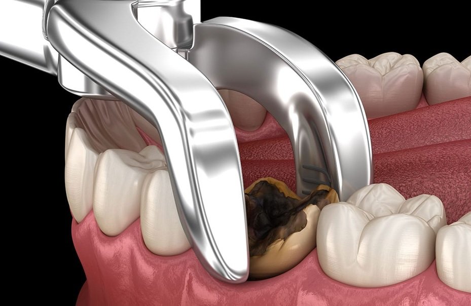
A tooth is indicated for extraction when it is no longer possible to restore or maintain in acceptable conditions considering its health, function and/or aesthetics. The extraction of a tooth triggers a series of events that further result in the decrease of height and width of the alveolar process, particularly on the buccal side and horizontally. After extraction, this resorptive event occurs during the first three months of healing until one year, with potential aesthetic and functional consequences for prosthetic rehabilitation.1
BIOLOGICAL EFFECT OF A TOOTH EXTRACTION
Tooth extraction sets off a series of biological events, with a local inflammatory response and an irreversible structural transformation of the periodontium.3 In terms of hard tissues, as previously stated, it can be expected a bone resorption, mainly in the first three months, causing both vertical and horizontal changes in the alveolar process.5,10
These anatomical changes are more buccally and horizontally pronounced3-7, with an average horizontal reduction of 3.79–3.87 mm and an average vertical reduction on the buccal side of 0.64–1.24 mm.5,7
Concerning the soft tissues, the socket defect will determine the healing process through secondary intention resulting in cell proliferation, whereas the gingival form mostly depends on the external shape of the alveolar bone.5
In order to diminish the biological effect of a tooth extraction, an appropriate treatment plan and technique are central.3 As for the surgical technique, a flapless approach is considered a simple, atraumatic and conservative method, being the usual choice to reduce post-surgery healing period, discomfort and inflammation.4 This surgical approach is characterized by the nondetachment of the periosteum, preserving the blood supply to the buccal bone, which, as mentioned before, suffers a more pronounced resorption.11
HEALING OF ALVEOLAR BONE AFTER TOOTH EXTRACTION
Osseous deformities of the alveolar ridge, including both width and height reduction of the residual ridge, are typically caused by tooth extraction and the following healing of the socket.
Healing pattern of socket alveolus:
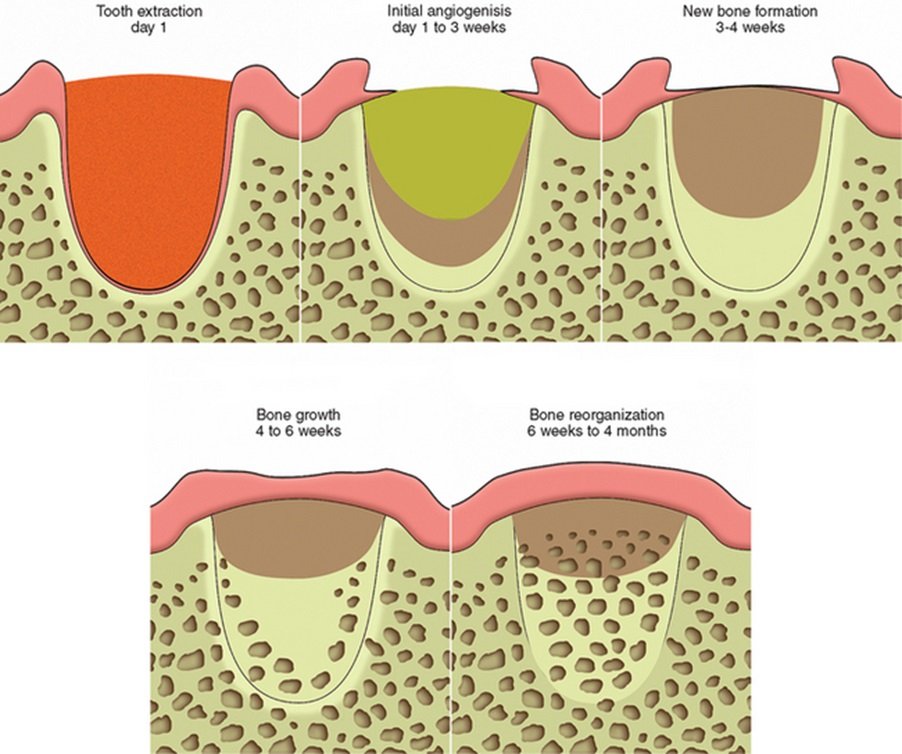
Residual ridge remodelling, begins with a cascade of inflammatory reactions that is activated immediately after tooth extraction. Healing of extraction sockets can be explained in five different stages:
- GRANULATION STAGE: The granulation stage lasts around five days. The early granulation tissue first appears at the bottom of the socket and spreads laterally, up the socket walls.
- ANGIOGENIC STAGE: The second phase, the initial angiogenic stage, happens within the first week of granulation stage. New trabeculae have begun to appear at the bottom of the socket, and the blood clot in the center has begun to shrink.
- NEW BONE FORMATION STAGE: The third phase begins two weeks after the tooth extraction. Sinusoid formations that began in the earlier phases yield bone trabeculae. At this point osteoid, can be detected. The bone formation follows the pattern of the sinusoid formations, which have mostly formed vertically starting in the apical region.
- BONE GROWTH STAGE: The bone growth stage which is characterized by well developed, thickened trabeculae that now fill 2/3rds of the socket. This happens 4-5 weeks after extraction. This woven bone is referred to as spongiosa. Some sinusoids are still forming in the coronal portion of the socket, while the apical portion appears to be more mature with less sinusoids.
- BONE REORGANIZATION STAGE: The final stage is the bone reorganization stage which is usually evident around six weeks after extraction. The primary spongiosa now develops into secondary spongiosa, or more lamellar bone, and is present in higher percentages towards the apical end of the residual socket.6
PROCEDURES TO BE OBSERVED TO PRESERVE THE ALVEOLAR SOCKET
TOOTH EXTRACTION: Atraumatic extraction techniques should be used to minimize damage to the alveolar bone during tooth extraction. The Easy-X-tractor is a device designed to extract single-rooted teeth with minimal trauma and does not require flap elevation. If the tooth structure is too broken down, the roots can be carefully sectioned into fragments and extracted without placing pressure on the alveolus. Once the tooth is extracted, the alveolus should be thoroughly debrided, removing all of the granulation tissue.
FLAP DESIGN: The extraction of a tooth results in a decrease in height of the papillae. The loss of papillae height is increased with the elevation of buccal and lingual flaps.4 In areas of esthetic concern, consideration should be given to extracting the tooth and augmenting the socket without elevating a flap or the use of a mini-flap on the buccal side preserving the papillae
POST-OPERATIVE MANAGEMENT: The most important step in the post-operative management of augmented extraction sockets is the placement and shaping of the temporary tooth that is placed over the extraction area. The loss of papillae height from extraction can be largely restored with the use of removable or fixed provisional appliances.6
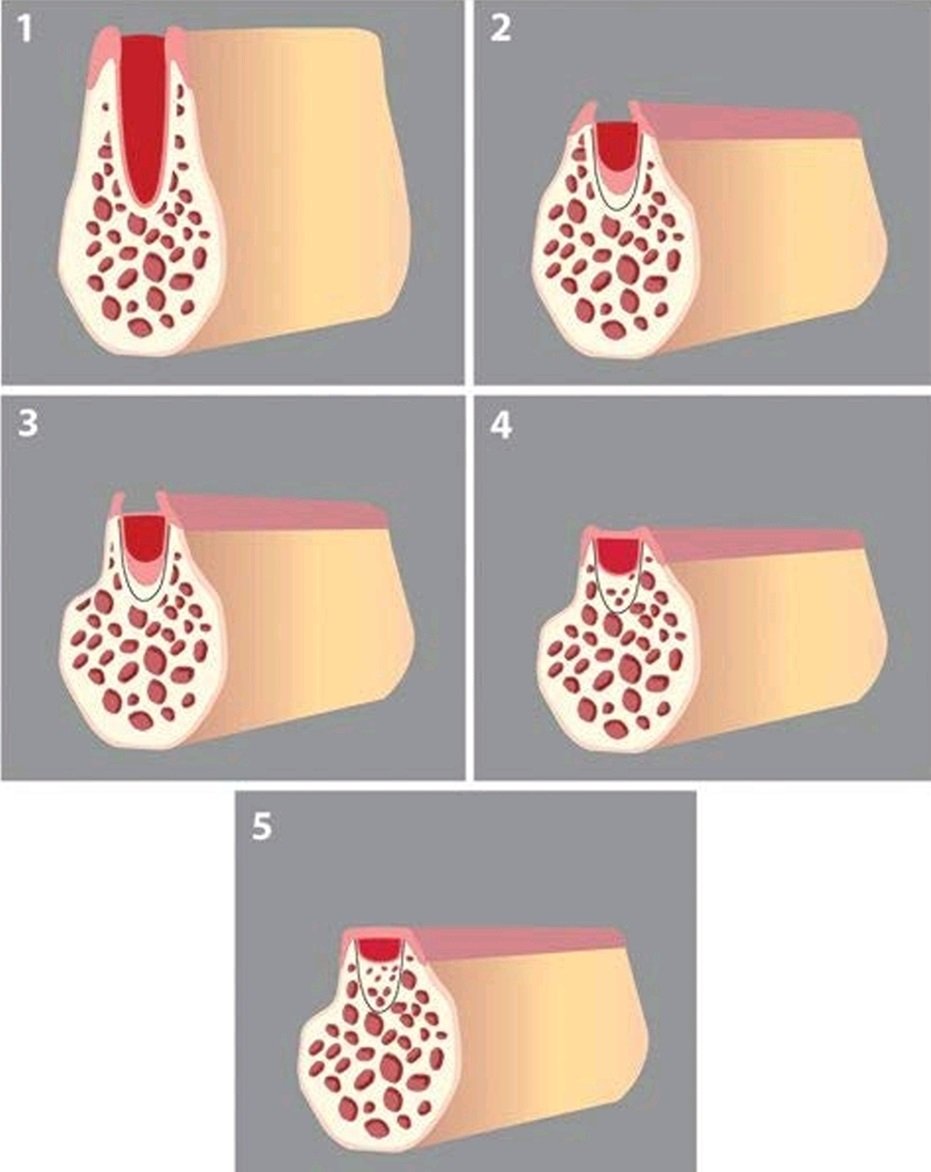
Bone resorption will result in a reduction of socket height in an apico-coronal direction and socket width in a bucco-lingual direction. Healing of an extraction socket is characterized by internal changes that lead to formation of bone within the socket, and, and external changes that lead to loss of alveolar ridge width and height.
DIMENSIONAL CHANGES FOLLOWING TOOTH EXTRACTION
Resorption of the external buccal and lingual socket walls results in a change in the dimensions of the ridge. Studies have shown that there exists a wide variation between subjects in the dimensional changes both clinically and radiographically following removal of teeth, characterized by very rapid reduction in both height and width.14-15
Several components may impact the changes of bone dimensions after tooth extraction, for example the tooth position in the dental arch, the number and proximity of teeth to be extracted, the condition of the socket before and after extraction, and the tissue biotype. Thin biotype with highly scalloped hard and soft tissues is more prone to display hard tissue resorption and soft tissue recession than the thick biotype.
The severity of the healing pattern may establish a problem for the clinician such as an aesthetic problem in the manufacture of an implant-supported restoration, an orthodontic tooth movement into extraction site, etc. In order to eliminate or minimize extensive hard and soft tissue regenerative surgical procedures, socket preservation can be carried out at the time of tooth extraction.
Socket preservation is a procedure at the time of tooth extraction to control bone resorption. It aims to preserve the bone volume and soft tissue position of the alveolar ridge, to reduce post-extraction dimensional changes and to eliminate future bone regeneration that required for ideal implant placement.8
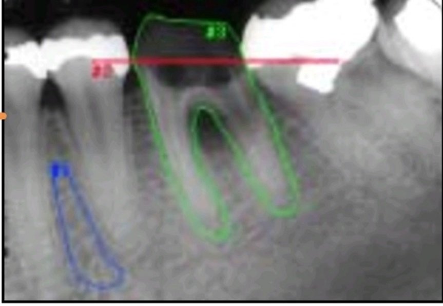
Linear radiographic measurements from reference line (#2) to crestal bone levels: images taken before tooth extraction (IBX). The contour of the tooth (#3) has been drawn in this image. #1= ROC.
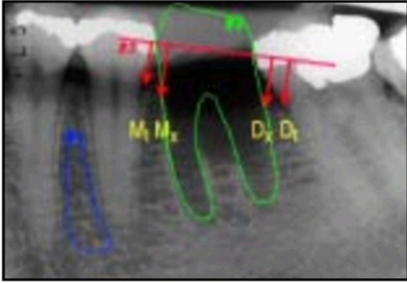
Image taken immediately after tooth extraction (I BASE). The tooth contour has been transferred to this image. Dt=mesial aspect of tooth distal to extraction site; Mt=distal aspect of tooth mesial to extraction site; Mx=level of alveolar bone crest at mesial aspect of extraction socket; Dx=level of alveolar bone crest at distal aspect of extraction socket.
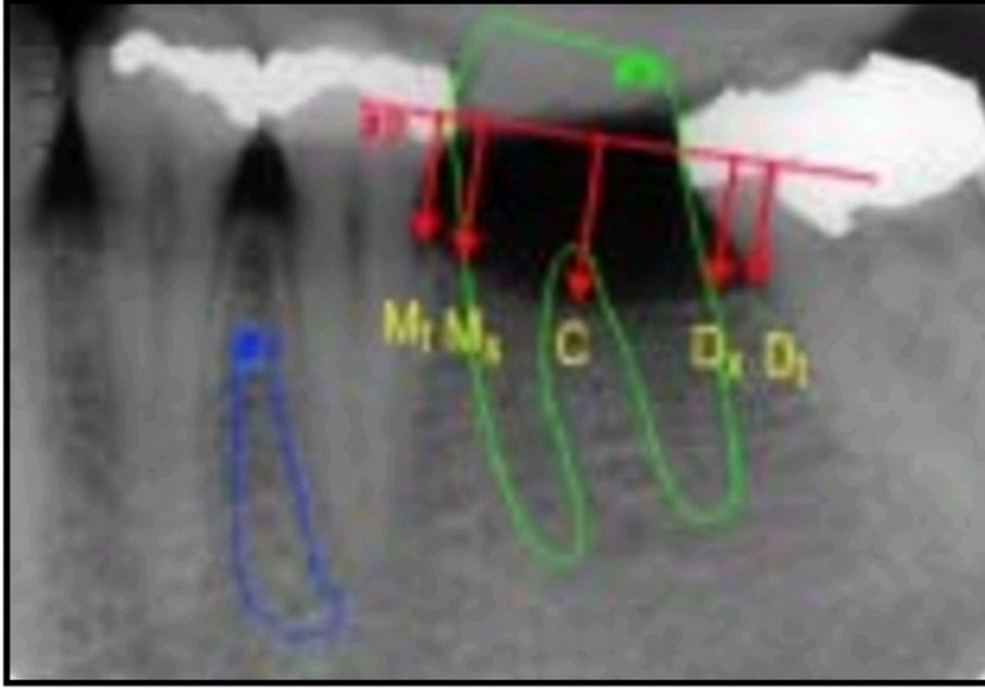
Image taken 12 months after tooth extraction (I12). C=most apically situated point between Mx and Dx.
INTERNAL CHANGES
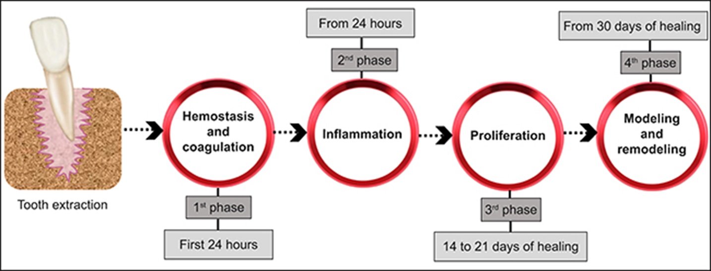
When a tooth is removed, there is haemorrhage followed by formation of a blood clot that fills the entire socket. With this is an inflammatory reaction that stimulates recruitment of cells to form granulation tissue. Within 48 to 72 hours after extraction the clot starts to breakdown as granulation tissue begins to infiltrate the clot especially at the base of the socket. By four days the epithelium proliferates along the socket periphery and immature connective tissue is apparent.
After seven days the granulation tissue has completely infiltrated and replaced by the clot. At this stage, osteoid is evident at the base of the socket as uncalcified bony spicules. Over the next 2–3 weeks (3–4 weeks after extraction) this begins to mineralize from the base of the socket coronally. This is accompanied by continued re-epithelialization which completely covers the socket by six weeks post-extraction. Further infill of bone takes place with maximum radiographic density at around 100 days.
A number of factors may affect the healing of undisturbed sockets. The size of the socket is important with wider sockets requiring more time to bridge the defect compared with narrower sockets; it takes longer period for bone formation at molar sites compared to single-rooted sites.
The sockets of teeth with horizontal bone loss heal more quickly as the lower level of the alveolar bone means less infill is required. It should be noted that bone does not regenerate to a level coronal to the horizontal level of the bone crest or to the level of the neighbouring teeth (i.e., 100 per cent socket fill does not occur).12
EXTERNAL CHANGES
A recent study by Araujo and Lindhe showed that in the first eight weeks following extraction in a dog model there is marked osteoclastic activity resulting in the resorption of the buccal and lingual crestal walls. They noted that the reduction of the height was more pronounced at the buccal wall and was accompanied by a horizontal loss on both buccal and lingual walls. This is an important finding because an adequate width and height of buccal bone is important for optimal implant aesthetics, and this study suggests that loss of buccal bone may result in poorer, suboptimal aesthetics.13
SUMMARY
There are definite indications for extraction of teeth. The approach to treatment is conservative, but when the outlook is hopeless, a long period of fruitless therapy should not be considered. The final decision regarding the particular type of treatment is made by the patient, and he is entitled to receive the best professional judgment of the dentist to help him make the correct decision. The recommendation of the dentist is only one of many factors which will be considered, most of which are nondental.
Dentures should be recommended when they are indicated, and emphasis should be placed on the service they will render. There are some good alternatives to extraction and they should be used whenever possible. Careful planning of dental treatment and making the best possible care available can provide many happy and useful years for the patient.9
REFERENCES
- Clinical Orthopaedics and Related Research 1987(225):7-16
- https://www.orasurgery.com/consequences-of-tooth-loss/
- Avila-Ortiz, G.; Chambrone, L.; Vignoletti, F. Effect of alveolar ridge preservation interventions following tooth extraction: A systematic review and meta-analysis. J. Clin. Periodontol. 2019, 46, 195–223. [CrossRef]
- Jambhekar, S.; Kernen, F.; Bidra, A.S. Clinical and histologic outcomes of socket grafting after flapless tooth extraction: A systematic review of randomized controlled clinical trials. J. Prosthet. Dent. 2015, 113, 371–382. [CrossRef]
- Tan, W.L.; Wong, T.L.T.; Wong, M.C.M.; Lang, N.P. A systematic review of post-extractional alveolar hard and soft tissue dimensional changes in humans. Clin. Oral Implants Res. 2012, 23, 1–21. [CrossRef]
- SOCKET PRESERVATION- “HEALER OF THE EMPTY SOCKET”- AN OVERVIEW DOI:10.37841/jidam_2020_V7_I2_03
- Van Der Weijden, F.; Dell’Acqua, F.; Slot, D.E. Alveolar bone dimensional changes of post-extraction sockets in humans: A systematic review. J. Clin. Periodontol. 2009, 36, 1048–1058. [CrossRef] [PubMed]
- Allen EP, Gainza CS, Farthing GG, Newbold DA. Improved technique for localized ridge augmentation. A report of 21 cases. J Periodontol. 1985; 56(4): 195-9.
- prosthodontic considerations in tooth removal author links open overlay panelcharles c. swoope d.d.s., m.s.d. https://doi.org/10.1016/s0011-8532(22)01608-1
- Bassir, S.; Alhareky, M.; Wangsrimongkol, B.; Jia, Y.; Karimbux, N. Systematic Review and Meta-Analysis of Hard Tissue Outcomes of Alveolar Ridge Preservation. Int. J. Oral Maxillofac. Implants 2018, 33, 979–994. [CrossRef] [PubMed]
- Barone, A.; Borgia, V.; Covani, U.; Ricci, M.; Piattelli, A.; Lezzi, G. Flap versus flapless procedure for ridge preservation in alveolar extraction sockets: A histological evaluation in a randomized clinical trial. Clin. Oral Implants Res. 2015, 26, 806–813. [CrossRef]
- Schropp L, Wenzel A, Kostopoulos L, Karring T. Bone healing changes and soft tissue contour changes following single-tooth extraction: a clinical and radiographic 12-month prospective study. Int J Periodontics Restorative Dent 2003;23:313–323.
- Araujo MG, Lindhe J. Dimensional ridge alterations following tooth extraction. An experimental study in the dog. J Clin Periodontol. 2005; 32(2): 212-8.
- Johnson K. A study of the dimensional changes occurring in the maxilla following tooth extraction. Aust Dent J 1969;14:241– 244.
- Atwood DA. Post-extraction changes in adult mandible as illustrated by microradiograph of midsagittal section and serial cephalometric roentogenograms. J Prosthet Dent 1963;13:810– 824.
Disclaimer from Guident
The views and opinions expressed in articles published in Guident are solely those of the authors and do not necessarily reflect the views of the editors, the editorial board or the publisher, Ivory India. Guident does not endorse any product, technique or treatment described. Clinical content is intended for qualified dental and medical professionals and is not a substitute for individual clinical judgement. Guident, its editors and the publisher accept no liability for any consequence arising from the use of information published in the journal.
How to cite this article
Dr. P. R. Vinutna, Dr. Sunil Dhaded. Exodontia- Events and Biological Effects on Alveolus. Guident. 2023 May;16(6). Available from: https://guident.in/articles/general/exodontia-events-and-biological-effects-on-alveolus.html
