Successful Management of Large Periapical Lesion Associated with Maxillary Central Incisors by Combined Endodontic and Surgical Intervention: A Case Report
Department of Conservative Dentistry and Endodontics, D.J. College of Dental Sciences & Research, Modinagar, U.P
ABSTRACT
The success of endodontic therapy depends on complete elimination of microorganisms from the root canal system and prevention of reinfection. However, persistent periapical lesions may remain even after conventional root canal treatment due to persistent intra-radicular infection, extra-radicular infection, apical biofilm, or inadequate apical seal. Surgical endodontic intervention becomes necessary when nonsurgical retreatment fails to achieve healing. This case report describes the management of a persistent periapical lesion associated with maxillary central incisors (11 and 21). Tooth 11 underwent primary root canal treatment, while tooth 21 underwent nonsurgical retreatment. Both teeth were obturated using mineral trioxide aggregate (MTA) as a sealer. Persistent periapical pathology necessitated surgical intervention with apicoectomy and retrograde MTA filling. Follow-up radiographic evaluation demonstrated satisfactory healing and reduction in periapical radiolucency. This case highlights the importance of peri-radicular surgery as a predictable treatment option in cases of persistent periapical pathology.
Keywords: Apicoectomy, periapical lesion, retrograde filling, MTA, surgical endodontics, maxillary incisors
INTRODUCTION
The primary objective of endodontic therapy is the elimination of microorganisms from the root canal system and prevention of reinfection through three-dimensional obturation. Successful endodontic treatment depends upon adequate cleaning, shaping, disinfection, and obturation of the entire root canal system.
Despite remarkable advances in endodontic materials, instruments, irrigation systems, and obturation techniques, failures of root canal therapy continue to occur in clinical practice.
Endodontic failure is most commonly associated with persistent intraradicular infection. Microorganisms may survive within inaccessible portions of the root canal system such as lateral canals, apical deltas, isthmuses, dentinal tubules, and ramifications, which are difficult to clean and disinfect completely using conventional endodontic procedures. Inadequate instrumentation, missed canals, coronal leakage, insufficient obturation, procedural errors, secondary contamination, and extraradicular infection are also important contributing factors responsible for persistent apical pathology.
Periapical lesions develop as an inflammatory response of peri-radicular tissues to microbial toxins and irritants originating from infected root canals. Chronic periapical lesions usually present radiographically as radiolucency associated with the root apex and histologically may represent granulomas, radicular cysts, or abscesses. These lesions are characterized by inflammatory cell infiltration, bone resorption, and destruction of periodontal ligament structures. Although most periapical lesions heal following adequate nonsurgical endodontic treatment, certain lesions persist despite apparently satisfactory treatment.
Nonsurgical retreatment is generally considered the first treatment option for failed endodontic cases because it provides an opportunity to eliminate residual infection, improve canal disinfection, and correct technical inadequacies of the previous treatment. However, retreatment alone may not always result in healing, especially in cases involving persistent apical biofilm, true cysts, apical transportation, blocked canals, extraradicular infection, or complex apical anatomy. In such situations, surgical endodontic intervention becomes necessary to preserve the natural dentition.
Endodontic surgery, also known as peri-radicular surgery, involves surgical access to the periapical region with removal of pathological tissues, root-end resection (apicoectomy), retrograde cavity preparation, and placement of a root-end filling material to achieve an apical seal. The purpose of apicoectomy is to eliminate apical ramifications and lateral canals that may harbor persistent microorganisms. Kim and Kratchman (2006) reported that resection of approximately 3 mm of the root apex removes nearly 98% of apical ramifications and 93% of lateral canals, thereby significantly reducing the risk of reinfection.
The success of surgical endodontics has improved considerably with the introduction of magnification systems, ultrasonic retropreparation, microsurgical instruments, and advanced root-end filling materials. Among the various retrograde filling materials available, mineral trioxide aggregate (MTA) has gained widespread acceptance because of its excellent biological and physical properties. Introduced by Torabinejad et al. in 1993, MTA demonstrates superior sealing ability, biocompatibility, moisture tolerance, low solubility, antibacterial properties due to alkaline pH, and the ability to stimulate Cementogenesis and osteogenesis. These characteristics make MTA the material of choice for root-end filling during apical surgery.
MTA-based sealers have also shown favourable outcomes when used in conventional orthograde obturation because of their bioactivity and ability to promote periapical healing. Studies have demonstrated that MTA induces hard tissue formation and provides an effective seal against bacterial microleakage. Consequently, MTA is increasingly used both as a root-end filling material and as a sealer in complex endodontic cases.
Maxillary anterior teeth are frequently affected by trauma, discoloration, and pulpal necrosis, often leading to development of chronic periapical lesions. Preservation of these teeth is particularly important because of their aesthetic and functional significance. Surgical endodontic treatment offers a conservative alternative to extraction and implant placement, especially in cases where nonsurgical treatment has failed.
The present case report describes the successful management of a persistent large periapical lesion associated with maxillary central incisors (11 and 21). Tooth 11 underwent primary root canal treatment, while tooth 21 required nonsurgical retreatment. Both teeth were obturated using MTA-based sealer. Due to persistence of the periapical lesion, surgical intervention with apicoectomy and retrograde MTA filling was performed. The case highlights the role of combined nonsurgical and surgical endodontic therapy in achieving successful healing and preservation of natural teeth.
AIM
To manage a large periapical lesion associated with maxillary central incisors using combined nonsurgical and surgical endodontic treatment with retrograde MTA filling.
OBJECTIVES
- To eliminate infection from teeth 11 and 21 through RCT and retreatment respectively.
- To perform apicoectomy and retrograde MTA filling for periapical healing.
- To preserve the affected anterior teeth and promote bone regeneration.
CASE REPORT
A 24-year-old male patient reported to the Department of Conservative Dentistry and Endodontics with a chief complaint of intermittent pain and discomfort in the upper anterior region for the past few months, which was aggravated during mastication. The patient gave a history of trauma due to a road traffic accident approximately 7–8 years earlier involving the maxillary anterior teeth. Following the traumatic injury, the patient noticed fracture and discoloration in relation to the upper front teeth but did not seek immediate treatment. The patient had undergone root canal treatment in tooth 21 approximately 2–3 years earlier. However, intermittent pain and discomfort persisted in the affected region and gradually increased in intensity over time.
The medical history was non-contributory and the patient was otherwise healthy.
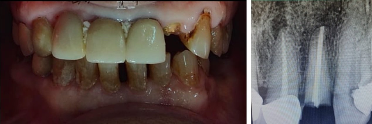
Clinical examination revealed an Ellis Class III fracture in tooth 21 associated with crown discoloration (Fig. 1). Tooth 11 did not show any visible fracture or discoloration but was tender on percussion. Teeth 12 and 22 were also tender on percussion, although no swelling, sinus tract, or abnormal mobility was present. Periodontal probing depths were within normal limits.
Pulp vitality testing revealed a non-vital response in tooth 11, while tooth 21 was previously root canal treated. Teeth 12 and 22 exhibited delayed and questionable responses to vitality testing along with tenderness on percussion, suggestive of pulpal degeneration secondary to previous traumatic injury.
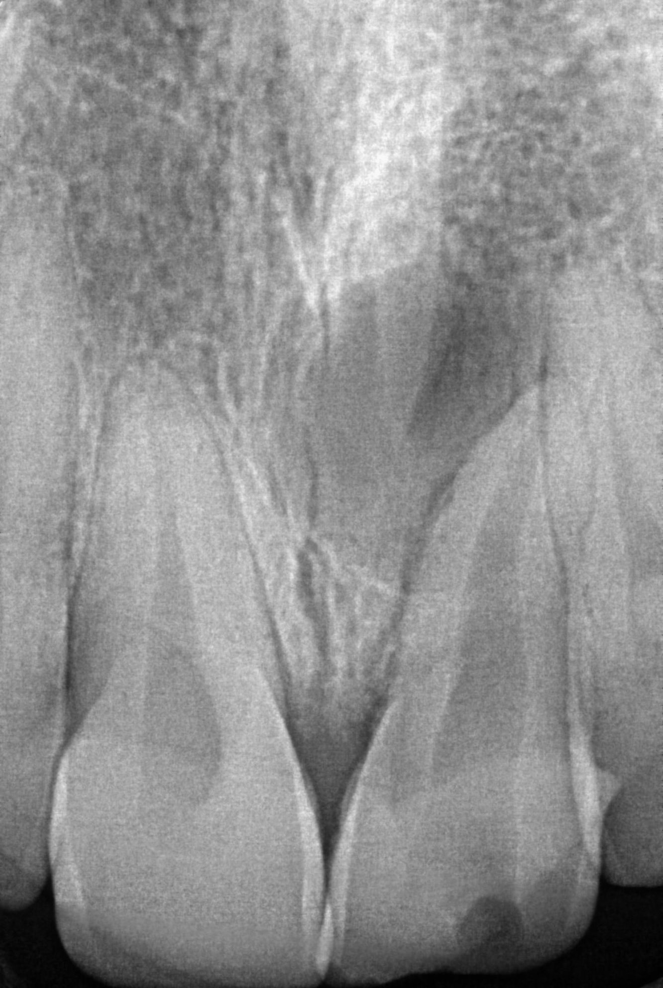
Radiographic examination using an intraoral periapical radiograph revealed a large well-defined periapical radiolucency involving the apices of teeth 11 and 21, associated with loss of lamina dura and widening of the periodontal ligament space (Fig. 2). Tooth 21 showed evidence of previous inadequate endodontic treatment, whereas tooth 11 demonstrated pulpal necrosis associated with chronic apical periodontitis. Teeth 12 and 22 did not exhibit significant periapical radiolucency; however, widening of the periodontal ligament space was observed in relation to both teeth.
Based on the clinical and radiographic findings, the diagnosis was established as follows:
- Tooth 11 – Pulp necrosis with chronic apical periodontitis
- Tooth 21 – Previously treated tooth with persistent chronic apical periodontitis associated with Ellis Class III fracture and discoloration
- Teeth 12 and 22 – Symptomatic apical periodontitis secondary to traumatic injury
Considering the extent of the lesion and the compromised apical architecture, a combined nonsurgical and surgical endodontic treatment approach was planned. Root canal treatment was initiated for tooth 11, while nonsurgical retreatment was performed for tooth 21 under rubber dam isolation. Root canal treatment was also performed for teeth 12 and 22 because of persistent tenderness on percussion and suspected pulpal involvement following trauma.
METHODOLOGY :
A 24-year-old male patient reported to the Department of Conservative Dentistry and Endodontics with a chief complaint of intermittent pain and discomfort in the upper anterior region. The patient gave a history of traumatic injury due to a road traffic accident approximately 7–8 years earlier. Clinical examination revealed an Ellis Class III fracture with discoloration in relation to tooth 21, while tooth 11 appeared clinically intact but was tender on percussion. Teeth 12 and 22 were also tender on percussion. Radiographic examination revealed a large periapical radiolucency involving the apices of teeth 11 and 21. Tooth 21 showed evidence of previous inadequate root canal treatment.
Based on the clinical and radiographic findings, the diagnosis of pulp necrosis with chronic apical periodontitis in tooth 11 and previously treated tooth with persistent chronic apical periodontitis in tooth 21 was established. Teeth 12 and 22 were diagnosed with symptomatic apical periodontitis secondary to traumatic injury.
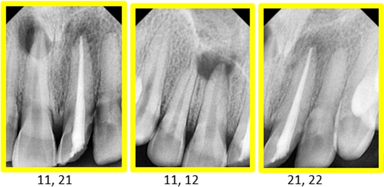
FIG 1: PREOPERATIVE RADIOGRAPH

FIG 2: RCT IRT 11,12,22 AND RE-RCT IRT 21: WORKING LENGTH DETERMINATION
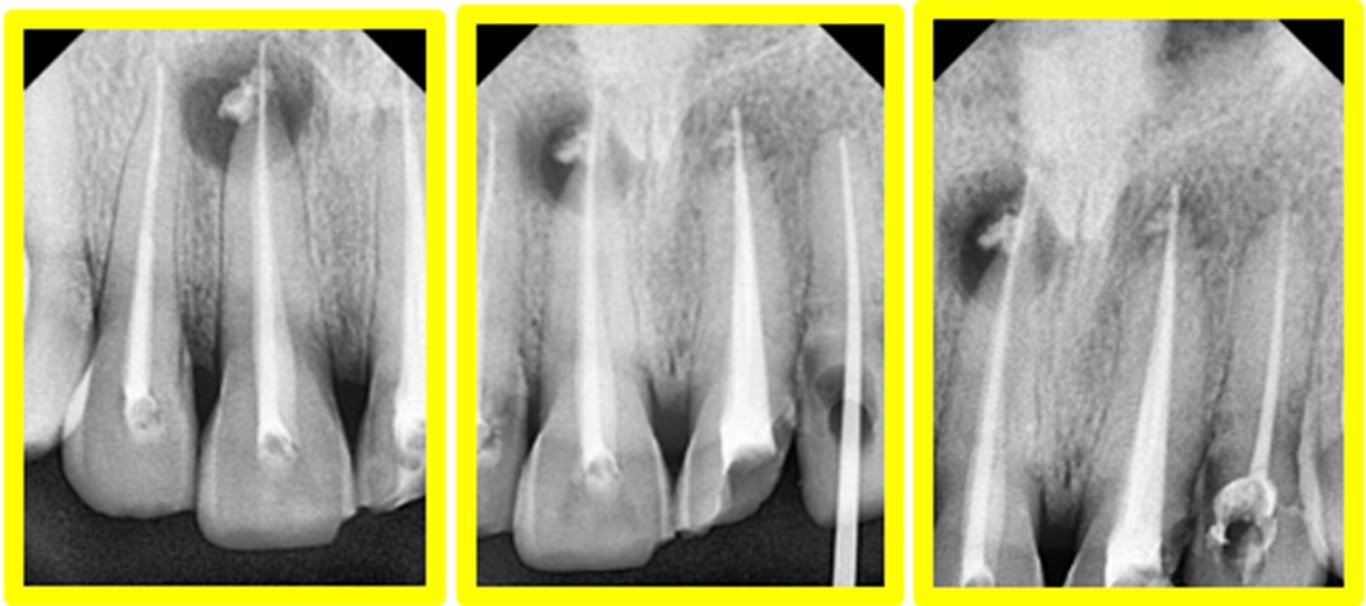
FIG 3: OBTURATION DONE WITH MTA AS A SEALER IRT 11,12,21,22: APICAL EXTRUSION OF GP IRT 11,21
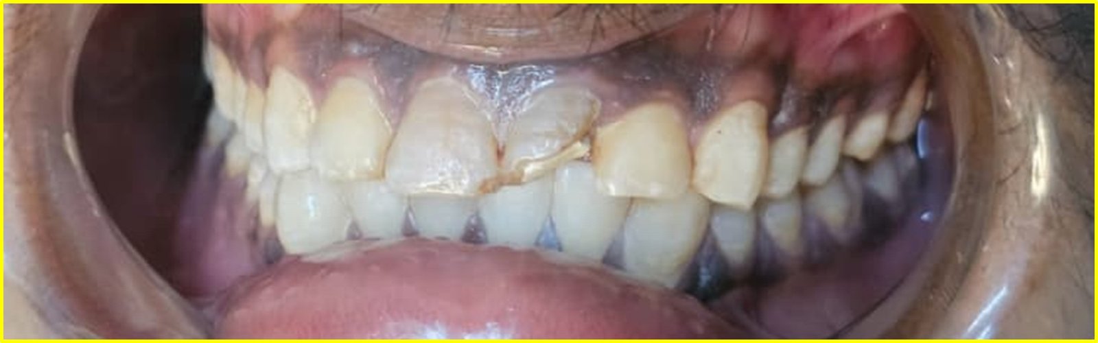
FIG 4: PREOPERATIVE
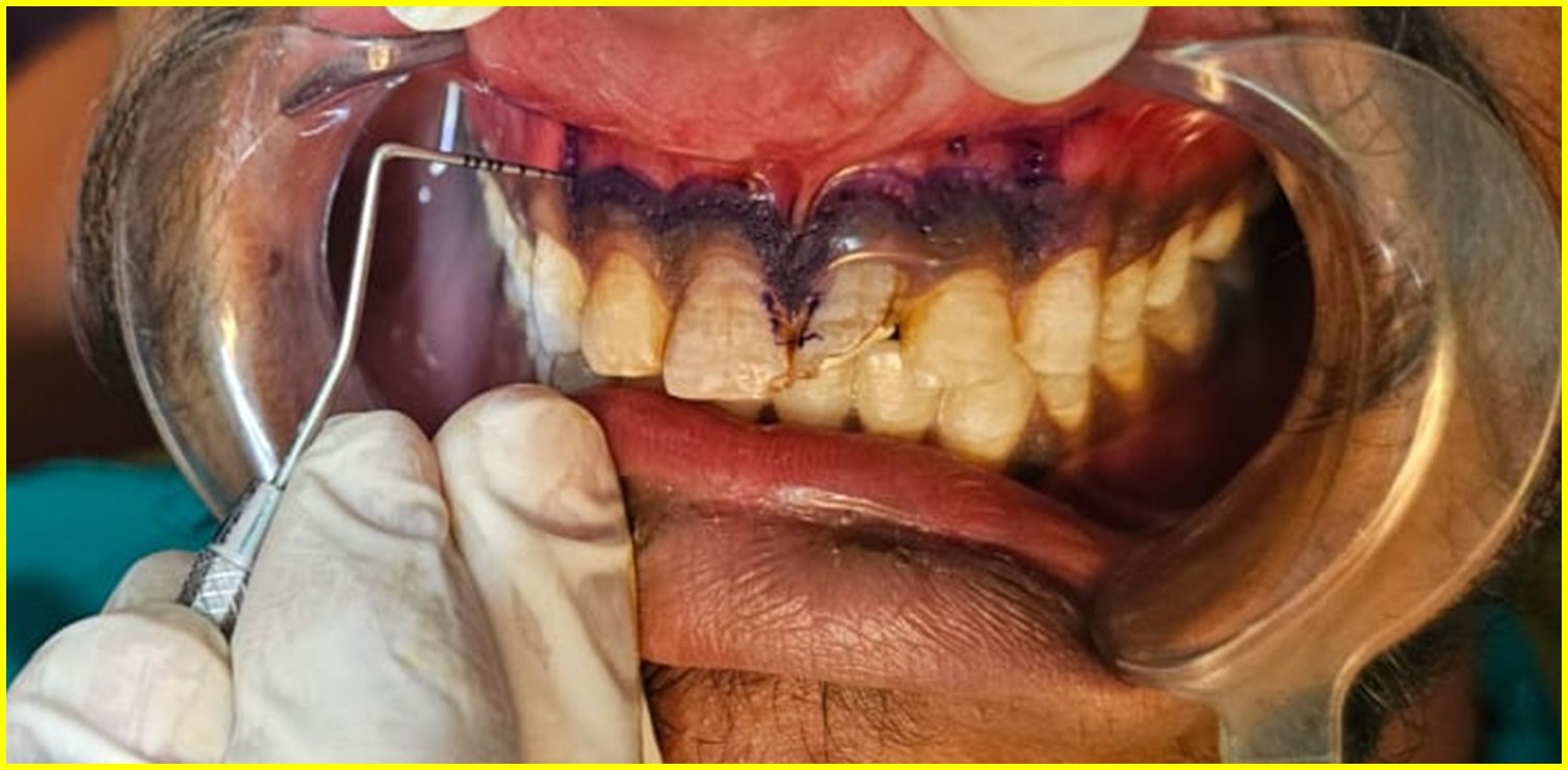
FIG 5: BLEEDING POINT MARKED
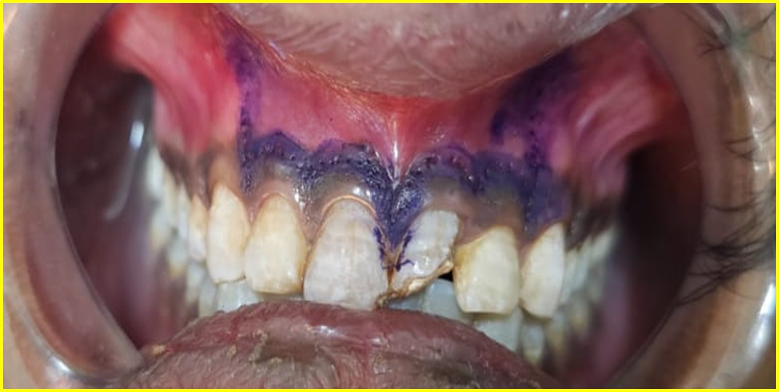
FIG 6: FLAP DESIGN (Luebke-Ochsenbein flap) INCORPORATING THE LABIAL FRENUM
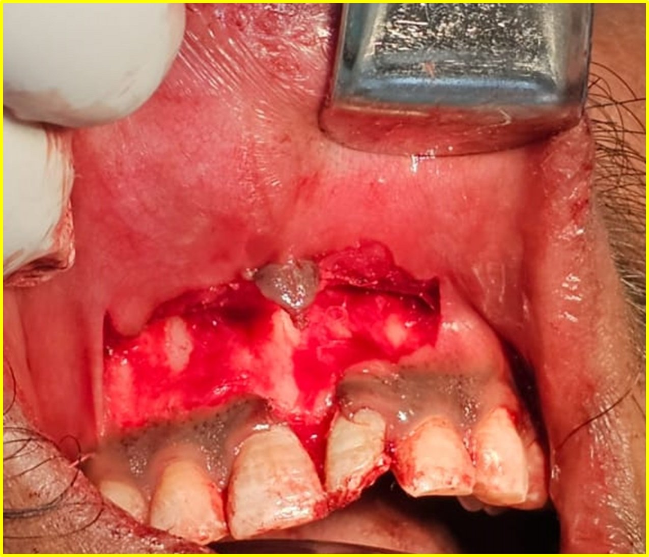
FIG 7: FLAP RAISED
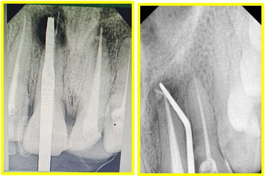
FIG 8: RADIOGRAPHIC LOCALIZATION OF OSSEOUS DEFECT
After administration of local anesthesia and rubber dam isolation, root canal treatment was initiated in teeth 11, 12, and 22, while nonsurgical retreatment was performed in tooth 21. Working length determination was carried out using an electronic apex locator and confirmed radiographically. Cleaning and shaping procedures were completed using hand and rotary instruments with copious irrigation using 3% sodium hypochlorite, 17% EDTA, and normal saline.
Following biomechanical preparation, obturation was performed using gutta-percha along with a mineral trioxide aggregate (MTA)-based sealer. Intentional slight extrusion of gutta-percha beyond the apex was carried out in teeth 11 and 21 to facilitate localization of the root apex during the surgical procedure because of the extensive periapical lesion and compromised apical constriction. Teeth 12 and 22 were obturated using conventional gutta-percha obturation techniques.
Following completion of endodontic therapy, surgical endodontic treatment was planned for teeth 11 and 21. Under local anesthesia, a Luebke–Ochsenbein flap incorporating the labial frenum was designed and elevated, extending from the maxillary right lateral incisor to the maxillary left lateral incisor to achieve adequate access to the extensive periapical lesion while maintaining the integrity of the marginal gingival tissues. The flap was carefully reflected, bleeding points were identified, and radiographic localization of the osseous defect was performed. Osteotomy was carried out using a surgical bur under copious saline irrigation until the periapical defect and root apices became clearly visible.
Periapical curettage was then performed to remove the pathological tissue, followed by root-end resection of approximately 3 mm of the apices of teeth 11 and 21. Retrograde cavity preparation was carried out using ultrasonic retro tips, and retrograde filling was performed using mineral trioxide aggregate (MTA). Following complete debridement of the defect, bone graft material was placed into the osseous cavity to promote healing and bone regeneration. The surgical site was irrigated thoroughly and sutured appropriately.
Postoperative instructions and medications were prescribed, and the patient was kept under periodic follow-up. Clinical evaluation after one week demonstrated satisfactory soft tissue healing with reduction in postoperative discomfort. One-month follow-up showed satisfactory healing with progressive reduction in periapical radiolucency and evidence of early bone regeneration. Further periodic follow-up was advised to assess long-term periapic healing and treatment success.

FIG 9: DEFECT VISIBLE FOLLOWING OSTEOTOMY
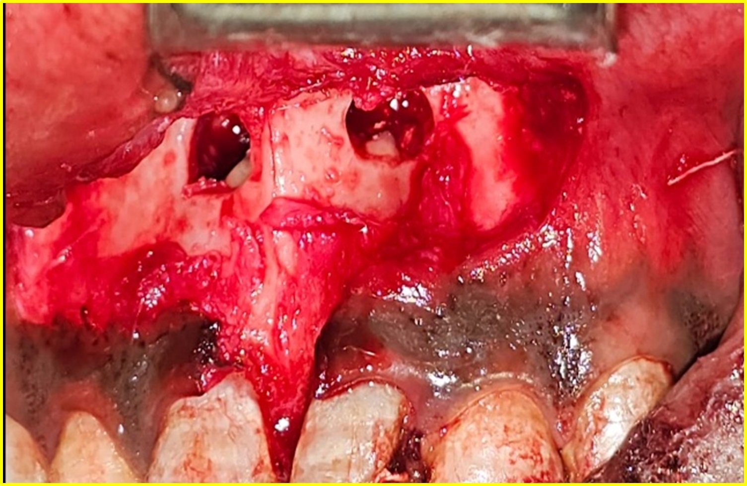
FIG 10: OSTEOTOMY WITH EXPOSED ROOT APEX FOLLOWING ROOT- END RESECTION

FIG 11: BONE GRAFT PLACEMENT

FIG 12: SUTURES GIVEN
RESULT
The patient tolerated both the endodontic and surgical procedures well, and the postoperative period was uneventful. Clinical evaluation at the one-week follow-up revealed satisfactory soft tissue healing with no evidence of swelling, infection, wound dehiscence, or other postoperative complications. Resorbable sutures had been used for flap closure, eliminating the need for suture removal and contributing to improved patient comfort and compliance during the healing period. The patient reported significant relief from pain and discomfort compared with the preoperative condition.
At the one-month follow-up, the patient remained asymptomatic, with absence of tenderness on percussion and palpation in relation to teeth 11, 12, 21, and 22. Clinical examination demonstrated satisfactory healing of the surgical site with healthy surrounding soft tissues and no signs of inflammation or recurrence of symptoms. Radiographic evaluation revealed early evidence of periapical healing characterized by a reduction in the size of the periapical radiolucency and initial bone fill within the osseous defect. The retrograde MTA fillings remained well adapted, and the bone graft material appeared stable within the defect area.

1 WEEK FOLLOW UP AND 1 MONTH FOLLOW UP
At the three-month follow-up, the patient continued to remain symptom-free with normal masticatory function. Clinical examination showed healthy soft tissues with no tenderness, swelling, mobility, or sinus tract formation. Radiographic assessment demonstrated progressive reduction of the periapical radiolucency with increased trabecular bone formation and continued integration of the bone graft material.
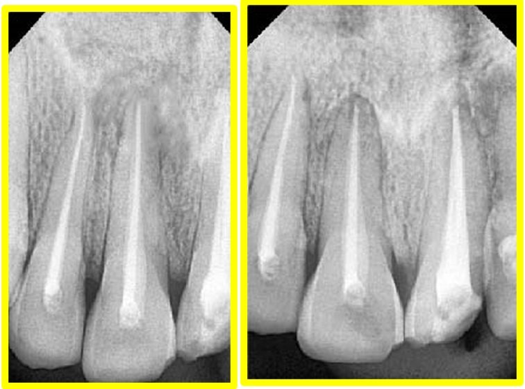
3 MONTHS FOLLOW UP AND 6 MONTHS FOLLOW UP
At the six-month follow-up, complete clinical healing was observed. The patient remained asymptomatic with no tenderness on percussion or palpation. Radiographic examination revealed substantial bone regeneration with significant resolution of the periapical radiolucency and re-establishment of normal periapical architecture. The retrograde MTA fillings remained intact and functional.
These findings indicated a favorable outcome following the combined nonsurgical and surgical endodontic management. Preservation of the involved maxillary anterior teeth was successfully achieved, with resolution of clinical symptoms and progressive radiographic evidence of healing. The patient was advised periodic follow-up to monitor continued bone regeneration and assess the long-term success of the treatment.
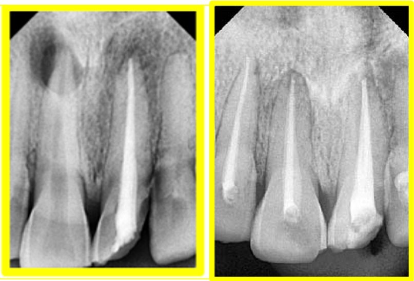
PREOPERATIVE AND POSTOPERATIVE
DISCUSSION
Nonsurgical retreatment is generally considered the first treatment option in cases of post-treatment apical periodontitis because it provides an opportunity to eliminate residual intracanal infection. However, when extensive periapical pathology exists in conjunction with loss of apical constriction and previous endodontic failure, surgical intervention may be required to achieve predictable healing while preserving the natural dentition. In the present case, the presence of a large periapical lesion involving teeth 11 and 21, along with the history of failed endodontic treatment in tooth 21, justified the decision to perform peri radicular surgery immediately following completion of orthograde endodontic treatment.
Kim and Kratchman (2006) reported that resection of 3 mm of the root apex eliminates approximately 98% of apical ramifications and 93% of lateral canals, thereby reducing the possibility of persistent infection. Accordingly, a 3-mm root-end resection was performed in the present case.
Mineral trioxide aggregate (MTA) was selected both as a root canal sealer and as the retrograde filling material because of its excellent sealing ability, biocompatibility, moisture tolerance, and bioactive properties. Torabinejad et al. demonstrated that MTA promotes Cementogenesis and facilitates regeneration of the periodontal apparatus. Intentional slight extrusion of gutta-percha beyond the apex in teeth 11 and 21 aided intraoperative localization of the root apices during surgery, particularly in the presence of extensive osseous destruction and loss of apical constriction.
Bone graft placement was performed following curettage and root-end management to support osseous healing and regeneration within the large periapical defect. The use of a Luebke–Ochsenbein flap incorporating the frenum provided adequate access to the surgical site while preserving the marginal gingival tissues and interdental papillae, thereby contributing to favourable esthetic outcomes in the anterior region.
The sequential follow-up findings in the present case demonstrated progressive clinical and radiographic healing, with resolution of symptoms and evidence of gradual bone regeneration. These findings support previous reports that a combined nonsurgical and surgical endodontic approach can be a predictable treatment modality for managing extensive periapical lesions associated with traumatized anterior teeth.
CONCLUSION
Within the limitations of this case report, the combined approach of orthograde endodontic treatment followed by periradicular surgery proved to be an effective treatment option for the management of a large periapical lesion associated with traumatized maxillary anterior teeth. Root-end resection with retrograde MTA filling, supplemented with bone graft placement, resulted in favorable clinical and radiographic healing. Careful case selection, appropriate surgical technique, and the use of bioactive materials such as MTA can facilitate preservation of natural teeth that might otherwise be considered for extraction.
REFERENCES
- Nair PNR. Pathogenesis of apical periodontitis and the causes of endodontic failures. Critical Reviews in Oral Biology and Medicine. 2004;15(6):348–381.
- Kim S, Kratchman S. Modern endodontic surgery concepts and practice: A review. Journal of Endodontics. 2006;32(7):601–623.
- Torabinejad M, Watson TF, Pitt Ford TR. Sealing ability of a mineral trioxide aggregate when used as a root-end filling material. Journal of Endodontics. 1993;19(12):591–595.
- Torabinejad M, Chivian N. Clinical applications of mineral trioxide aggregate. Journal of Endodontics. 1999;25(3):197–205.
- Torabinejad M, Parirokh M. Mineral trioxide aggregate: A comprehensive literature review—Part II: Leakage and biocompatibility investigations. Journal of Endodontics. 2010;36(2):190–202.
- Parirokh M, Torabinejad M. Mineral trioxide aggregate: A comprehensive literature review—Part I: Chemical, physical, and antibacterial properties. Journal of Endodontics. 2010;36(1):16–27.
- European Society of Endodontology. Quality guidelines for endodontic treatment: Consensus report of the European Society of Endodontology. International Endodontic Journal. 2006;39(12):921–930.
- Von Arx T, AlSaeed M. The use of regenerative techniques in apical surgery: A literature review. Saudi Dental Journal. 2011;23(3):113–127.
- Taschieri S, Del Fabbro M, Testori T, Weinstein R. Endodontic surgery using 2 different magnification devices: Preliminary results of a randomized controlled study. Journal of Oral and Maxillofacial Surgery. 2006;64(2):235–242.
- Tsesis I, Rosen E, Schwartz-Arad D, Fuss Z. Retrospective evaluation of surgical endodontic treatment: Traditional versus modern technique. Journal of Endodontics. 2006;32(5):412–416.
- Almansour MI. Surgical endodontic treatment of maxillary incisors: Case report. Clinical Case Reports. 2023;11:e6860.
- Gutmann JL, Harrison JW. Surgical Endodontics. Boston: Blackwell Scientific Publications; 1991.
- Torabinejad M, Corr R, Handysides R, Shabahang S. Outcomes of nonsurgical retreatment and endodontic surgery: A systematic review. Journal of Endodontics. 2009;35(7):930–937.
- Abbott PV. Prevention and management of external inflammatory resorption following trauma to teeth. Australian Dental Journal. 2016;61(Suppl 1):82–94.
Disclaimer from Guident
The views and opinions expressed in articles published in Guident are solely those of the authors and do not necessarily reflect the views of the editors, the editorial board or the publisher, Ivory India. Guident does not endorse any product, technique or treatment described. Clinical content is intended for qualified dental and medical professionals and is not a substitute for individual clinical judgement. Guident, its editors and the publisher accept no liability for any consequence arising from the use of information published in the journal.
How to cite this article
Dr. Subia, Dr. Anil Dhingra. Successful Management of Large Periapical Lesion Associated with Maxillary Central Incisors by Combined Endodontic and Surgical Intervention: A Case Report. Guident. 2026 Aug;19(9). Available from: https://guident.in/articles/endodontics/successful-management-of-large-periapical-lesion-associated-with-maxillary-central-incisors-by-combined.html