Endodontic Surgical Management of Periapical Lesions with MTA and Bone Graft: A Case Series
Department of Conservative Dentistry and Endodontics, D.J. College of Dental Sciences & Research, Modinagar, U.P
ABSTRACT
Periapical lesions that persist after conventional endodontic treatment present a clinical challenge and often necessitate surgical intervention. This case series describes the management of three such cases using periapical surgery in combination with Mineral Trioxide Aggregate (MTA) and bone grafting. In all cases, Safe Endo MTA was used as a retrograde filling material, and Osstem bone graft was used to promote osseous regeneration. The surgical protocol involved flap elevation, lesion curettage, retrograde MTA placement, bone grafting, and suturing. Post-operative follow-up demonstrated favorable clinical and radiographic healing, with reduction in periapical radiolucency and absence of complications. The combined use of MTA and bone graft material proved to be an effective and predictable approach for the management of persistent periapical lesions.
INTRODUCTION
Periapical surgery is indicated when conventional root canal therapy fails to resolve periapical pathology or when retreatment is not feasible. The primary objective of surgical endodontics is the elimination of pathological tissue and the establishment of a hermetic apical seal to prevent reinfection. Mineral Trioxide Aggregate has emerged as a material of choice for retrograde filling due to its excellent sealing ability, biocompatibility, and capacity to induce hard tissue formation. Additionally, the use of bone grafts in periapical defects enhances healing by providing an osteoconductive scaffold that supports new bone formation. This case series aims to highlight the clinical effectiveness of Safe Endo MTA in conjunction with Osstem bone graft in the surgical management of persistent periapical lesions.
MATERIALS
The materials used in all three cases included Safe Endo MTA as the retrograde filling material and Osstem bone graft for defect reconstruction. Standard surgical instruments were used for incision, flap reflection, and curettage, and sutures were employed for wound closure following the procedure.
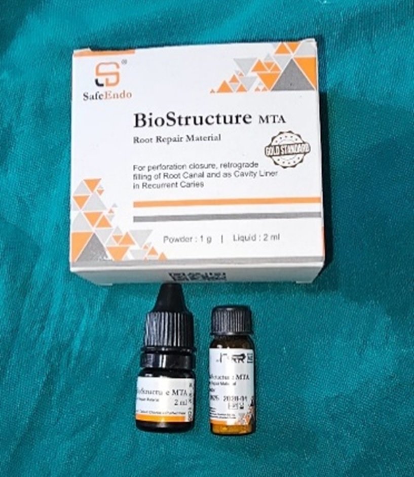
MTA
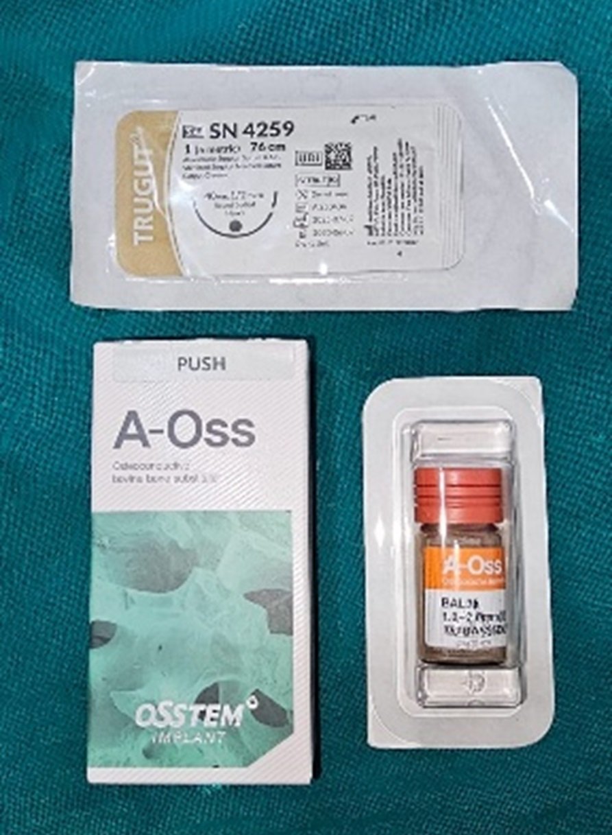
BONE GRAFT & SUTURES
CASE-1
Procedure
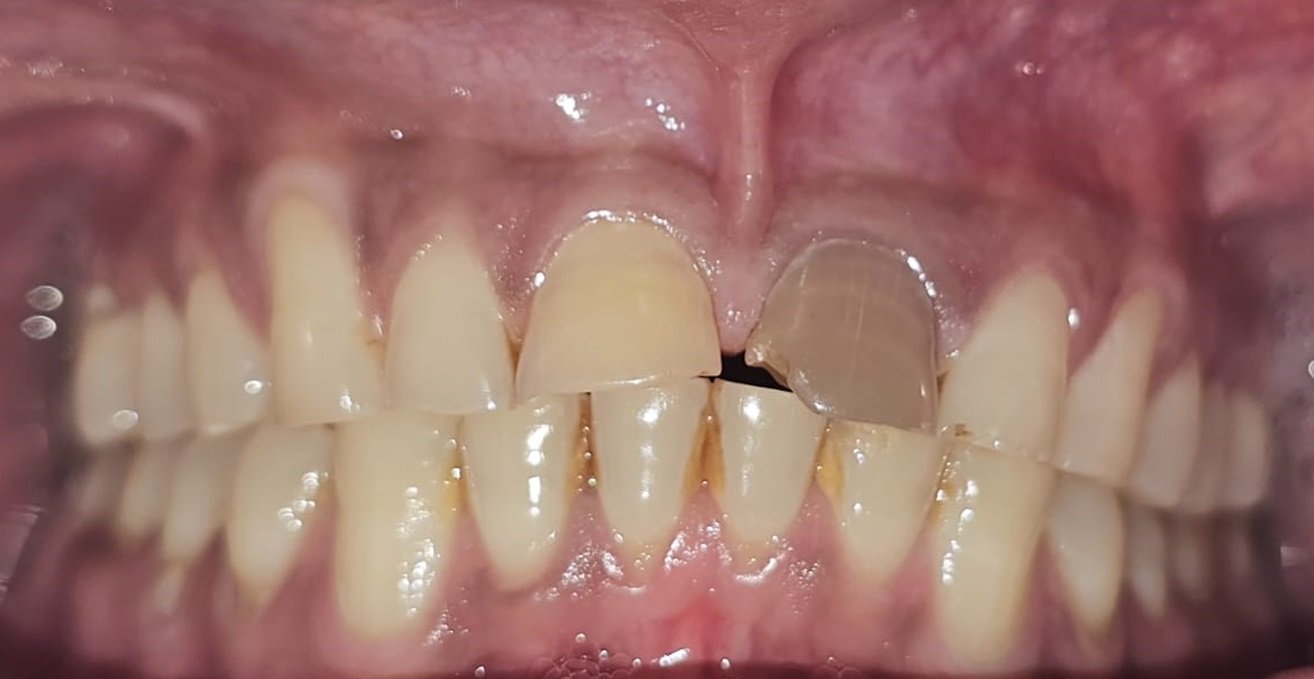
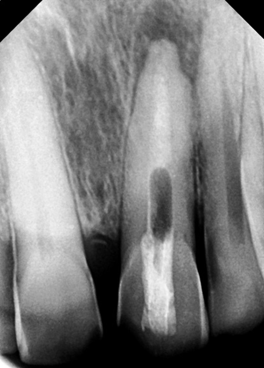
In the first case, a 33-year-old male patient presented with a persistent periapical lesion associated with a previously treated tooth. Pre-operative intraoral periapical radiographs revealed a well-defined radiolucency. After administration of local anesthesia, an incision was made and a full-thickness mucoperiosteal flap was raised to expose the surgical site. The lesion was thoroughly curetted, and an MTA plug was prepared and placed, followed by retrograde MTA filling to achieve an effective apical seal. The osseous defect was then filled with Osstem bone graft, and the flap was repositioned and sutured. Immediate post-operative radiographs were obtained, and follow-up at one and three months showed progressive healing and reduction in radiolucency.

PRE-OPERATIVE IOPA
INTRAORAL IMAGE
MTA PLUG WAS MADE

INCISION WAS GIVEN AND FLAP WAS RAISED And RETROGRADE MTA WAS PLACED

BONE GRAFT WAS PLACED FOLLOWED BY SUTURES

IMMEDIATE POSTOPERATIVE RADIOGRAPH, 1 MONTH FOLLOW UP, 3 MONTH FOLLOW UP
CASE-2
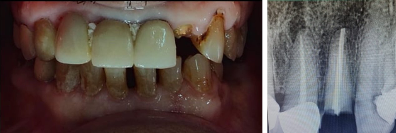
In the second case, a 20-year-old female patient underwent working length determination and obturation with MTA prior to surgical intervention. A similar surgical approach was followed, wherein an incision was given, the flap was raised, and the lesion was identified and curetted. Retrograde MTA placement was performed, followed by placement of bone graft material and suturing. Post-operative evaluation and follow-up revealed satisfactory healing without any complications.

PRE-OPERATIVE IOPA & INTRA ORAL IMAGE
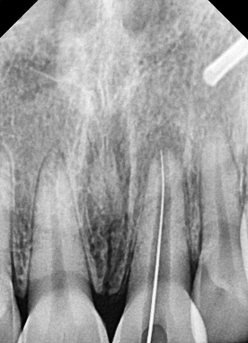
WL DETERMINATION

CANAL IS OBTURATED WITH MTA

INCISION WAS GIVEN, FLAP RAISED & LESION WAS LOCATED
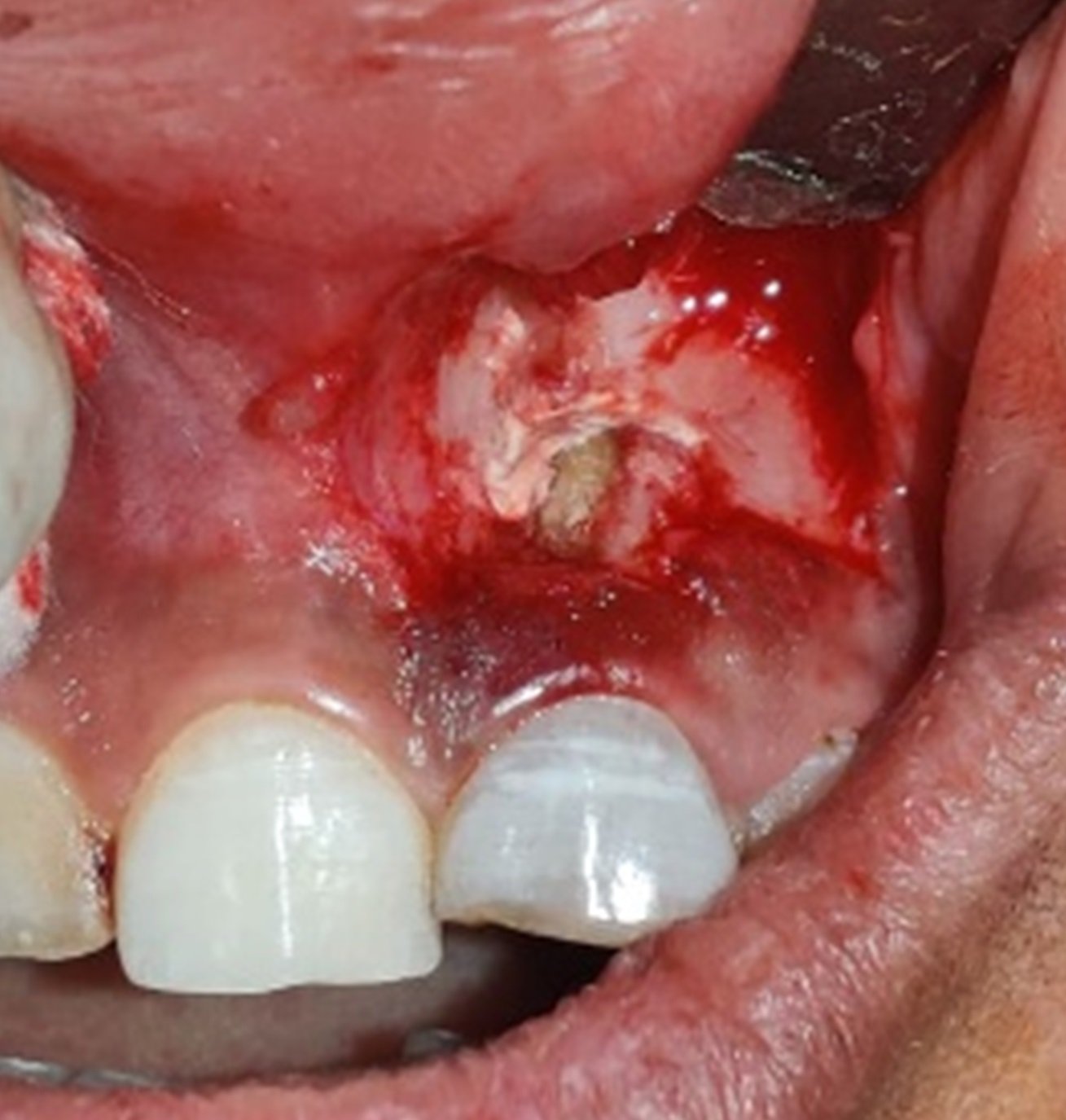
MTA PLACED

BONE GRAFT WAS PLACED FOLLOWED BY SUTURES
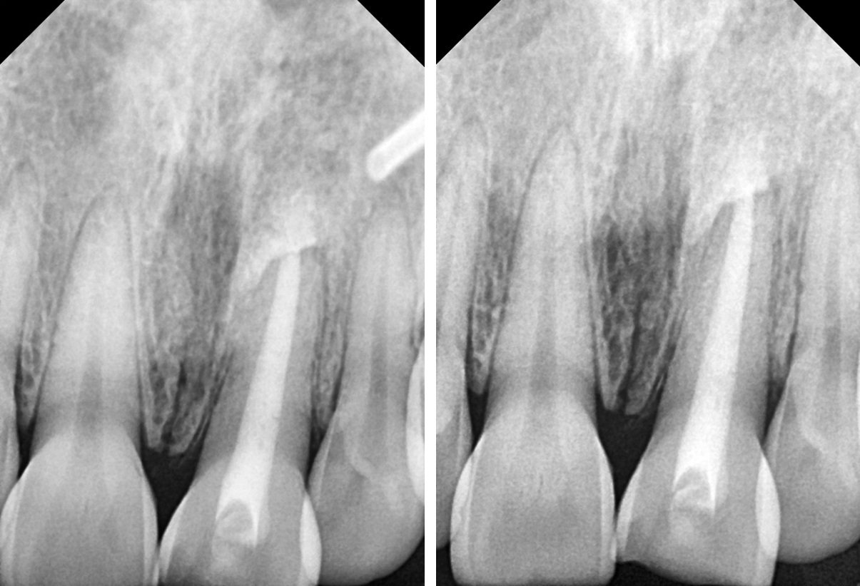
IMMEDIATE POST-OPERATIVE RADIOGRAPH AND 1 MONTH FOLLOW UP
CASE-3
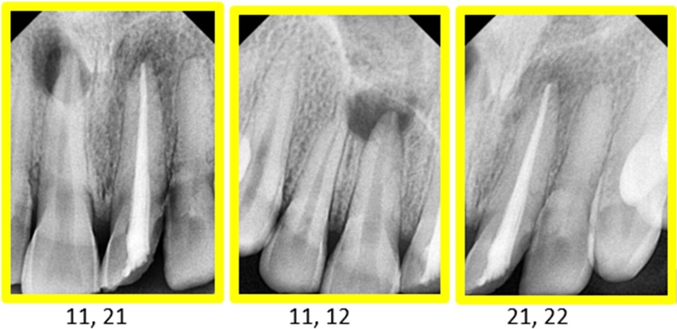
In the third case, a 25-year-old female patient underwent obturation followed by surgical management. Bleeding points were identified to guide incision placement, and a flap was raised to access the lesion. After complete debridement, MTA was placed as a retrograde filling material, and the defect was filled with bone graft before suturing. The clinical and radiographic outcomes in this case were also favorable, demonstrating effective healing.
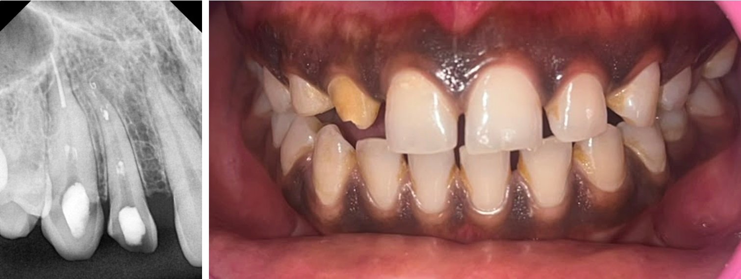
PRE-OPERATIVE IOPA AND INTRAORAL IMAGE

OBTURATION DONE

BLEEDING POINTS MARKED AND INCISION GIVEN
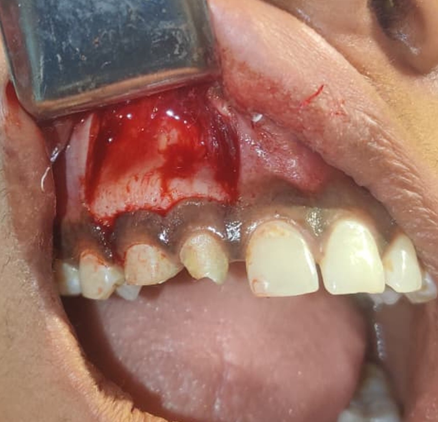
FLAP RAISED
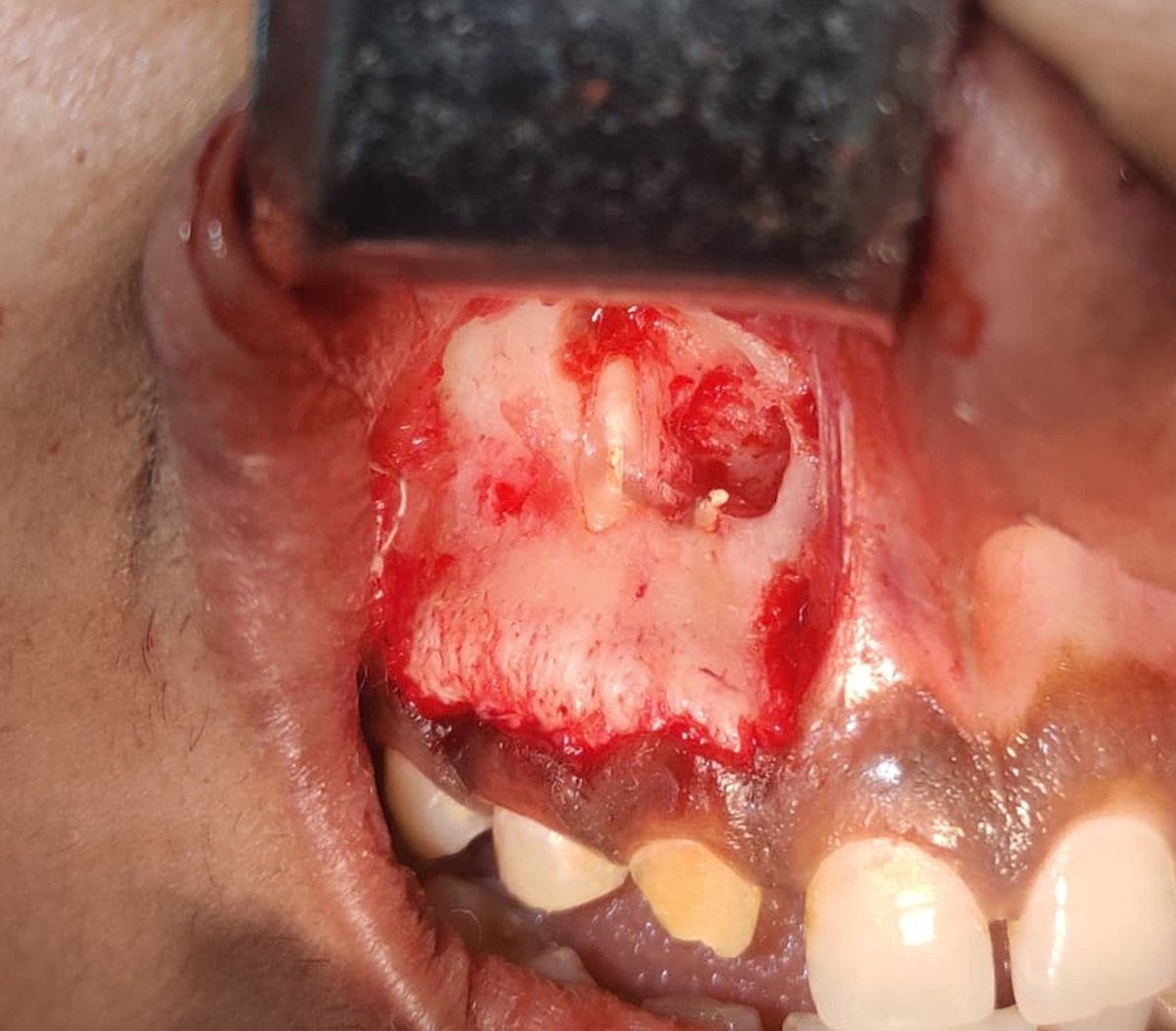
LESION LOCATED
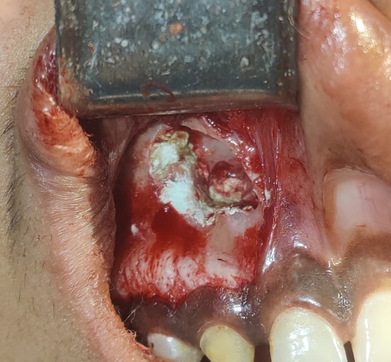
MTA PLACED

BONE GRAFT PLACED
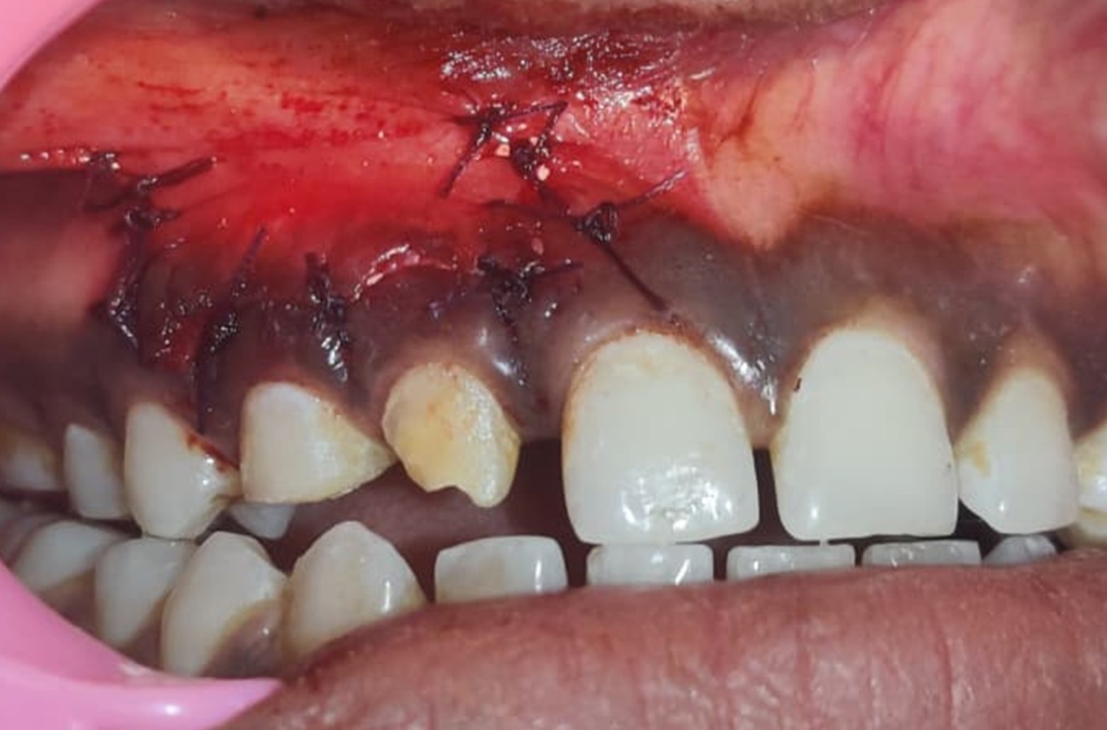
SUTURES PLACED
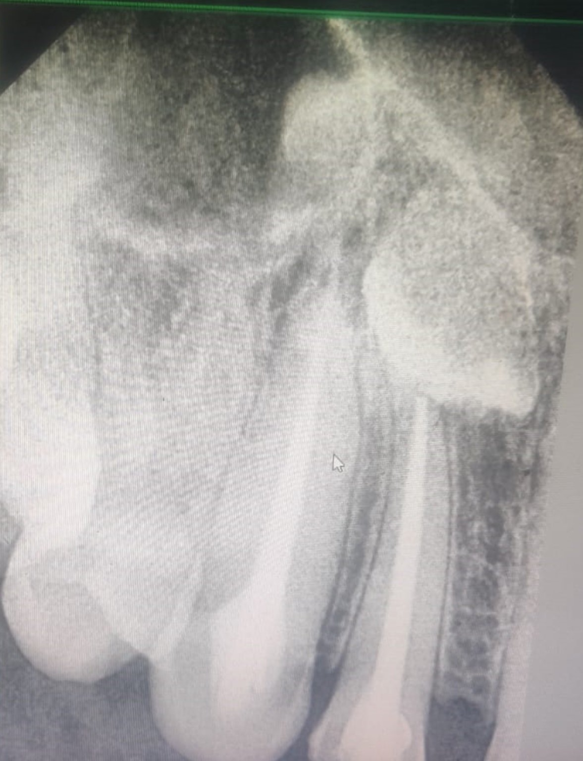
IMMEDIATE POST OPERATIVE
RESULTS
All three cases demonstrated satisfactory clinical and radiographic outcomes. There was a noticeable reduction in periapical radiolucency over the follow-up period, along with good soft tissue healing and absence of post-operative complications such as pain, swelling, or infection. The patients reported improvement in symptoms, and the treated teeth remained functional.
DISCUSSION
The successful outcomes observed in this case series can be attributed to the combined biological and physical properties of Mineral Trioxide Aggregate and the regenerative potential of bone graft materials. MTA has been widely documented in endodontic literature for its superior sealing ability, which minimizes microleakage and prevents bacterial ingress at the apical region. Its alkaline pH and release of calcium ions contribute to the formation of hydroxyapatite, thereby promoting hard tissue deposition and enhancing periapical healing. Furthermore, MTA exhibits excellent biocompatibility, reducing the risk of inflammatory response and ensuring favorable interaction with periapical tissues.
The incorporation of Osstem bone graft in these cases played a significant role in enhancing osseous regeneration. Bone grafts act as an osteoconductive matrix, facilitating the migration and proliferation of osteogenic cells into the defect site. This not only accelerates the healing process but also improves the structural integrity of the bone surrounding the root apex. The combination of thorough surgical debridement, effective apical sealing with MTA, and defect reconstruction with bone graft creates an optimal environment for tissue repair and regeneration.
Another important aspect contributing to the success of these cases is the adherence to a standardized surgical protocol, including precise flap design, complete lesion curettage, and careful placement of materials. The absence of post-operative complications and the progressive radiographic healing observed during follow-up further validate the effectiveness of this approach. These findings are consistent with previously reported studies, which emphasize the reliability of MTA as a retrograde filling material and the adjunctive benefit of bone grafting in large periapical defects.
CONCLUSION
Within the limitations of this case series, it can be concluded that periapical surgery using Safe Endo MTA in combination with Osstem bone graft is an effective and reliable treatment modality for persistent periapical lesions. The use of MTA ensures an excellent apical seal, while the bone graft enhances regenerative healing, resulting in predictable and successful clinical outcomes.
REFERENCES
- Torabinejad M, Watson TF, Pitt Ford TR. Sealing ability of a mineral trioxide aggregate when used as a root end filling material. J Endod. 1993;19(12):591–595.
- Torabinejad M, Chivian N. Clinical applications of mineral trioxide aggregate. J Endod. 1999;25(3):197–205.
- Parirokh M, Torabinejad M. Mineral trioxide aggregate: A comprehensive literature review—Part I: Chemical, physical, and antibacterial properties. J Endod. 2010;36(1):16–27.
- Parirokh M, Torabinejad M. Mineral trioxide aggregate: A comprehensive literature review—Part II: Leakage and biocompatibility investigations. J Endod. 2010;36(2):190–202.
- Kim S, Kratchman S. Modern endodontic surgery concepts and practice: A review. J Endod. 2006;32(7):601–623.
- Von Arx T, AlSaeed M. The use of regenerative techniques in apical surgery: A literature review. Saudi Dent J. 2011;23(3):113–127.
- Taschieri S, Del Fabbro M, Testori T, Weinstein R. Endodontic surgery using modern techniques: A prospective study on 30 cases. Int Endod J. 2007;40(6):412–418.
- Velvart P. Endodontic surgery in molars: A review. J Endod. 2002;28(11):798–801.
- Pitt Ford TR, Torabinejad M, Abedi HR, Bakland LK, Kariyawasam SP. Using mineral trioxide aggregate as a pulp capping material. J Am Dent Assoc. 1996;127(10):1491–1494.
- Sheikh Z, Sima C, Glogauer M. Bone replacement materials and techniques used for achieving vertical alveolar bone augmentation. Materials (Basel). 2015;8(6):2953–2993.
Disclaimer from Guident
The views and opinions expressed in articles published in Guident are solely those of the authors and do not necessarily reflect the views of the editors, the editorial board or the publisher, Ivory India. Guident does not endorse any product, technique or treatment described. Clinical content is intended for qualified dental and medical professionals and is not a substitute for individual clinical judgement. Guident, its editors and the publisher accept no liability for any consequence arising from the use of information published in the journal.
How to cite this article
Dr. Dipasha Sharma, Dr. Anil Dhingra, Dr. Ruchi Gupta. Endodontic Surgical Management of Periapical Lesions with MTA and Bone Graft: A Case Series. Guident. 2026 Sep;19(10). Available from: https://guident.in/articles/endodontics/endodontic-surgical-management-of-periapical-lesions-with-mta-and-bone-graft-a-case-series.html