Management of a Traumatised Maxillary Central Incisor with External Root Resorption Using Nonsurgical Root Canal Treatment and CAD-CAM Crown: A Case Report
Department of Conservative Dentistry and Endodontics, Seema Dental College And Hospital, Rishikesh, U.K
ABSTRACT
Traumatic dental injuries to anterior teeth may result in delayed pulpal and periapical pathologies, often remaining asymptomatic for several years. Such delayed sequelae may include chronic periapical infection and external root resorption, posing diagnostic and therapeutic challenges. This case report describes the management of a traumatised maxillary central incisor in a 43-year-old Male patient with a history of trauma, pus discharge, periapical pathology, and external root resorption. The tooth was successfully managed with nonsurgical root canal treatment using a mineral trioxide aggregate apical plug, followed by definitive rehabilitation with a CAD/CAM-fabricated crown. Clinical and radiographic evaluation at one-year follow-up demonstrated favourable healing and satisfactory esthetic and functional outcomes.
INTRODUCTION
Traumatic injuries to the permanent dentition are frequently encountered in clinical practice, with maxillary central incisors being the most commonly affected teeth due to their anterior position. The biological response of the pulp and periapical tissues following trauma is variable and may range from transient pulpitis to pulpal necrosis and progressive periapical pathology. In many cases, these sequelae may remain asymptomatic for prolonged periods, leading to delayed diagnosis and treatment.
External root resorption is a known complication of dental trauma and is often associated with chronic pulpal and periapical inflammation. Its diagnosis relies primarily on radiographic evaluation and careful clinical correlation. Management of such cases requires thorough disinfection of the root canal system and establishment of an adequate apical seal. Mineral trioxide aggregate (MTA) has been widely advocated in such situations due to its superior sealing ability and biocompatibility.
The present case report is unique as it highlights the successful nonsurgical management of a long-standing traumatised maxillary central incisor with external root resorption and chronic periapical infection, followed by esthetic rehabilitation using contemporary CAD/CAM technology.
PATIENT INFORMATION
A 43-year-old Male patient reported to the Department of Conservative Dentistry and Endodontics with the chief complaint of a decayed and fractured tooth in the upper front region. The patient gave a history of trauma to the maxillary anterior region approximately six years earlier. She also reported intermittent pus discharge from the same region in the past. There was no history of pain or sensitivity at the time of presentation.
The patient was medically fit with no significant medical, family, or psychosocial history. No prior dental treatment had been undertaken for the affected tooth.
CLINICAL FINDINGS
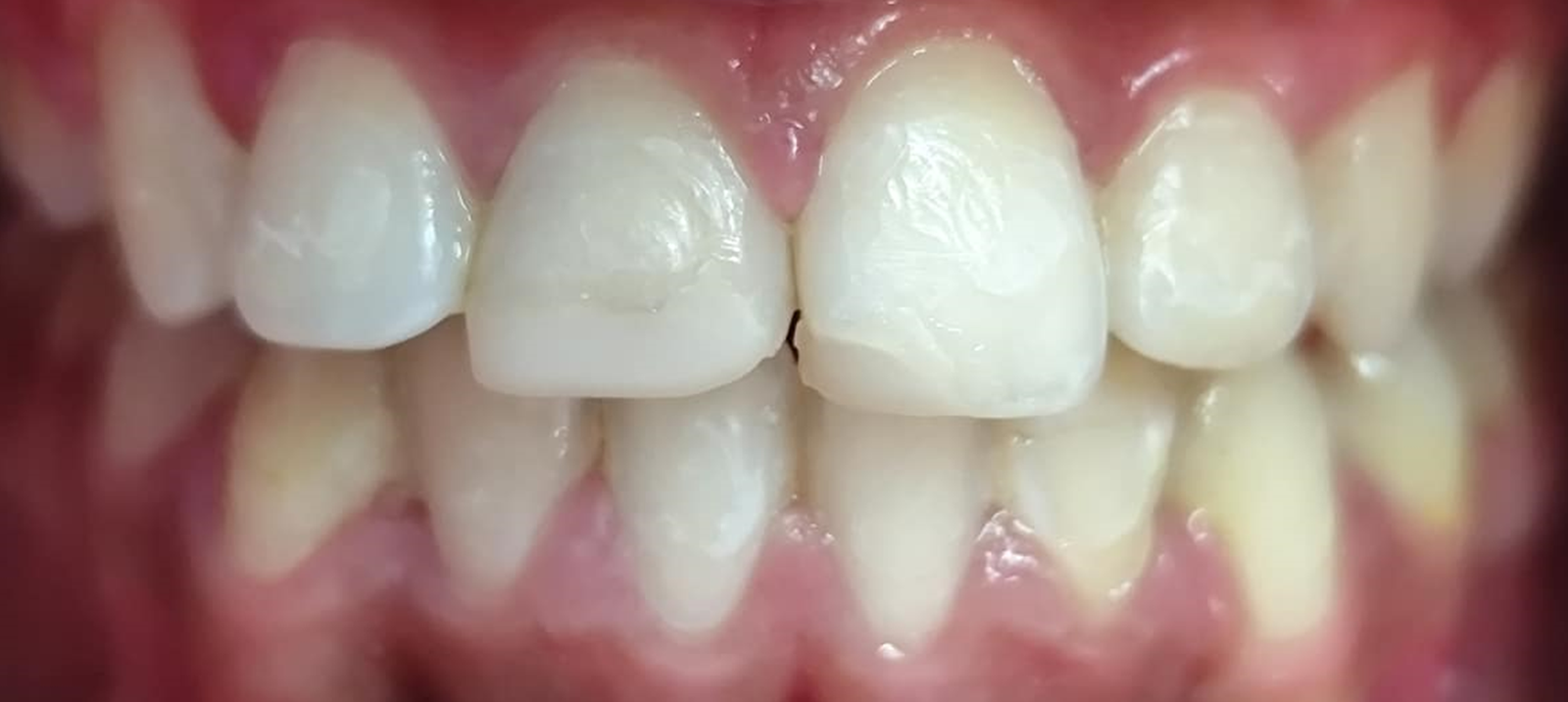
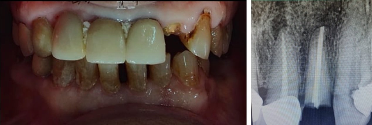
Intraoral examination revealed an Ellis Class III fracture involving the maxillary left central incisor (tooth 21), with loss of coronal tooth structure and discoloration. The tooth was non-tender to percussion and palpation. Periodontal probing depths were within normal limits, and no abnormal mobility was detected.
Timeline
- Six years prior: History of trauma to maxillary anterior region
- Present visit: Clinical and radiographic examination
- First visit: Access cavity preparation and working length determination
- Subsequent visit: Placement of MTA apical plug and obturation
- Post-endodontic phase: Tooth preparation and CAD/CAM crown fabrication
- One-year follow-up: Clinical and radiographic evaluation
DIAGNOSTIC ASSESSMENT
Radiographic examination revealed radiolucency involving enamel, dentin, and pulp with an associated periapical radiolucency in relation to tooth 21. Evidence of external root resorption was also noted. Based on the clinical and radiographic findings, a diagnosis of asymptomatic irreversible pulpitis with apical periodontitis was established.
Differential diagnoses considered included pulp necrosis with chronic apical abscess and previously treated tooth with persistent apical pathology; however, these were ruled out based on clinical findings and patient history. The prognosis was considered favourable due to adequate remaining root length and absence of periodontal involvement.
THERAPEUTIC INTERVENTION
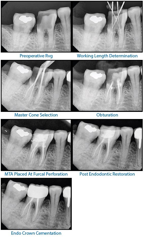
Considering the chronic nature of the infection and presence of external root resorption, a nonsurgical root canal treatment approach was planned. Following rubber dam isolation, access cavity preparation was performed. Working length was determined radiographically. Biomechanical preparation was carried out with copious irrigation. An MTA apical plug was placed to achieve an optimal apical seal in view of the resorptive defect. After confirmation of set, the canal was obturated and the access cavity was sealed.
Following successful endodontic therapy, the tooth was prepared for full-coverage restoration. A digital impression was recorded, and the crown was designed, milled, sintered, glazed, and cemented using CAD/CAM technology.
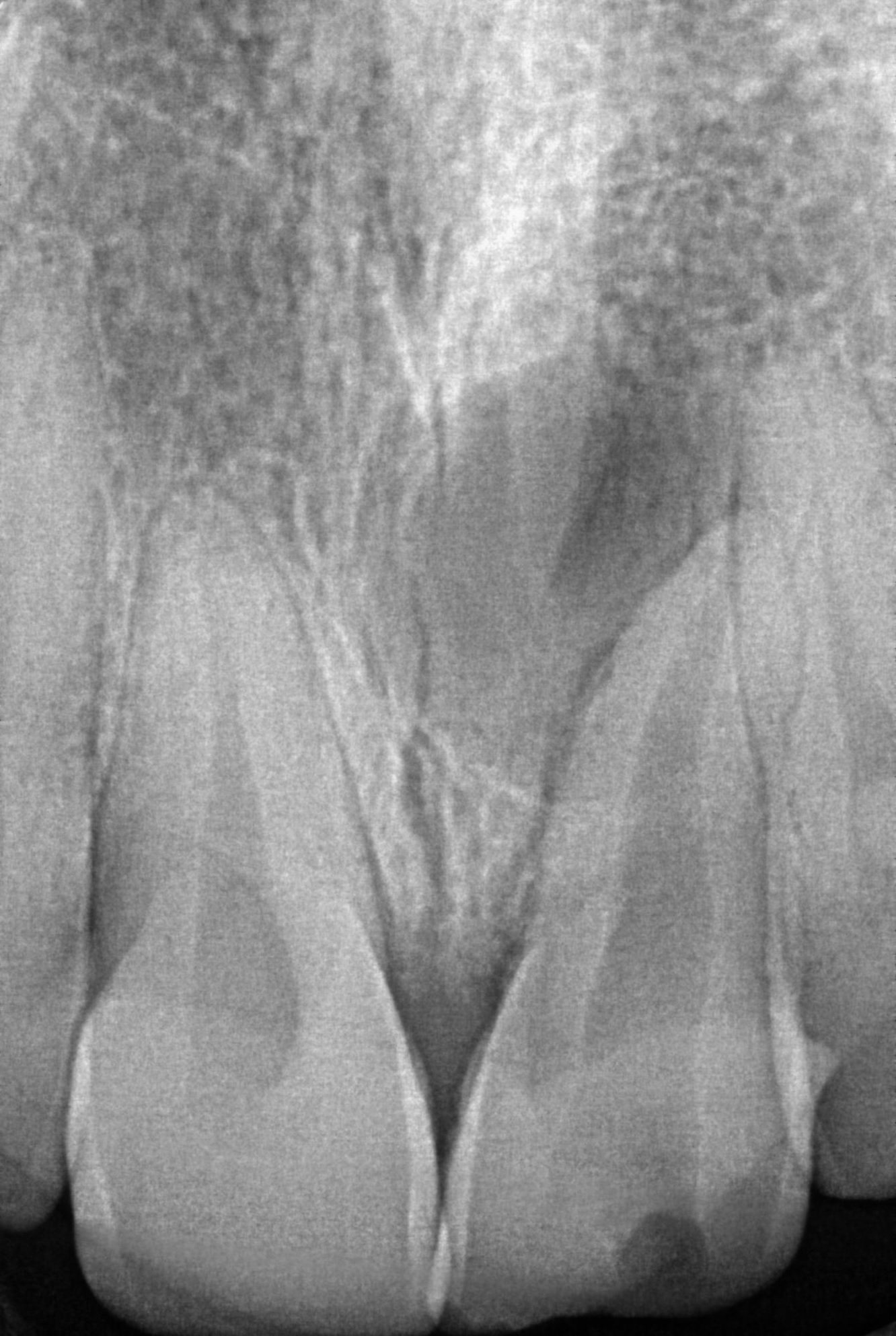
FIG 1 : PRE-OPERATIVE RADIOGRAPH
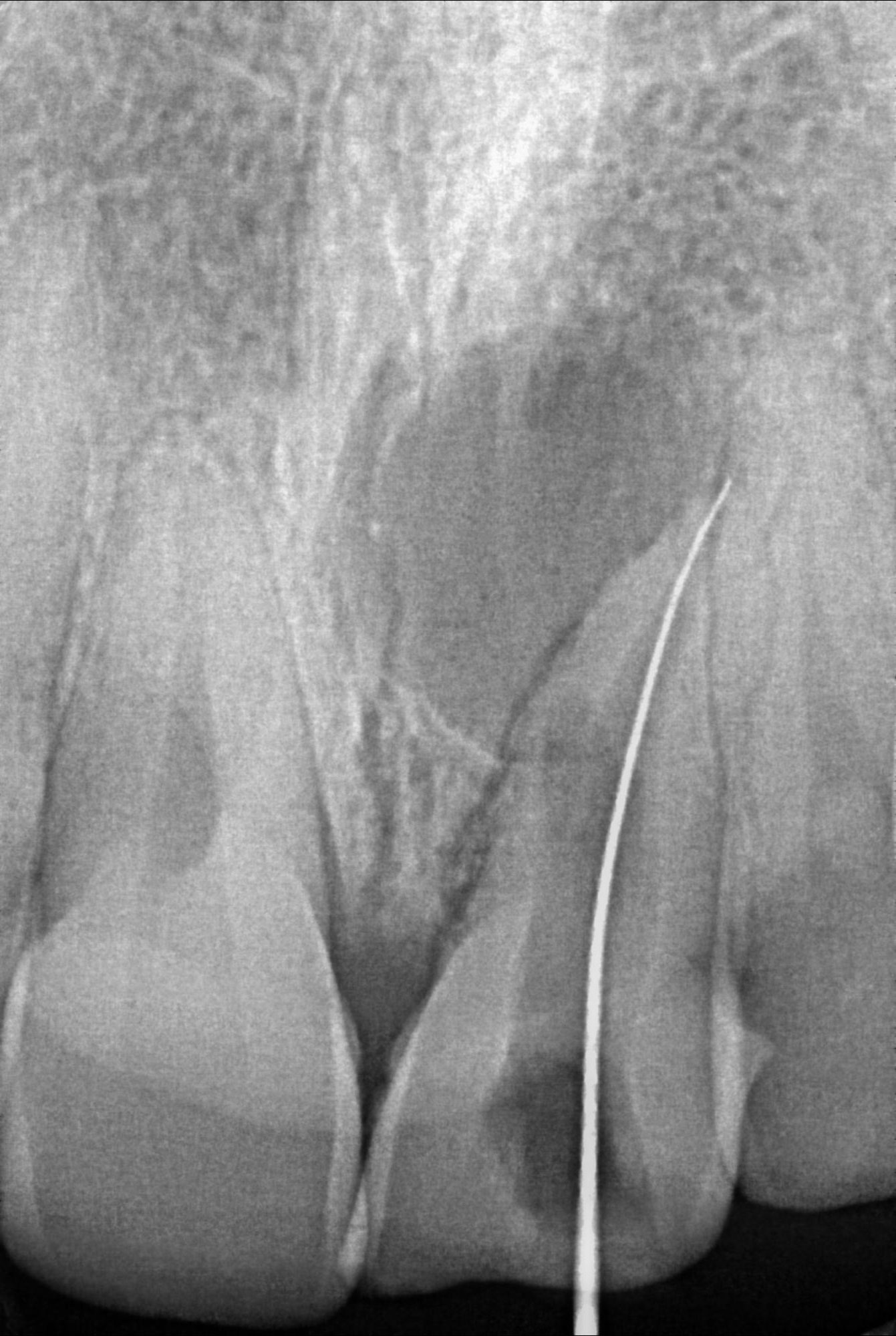
FIG 2 : WORKING LENGTH WAS DETERMINED
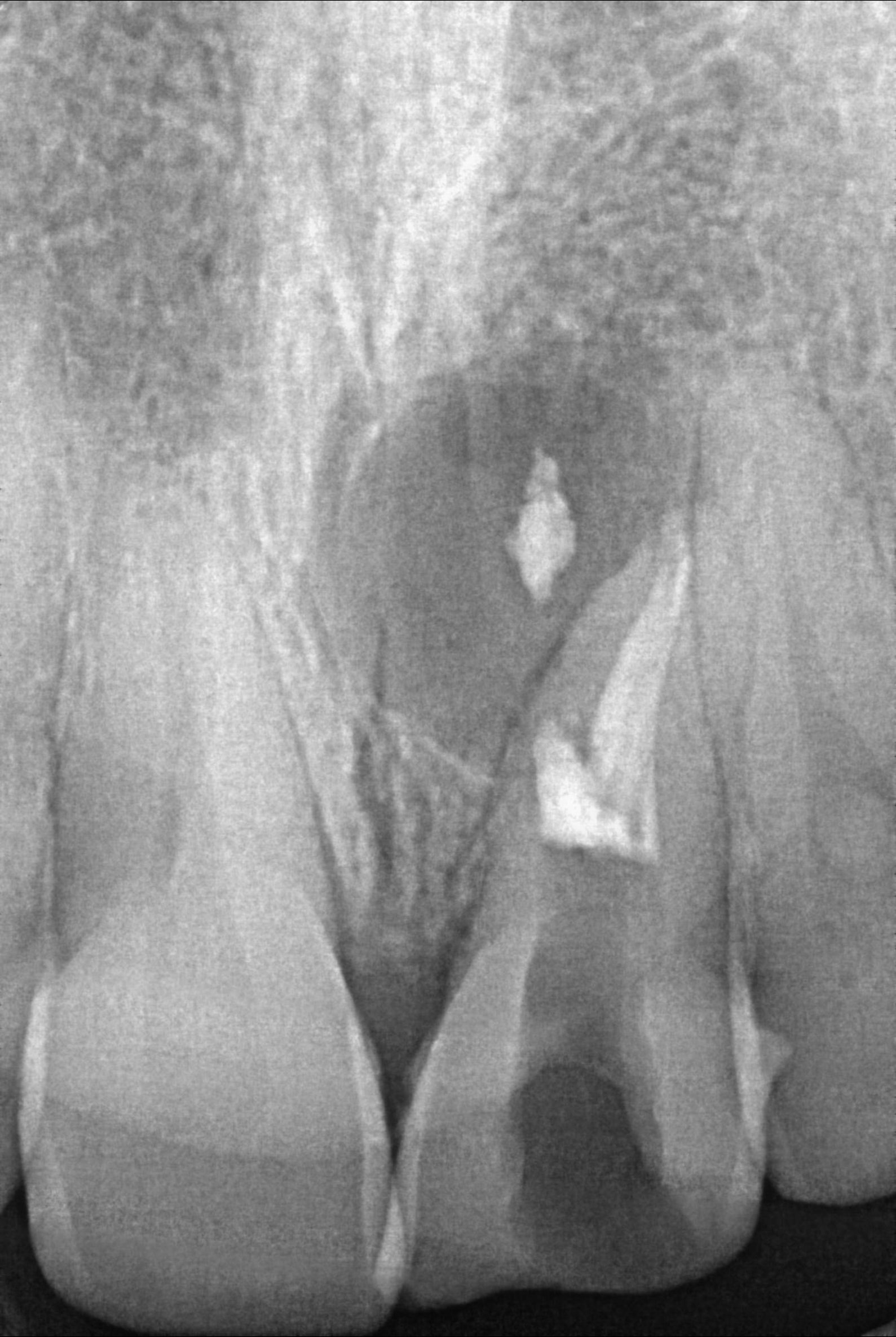
FIG 3 : MTA PLUG WAS MADE
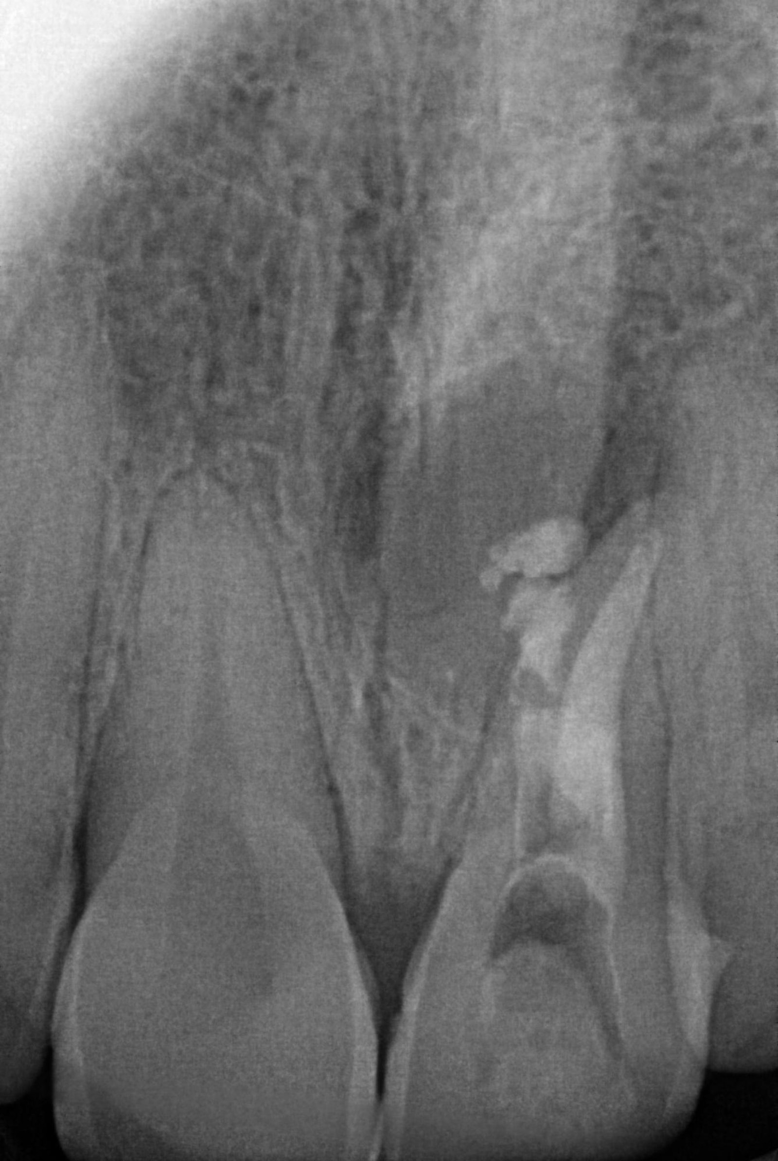
FIG 4 : POST OBTURATION RADIOGRAPH
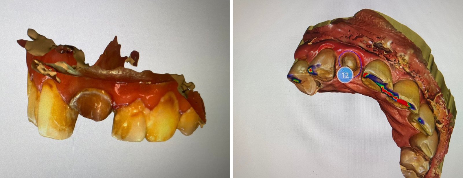
FIG 5 : DIGITAL IMPRESSION WAS TAKEN AND CROWN WAS DESIGNED
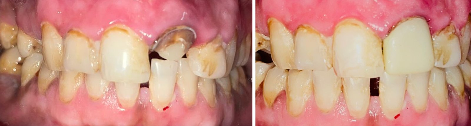
FIG 6 : CROWN WAS GIVEN
Follow-up and Outcomes
At the one-year follow-up visit, the patient was asymptomatic (FIG 7). Radiographic evaluation demonstrated reduction in periapical radiolucency, suggestive of healing. The CAD/CAM crown showed satisfactory marginal integrity, esthetics, and function. No adverse or unanticipated events were reported, and the patient demonstrated excellent compliance.
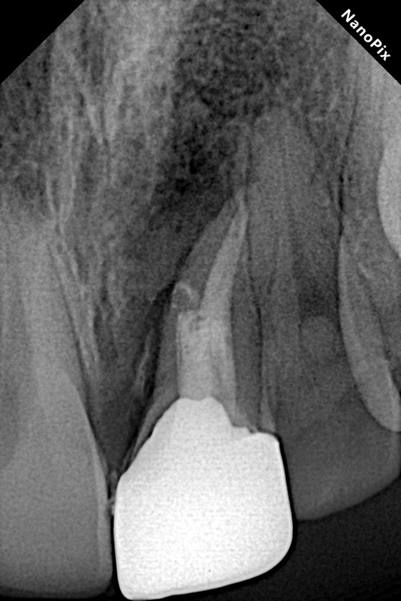
FIG 7 : ONE YEAR FOLLOW UP
DISCUSSION
Delayed presentation of traumatic dental injuries is a common clinical scenario, particularly when the teeth remain asymptomatic. In the present case, the history of trauma, pus discharge, and radiographic evidence of external root resorption indicated long-standing pulpal pathology. External root resorption is often initiated by damage to the protective cementum layer, followed by sustained inflammatory stimulus from an infected root canal system.
Nonsurgical root canal treatment remains the treatment of choice in such cases, provided adequate disinfection and apical sealing can be achieved. MTA was selected as the apical barrier material due to its excellent sealing ability, biocompatibility, and ability to promote periapical healing. Definitive rehabilitation with a CAD/CAM crown ensured optimal esthetics, precise marginal adaptation, and long-term durability.
A limitation of this case report is the relatively short follow-up period; however, the clinical and radiographic outcomes observed at one year are encouraging.
CONCLUSION
This case report highlights that traumatised anterior teeth with delayed pulpal and periapical sequelae, including external root resorption, can be successfully managed with meticulous nonsurgical root canal treatment and contemporary restorative approaches. Early diagnosis, appropriate material selection, and interdisciplinary planning are key to achieving predictable long-term outcomes.
PATIENT PERSPECTIVE
The patient reported satisfaction with the treatment outcome and expressed improvement in confidence due to the esthetic restoration of the anterior tooth.
Informed Consent
Written informed consent was obtained from the patient for publication of this case report and accompanying clinical images, with assurance of anonymity.
REFERENCES
- Andreasen JO, Andreasen FM. Textbook and Color Atlas of Traumatic Injuries to the Teeth. 4th ed. Wiley-Blackwell; 2007.
- Trope M. Root resorption due to dental trauma. Endodontic Topics. 2002;1:79–100.
- Fuss Z, Tsesis I, Lin S. Root resorption—diagnosis, classification and treatment choices. Endod Topics. 2003;7:175–189.
- Patel S, Kanagasingam S, Pitt Ford T. External cervical resorption: a review. J Endod. 2009;35:616–625.
- Patel S, Foschi F, Mannocci F, Patel K. External root resorption: a review. Int Endod J. 2018;51:663–680.
- Torabinejad M, Parirokh M. Mineral trioxide aggregate: a comprehensive literature review—Part I. J Endod. 2010;36:16–27.
- Torabinejad M, Parirokh M. Mineral trioxide aggregate: a comprehensive literature review—Part II. J Endod. 2010;36:190–202.
- Ng YL, Mann V, Gulabivala K. Outcome of secondary root canal treatment: a systematic review. Int Endod J. 2008;41:1026–1046.
- Mörmann WH. The evolution of the CEREC system. J Am Dent Assoc. 2006;137 Suppl:7S–13S.
- Øilo M, Gjerdet NR. Dental CAD/CAM materials. Dent Mater. 2017;33:e185–e197.
Disclaimer from Guident
The views and opinions expressed in articles published in Guident are solely those of the authors and do not necessarily reflect the views of the editors, the editorial board or the publisher, Ivory India. Guident does not endorse any product, technique or treatment described. Clinical content is intended for qualified dental and medical professionals and is not a substitute for individual clinical judgement. Guident, its editors and the publisher accept no liability for any consequence arising from the use of information published in the journal.
How to cite this article
Dr. Aashi Mittal, Dr Pulkit Gupta Dr. Deepti Kushwaha. Management of a Traumatised Maxillary Central Incisor with External Root Resorption Using Nonsurgical Root Canal Treatment and CAD-CAM Crown: A Case Report. Guident. 2026 Jul;19(8). Available from: https://guident.in/articles/endodontics/management-of-a-traumatised-maxillary-central-incisor-with-external-root-resorption-using-nonsurgical-root.html