Pink Tooth of Murmmery-Internal Resorption
Department of Conservative Dentistry and Endodontics, Seema Dental College And Hospital, Rishikesh
INTRODUCTION
According to the Glossary of the American Association of Endodontists, resorption is defined as “a condition associated with either a physiologic or a pathologic process resulting in the loss of dentin, cementum, or bone”. Bell first reported on internal resorption in 1830. Fothergil referred to the condition as “pink spot”. In 1920, Mummery published the first extensive study of pink spots, and Pritchard later showed histologically that internal resorption is comparable to a granuloma of the pulp.1 Root resorption is the loss of dental hard tissues as a result of clastic activities. It might occur as a “physiologic” or “pathologic” phenomenon. Root resorption in the primary dentition is a normal physiologic process except when the resorption occurs prematurely. The initiating factors involved in physiologic root resorption in the primary dentition are not completely understood, although the process appears to be regulated by cytokines and transcription factors that are similar to those involved in bone remodelling.3 Unlike bone that undergoes continuous physiologic remodeling throughout life, root resorption of permanent teeth does not occur naturally and is invariably inflammatory in nature. Thus, root resorption in the permanent dentition is a pathologic event; if untreated, this might result in the premature loss of the affected teeth.4PREVALENCE
Internal root resorption has been described as intraradicular or apical according to the location in which the condition is observed.4 The condition is more frequently observed in male than female subjects. Although intraradicular internal root resorption is a relatively rare clinical entity even after traumatic injury a higher prevalence of the condition has been associated with teeth that had undergone specific treatment procedures such as autotransplantation.11CLASSIFICATION OF TOOTH RESORPTION
Classification proposed by Lindskog subdivides resorption into 3 broad groups:1. Trauma induced tooth resorption
a. Surface Resorption
b. Transient Apical Internal Resorption
c. Pressure
d. Orthodontic
e. Replacement
2. Infection induced tooth resorption
a. Internal Inflammatory (Infective) Resorption
i. Apical
ii. Interradicular
b. External Inflammatory Resorption
c. Communicating External Inflammatory Resorption
3. Hyperplastic invasive tooth resorption
a. Internal (invasive) Replacement Resorption
b. Invasive Coronal Resorption
c. Invasive Cervical Resorption
d. Invacive Radicular Resorption
1. INTERNAL
a. Inflammatory
b. Replacement
2. EXTERNAL
a. Surfaceb. Inflammatory
c. Replacement
ETIOLOGY
Traditionally, internal resorption has been associated with a longstanding chronic inflammation in the pulp.6 The process is initiated by a variety of stimuli such as trauma, pulpotomy, extreme heat produced during cutting of dentin, chronic inflammation of the pulp following caries perpetuated by bacterial factors, cracked tooth, tooth transplantation, and orthodontic treatment.6,7 The literature also cites the association of herpes zoster with resorption and the degeneration of odontoblast due to systemic viral infection.12PATHOGENESIS

1. Osteoclasts are motile, multinucleated giant cells that are responsible for bone resorption. They are formed by the fusion of mononuclear precursor cells of the monocyte-macrophage lineage derived from the spleen or bone marrow, as opposed to osteoblasts and osteocytes that are derived from skeletal precursor cells.4
- Osteoclasts are recruited to the site of injury or irritation by the release of many proinflammatory cytokines.
- They attach themselves to the bone surface.
- On contact with mineralized extracellular matrices, the actin cytoskeleton of an actively resorbing osteoclast produces an organelle-free zone of sealing cytoplasm (clear zone) associated with the osteoclast’s cell membrane to enable it to achieve intimate contact with the hard tissue surface.
- The clear zone surrounds a series of finger-like projections (podosomes) of cell membrane known as the ruffled border.
- It is underneath this ruffled border that bone resorption occurs.
2. Odontoclasts resorb dental hard tissues and are morphologically similar to osteoclasts. Odontoclasts differ from osteoclasts by being smaller in size and having fewer nuclei and smaller sealing zones. Both resorb their target tissues in a similar manner. Both cells possess similar enzymatic properties, and both create resorption depressions termed Howship’s lacunae on the surface of the mineralized tissues.
3. The OPG/RANKL/RANK transcription factor system controls clastic functions during bone remodeling has also been identified in root resorption. The system is responsible for the differentiation of clastic cells from their precursors via complex cell-cell interactions with osteoblastic stromal cells.
INTERNAL INFLAMMATORY ROOT RESORPTION
Internal (inflammatory) root resorption can be characterized both as a well-known and poorly known disease entity destroying the dental hard tissue. It is well known in the sense that most dentists recognize the diagnosis ‘internal resorption.2 Internal inflammatory resorption is rare and often detected after its activity has ceased. Clinical symptoms and signs are usually not identified until the internal inflammatory resorption is advanced. The tooth may may be discoloured and respond negatively to sensitivity testing with advanced lesions.5 Internal inflammatory resorptions may be classified according to location as: apical and intraradicular.10INTERNAL REPLACEMENT (INVASIVE) RESORPTION
Internal root canal replacement resorption is characterized by an irregular radiographic enlargement of the pulp chamber, with discontinuity of the normal canal space. Because the resorption process is initiated within the root canal, the defect includes part of the canal space, and hence the outline of the original canal appears distorted. The enlarged canal space appeared radiographically to be obliterated by a fuzzy-appearing material of mild to moderate radiodensity.13 This type of resorption is relatively rare and may appear clinically as a pink area in the crown of the affected tooth.10 This form of resorption is typically asymptomatic, and the affected teeth might respond normally to thermal and/or electric pulp testing unless the resorptive process results in crown or root perforation.14 A variant of internal root canal replacement resorption has previously been reported as ‘‘internal tunnelling resorption’’CLINICAL FEATURES
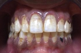
Internal resorption is usually asymptomatic and approximately only 2% shows clinical signs.It is more frequently observed in males than females.8 Usually found either in the mid or apical area of root. They are discovered by chance on routine radiographs or by the clinical sign of a “pink spot” on the crown. The pulp can either show partial or complete necrosis. In an actively progressing lesion, the tooth may be partially vital and may present symptoms typical of pulpitis.2 Pain may be a presenting symptom if there is complete perforation of the crown and the granulation tissue is exposed to oral environment. Another characteristic feature that gives an indication about the centre of origin of the resorption is the location of “the spot”: a color change from inflammatory internal resorption seen typically in the middle of the tooth in the mesiodistal direction (except in multirooted teeth), whereas in cervical resorption it is located eithermesially, centrally, or distally.2
DIFFERENTIAL DIAGNOSIS 1

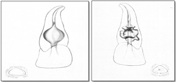
Internal resorption: normal outline of canal cannot be followed through lesion & External resorption: normal outline of canal can be followed through lesion.
Diagrammatic representations of midroot resorptive lesions (Based on drawings by Leppg)1
MANAGEMENT OF INTERNAL RESORPTION
1. Conservative Dental Treatments of Resorbed Teeth - Root canal treatment remains the treatment of choice of internal root resorption as it removes the granulation tissue and blood supply of the clastic cells. The access cavity preparation must be as conservative as possible to preserve tooth structure and avoid further weakening of the already compromised tooth. The workinglength determination with an apex locator is not possible in case of resorptive perforation. A great emphasis must be placed on the chemical dissolution of the vital and necrotic pulp tissue with sodium hypochlorite. The use of calcium hydroxide as an interappointment dressing maximizes the effect of disinfection procedures, helps to control the bleeding, and necrotizes residual pulp tissue. About the root canal filling, thermoplastic guttapercha techniques seem to give the best results when the canal walls are respected.162. Complete Root Canal Filling with Warm Gutta Percha - This option is for IRR with no perforation of the canal walls which is the most favorable situation in long-term prognosis. The treatment is performed in two sessions.16
3. Sealing of Internal Root Resorption with Bioactive Cements as MTA - This option is indicated in presence of a perforation of the canal walls giving a communication between the root canal system and the periapical tissue. In this clinical situation, the smaller the perforation size, the more predictable the prognosis of the tooth. The treatment is performed in two sessions.16
4. Surgical Treatment of Internal Root Resorption - Surgical approach is needed when it is not possible to get access to the lesion through the canal. Surgical treatment should always be performed in a second intention, after orthograde treatment (or retreatment) has been performed, the coronal part of the canal being filled. In these cases, because of the shape of the lesion, surgical approach allows to get direct access to the lesion and to perform a mechanical cleaning of the resorbed defect.16
REFERENCES
- Gartner, A. H., Mack, T., Somerlott, R. G., & Walsh, L. C. (1976). Differential diagnosis of internal and external root resorption. Journal of Endodontics, 2(11), 329–334. doi:10.1011/s0099-2399(76)80071-4
- HAAPASALO, M., & ENDAL, U. (2006). Internal inflammatory root resorption: the unknown resorption of the tooth. Endodontic Topics, 14(1), 6079. doi:10.1111/j.1601-1546.2008.00226.x
- Harokopakis-Hajishengallis, E. (2007). Physiologic root resorption in primary teeth: molecular and histological events. Journal of Oral Science, 49(1), 1 12.
- Patel, S., Ricucci, D., Durak, C., & Tay, F. (2010). Internal Root Resorption: A Review. Journal of Endodontics, 36(7), 1107–1121. doi:10.1016/j.joen.2010.03.014
- Patel, S., & Ford, T. P. (2007). Is the Resorption External or Internal? Dental Update, 34(4), 218–229. doi:10.12968/denu.2007.34.4.218
- Tronstad, L. (1988). Root resorption - etiology, terminology and clinical manifestations. Dental Traumatology, 4(6), 241–252. doi:10.1111/j.1600-9657.1988.tb00642.x
- Darcey, J., & Qualtrough, A. (2013). Resorption: part 1. Pathology, classification and aetiology. British Dental Journal, 214(9), 439–451. doi:10.1038/sj.bdj.2013.431
- Thomas, P., Krishna Pillai, R., Pushparajan Ramakrishnan, B., & Palani, J. (2014). An Insight into Internal Resorption. ISRN Dentistry, 2014, 1–7. doi:10.1155/2014/759326
- Shenoi, P.R., Morey, E., Sonarkar, S.S., Kubde, R.R., Badole, G.P., & Singare, P.G. (2015). Internal Root resorption (Pink tooth of Mummery) A Review.
- Heithersay, G. (2007). Management of tooth resorption. Australian Dental Journal, 52, S105–S121. doi:10.1111/j.1834-7819.2007.tb00519.x
- Goultschn J, Nitzan D, Azaz B. Root resorption: review and discussion. Oral Surg Oral Med Oral Pathol 1982;54:586–91.
- S. Seltzer and I. Bender, The Dental Pulp, J. B. Lippincott, Philadelphia, Pa, USA, 2nd edition, 1975.
- Oehlers FAC. A case of internal resorption following injury. Br Dent J 1951;90:13–6.
- Ne RF, Witherspoon DE, Gutmann JL. Tooth resorption. Quintessence Int 1999;30:9–25.
- Andreasen FM, Andreasen JO. Resorption and mineralization processes following root fracture of permanent incisors. Endod Dent Traumatol 1988;4:202–14.
- Nilsson, Elisabeth & Bonte, Eric & Bayet, François & Lasfargues, Jean-Jacques. (2013). Management of Internal Root Resorption on Permanent Teeth. International journal of dentistry. 2013. 929486. 10.1155/2013/929486.
Disclaimer from Guident
The views and opinions expressed in articles published in Guident are solely those of the authors and do not necessarily reflect the views of the editors, the editorial board or the publisher, Ivory India. Guident does not endorse any product, technique or treatment described. Clinical content is intended for qualified dental and medical professionals and is not a substitute for individual clinical judgement. Guident, its editors and the publisher accept no liability for any consequence arising from the use of information published in the journal.
How to cite this article
Dr. Annil Dhingra, Dr. Unnati Nautiyal. Pink Tooth of Murmmery-Internal Resorption. Guident. 2020 May;13(6). Available from: https://guident.in/articles/endodontics/pink-tooth-of-murmmery-internal-resorption.html