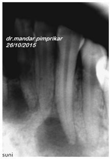
Mandibular Second Molar with Radix Paramolaris
Seema Dental College and Hospital, Rishikesh, Uttrakhand, India
INTRODUCTION
Success of endodontic therapy depends on the complete cleaning and shaping of the root canal systems, tridimensional obturation, and proper coronary sealing. For that purpose, the knowledge of the morphology of the root canal system as well as its variations is essential. The mandibular first molar is known to display several anatomical variations. The number of roots may also vary in mandibular molars, in which a third additional root, firstly mentioned in the literature by Carabelli1, is called radix entomolaris. This supernumerary root is located in distolingual position, mainly in the mandibular first molars. When located in the mesiobuccal surface, the anomaly is called radix paramolaris2. The frequency of radix entomolaris is less than 5% in Caucasian, African, Eurasian, and Indian populations3, whereas its bilateral occurrence is less than 2.19% 4. This extra root is typically smaller and more sharply curved than the distobuccal (DB) root, requiring special attention when root canal treatment is being considered.
CASE 1
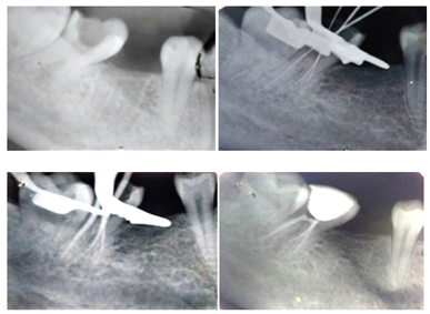
21-year-old female patient reported with the chief complaint of pain in lower left back region since two weeks. The pain was aggravated on mastication.Theclinical examination revealed grossly decayed tooth w.r.t 37. Intraoral periapical radiograph of 36 revealed deep carious lesion involving the pulp with periapical radiolucency at the root apices and presence of an extra root in 37. Based on clinical and radiographic examination, diagnosis of symptomatic irreversible pulpitis with apical periodontitis was established. After adequate anesthesia and isolation with rubber dam, access cavity was prepared. Working length was determined using #15 k files followed by Cleaning and shaping till # 20/0.6 in mesial canals and #30/0.6 in distal canal. All the canals were irrigated with 2.5% sodium hypochlorite and 17% EDTA after use of each file. In the subsequent appointment canals were dried using paper points and obturation was done using gutta percha and MTA Fillapex sealer. Later, the access cavity was sealed with composite.
CASE 2
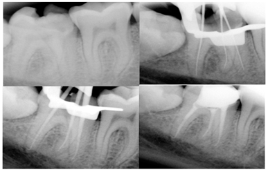
18-year-old male patient reported to the dental office with the chief complaint Pain in lower right back region of the mouth since 2-3 days. Clinical examination revealed unusual crown anatomy with and deep mesio occlusal caries with 47.Intraoral periapical radiograph of 47 revealed deep carious lesion approaching the pulp with thickened periodontal ligament space and presence of an extra root in 47. Based on clinical and radiographic examination, diagnosis of symptomatic irreversible pulpitis with apical periodontitis was established. After adequate anesthesia and isolation with rubber dam, access cavity was prepared. Working length was determined using #15 k files followed by Cleaning and shaping till # 25/0.4 in all the canals. All the canals were irrigated with 2.5% sodium hypochlorite and 17% EDTA after use of each file. In the subsequent appointment canals were dried using paper points and obturation was done using gutta percha and MTA Fillapex sealer. Later, the access cavity was sealed with composite
DISCUSSION
Accurate diagnosis of supernumerary roots like RP can contribute to the successful outcome of endodontic treatment. The presence of Radix Paramolaris in the mandibular first molar has been extensively evaluated in the literature, but only a few studies have evaluated the morphology of RP in mandibular second molars5-6.The etiology behind the formation of RP remains unknown. This unusual morphology of mandibular molars could be related to the penetrance of an atavistic gene or polygenetic system or due to external factors during odontogenesis7.
The mandibular molars usually have two roots with two canals in mesial root and one canal in distal root. It is more common that mandibular molars having four canals with two in mesial and distal root viz. the first molar8. The isthmus connecting the mesiobuccal and lingual or distobuccal and lingual is debrided and left uninstrumented in most of the cases. In these cases there are two mesial canals, both files and gutta percha points went until full working length independently without any fusion at the apex to exit as one. Radix Paramolaris is an extra third root in mandibular molars, which is shorter than the mesial and distal roots. In this case, the extra root was seen in buccal version.
The Radix Paramolaris occurrence rate is about 0%, 2%, and 5% in mandibular first, second and third molars respectively. Carlsen and Alexandersen described two different types: Type A: Radix Paramolaris in which the cervical part is located on the mesial root complex. Type B: Radix Paramolaris in which the cervical part is located centrally, between the mesial and distal root complexes, an additional cusp is present on the buccal side9. In the present case series Case 1 belongs to Type A of Radix Paramolaris and Case 2 belongs to Type B Radix Paramolaris.
CONCLUSION
The complex root canal anatomy stresses the operator to observe the canal morphology with care without underestimating it and missing the extra root and roots canals.Thus, the success of endodontics lies in the careful exploration of the canals and debridement of it.
“Expect the unexpected though it is rare.”
REFERENCES
- G. Carabelli, Systematisches Handbuch der Zahnheilkunde, Braumuller und Seidel, Viena, Austria, 2 edition, 1844.
- F. L. Calberson, R. J. DeMoor, andC. A. Deroose. The radix entomolaris and paramolaris: clinical approach in endodontics. J Endod. 2007; 33(1)58–63.
- M. E. Curzon. Three-rooted mandibular permanent molars in English Caucasian. Journal of Dental Research.1973;52(1)181, 197.
- C. L. Sabala, F.W. Benenati, and B. R. Neas. Bilateral root or root canal aberrations in a dental school patient population. J Endod.1994(20)38–42.
- Manning SA. Root canal anatomy of mandibular second molars. Part I. Int Endod J 1990;23:34-39.
- Reichart PA, Metah D. Three-rooted permanent mandibular first molars in the Thai. Community Dent Oral Epidemiol 1981;9:191-192.
- Neelakantan P, Subbarao C, Subbarao CV, Ravindranath M. Root and canal morphology of mandibular second molars in an Indian population. J Endod 2010;36:1319- 1322.
- N. Ragavendran, Ganesh T. Bhat, Mithra N. Hegde Mandibular second molar with 3 mesial canals and a radix paramolaris Journal of Pharmacy and Bioallied Sciences July 2014 Vol 6 Supplement 1.
- Carlsen O, Alexandersen V. Radix entomolaris: Identification and morphology. Scand J Dent Res 1990;98:363‑73.
Disclaimer from Guident
The views and opinions expressed in articles published in Guident are solely those of the authors and do not necessarily reflect the views of the editors, the editorial board or the publisher, Ivory India. Guident does not endorse any product, technique or treatment described. Clinical content is intended for qualified dental and medical professionals and is not a substitute for individual clinical judgement. Guident, its editors and the publisher accept no liability for any consequence arising from the use of information published in the journal.
How to cite this article
Dr. Annil Dhingra Dr. Gazal Bisht Department of Conservative Dentistry and Endodontics. Mandibular Second Molar with Radix Paramolaris. Guident. 2017 Dec;11(1). Available from: https://guident.in/articles/endodontics/mandibular-second-molar-with-radix-paramolaris.html