Anatomy of the Dental Operating Microscope
Director, Upskill Dental Academy, Mumbai, India
DENTAL OPERATING MICROSCOPE

Pic courtesy: Sanma Medineers
Operating microscopes have been used for more than eighty years in various medical specialties as ophthalmology, otolaryngology, neurology or vascular and cardiac surgery and its use in dentistry began thirty years ago . Traditionally the reading of the pulp canal chamber was based on a mental image of the most common anatomies associated with a tactile impression of the operator because of limited illumination and visibility of the procedure . The predictability of the endodontic therapy is related with the correcttissues removal and disinfection of the root canal system, if a canal cannot be found, it cannot be cleaned and shaped and becomes a potential cause of failure of endodontic treatment . This has changedwith the introduction of the magnification in endodontics. Superior lighting and magnification provides the clinician with a superior ability of treating cases that in the past were considered untreatable or with an inferior prognosis. But the clinical better visualization, allowed by the microscope, must be associated with a user friendly performance of the instrument. Ergonomic design, compact in office space, support for documentation, capacity of receiving upgrades and experience of personal satisfaction are also factors mandatory for microscope1
The microscope may be divided in four main groups of components: magnification, illumination, mounting and accessories.
MAGNIFICATION COMPONENT

pic courtesy: Sanma Medineers
The magnification of a microscope is dependent on the binocular tube, objective lens and magnification changers.
Binocular Tube The binocular tube has two important factors that have direct impact on the magnification of the microscope, the focal length tube (FLT) and the eyepiece power (EP) . The eyepiece value is the one that may have more options depending on the manufacture, it may range from10x to12.5x . The end of each eyepiece has a rubber cup that can be turned down for clinicians who wear eyeglasses. Eyepieces also have adjustable diopter settings. Diopter settings range from -5 to +5 and are used to adjust for accommodation, which is the ability to focus the lens of the eyes. Diopter settings is particularly important when an assistant scope and documentation equipment are used, so that everything is uniformly in focus THE BINOCULARS: it contain the eyepieces and allow the adjustment of the interpupillary distance. They are aligned manually or with a small knob until the two divergent circles of light combine to effect a single focus. Once the diopter setting and interpupillary distance adjustments have been made, they should not have to be changed until the microscope is used by a surgeon with different optical requirements. Binoculars are available with 3 different kind of tubes i.e. (1) Straight: Straight tube binoculars are orientated so that the tubes are parallel to the head of the microscope. They are generally used in otology and are not well suited for dentistry. (2) Inclined: Inclined tubes are fixed at a 45° angle to the line of sight of the microscope. (3) Inclinable tubes: The inclinable tubes are adjustable through a range of angles and allow the clinician to always establish a very comfortable working position. It is therefore obvious that, even if more expensive, the inclinable binocular is always to be preferred .
MAGNIFICATION CHANGER
The magnification changer (MC) is the responsible for the zoom change. It consists of a group of lens with different magnification values that are lodged in interior of the body that may rotate through a dial. Manual step changers consist of lenses that are mounted on a turret that is connected to a dial located on the side of the microscope. The magnification is altered by rotating the dial. A power zoom changer is a series of lenses that move back and forth on a focusing ring to give a wide range of magnification factors. The advantage of the power zoom changers is that they avoid the momentary visual disruption or jump that occurs with manual step changers as the clinician rotates the turret and progresses up or down in magnification. The disadvantages are the following: the excursion from the minimum to the maximum magnification is quite slow, while it is must faster with the manual step changers; the number of lenses is much higher compared to the manual step changers, and this means a greater absorption of light; power zoom changer are much more expensive.
The objective lens is the final optical element, and its focal length determines the working distance between the microscope and the surgical field. The range of focal length varies from 100 mm to 400 mm. A 200 mm focal length allows approximately 20 cm (8 inches) of working distance, which is generally adequate for utilisation in endodontics.he objective lens, all have several layers of an anti-reflective coating on both surfaces, which reduces return light loss from normally 2% per lens surface to only 0.5% per lens surface.
OBJECTIVE LENS
The objective lens is the final lens of the microscope. It determines the working distance between the microscope and the operative area. This objective focal length (OFL) varies from 100mm to 400mm .
To calculate the total magnification value of a microscope the following formula has to be calculated Total Magnification = (FLT/OFL) x EP x MC On this formula the only value that may change, during the clinical endodontic procedures, affecting the total magnification is the dial of the magnification changer
MAGNIFICATION & DEPTH OF FIELD
One factor to take in consideration is the depth of a field of the different type of magnifications. The depth of a field is inversely proportional to the total magnification. If the total magnification increases, the depth of the field decreases . The same happens with the field of view. The higher the magnification, the smaller the field of view.
POINT TO NOTE
The quality of a microscope is not measured by its total higher magnification power, since high magnifications have small width and deep fields, but if it has acceptable magnifications compatible with clinical work. Different magnifications work better for different procedures but the most usual in routine endodontic procedures are between 6x and 8x
CO AXIAL ILLUMINATION
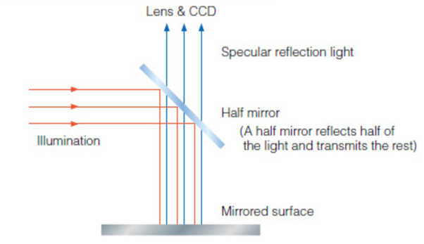
Pic: Internet source
illuminator sends the beam through the objective lens so that the line of illuminationis at par with viewers line of vision.the spot on focus will benefit with shadow free clear vision. The light of the microscope is emitted directly through the objective lens itself which means the paths of sight and light are parallel.
To get uninhibited illumination it is essential for the operator to adjust the head lamp of the chair repeatedly while at work.the solution to this problem would be the co axial illumination of a microscope.

Objective lens with lights off.
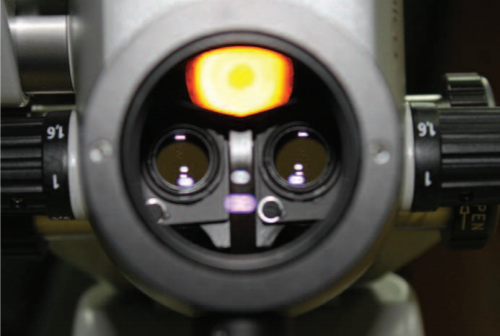
Objective lens with lights on1
The light comes out from the objective lens and takes a path parallel to the visual path. . Usually the lamps of the microscope are logged in a lamp modulewhich is part ofthe suspension system,the light ofthe lamp travels through a fiberoptic cable and enter the microscope body through a light guide socket , after the light crosses a series of prisms and gets out through the objective lens and illuminates the operating field, while the light illuminates the operating field the image is being transmitted back by a parallel path to the objective lens, magnification lens and finishes on the binocular eye pieces where it can be observed by the clinician.There are two light systems available, a halogen or a xenon illumination system . The xenon light is similar to day light, white and natural. The halogen light is more yellow and not so natural . The xenon light is the light of excellence, has a longer life time but is much more expensive than the halogen light. Itis possible to adjust the fiber optic light brightness through a control knob. It is recommended not to set the brightness to a higher level than the required to the operation .LED is the most preferred option among the sources of illumination.
MOUNTING
When using high power magnification the deep visual field is very small, so any movement may change the focus of the microscope. Both movements by the patient or by the microscope may change the focus. It is essential that the microscope becomes stable while in operation, but at the same time remaining maneuverable and easy to use even when using high power magnifications . The suspension systems play an important role in the whole stability while in operation. The mounting selection must be based on the dimension, decor and renovation cost of an already existing operatory. Options as cumbersome rolling floor models may be favorable in multi room offices where the microscope has to change room once in a while. Already existing operating room where office renovations are not desired may also favor this option.
For wall mounted and ceiling mounted microscopes the wall and the ceiling must have a structure enough strong to support the weight of the device. The distance between the suspension systems and the body of the microscope should be the smallest possible,the stability of the whole device increases with the decrease of the distance between the fixation point and the microscope body. All the built-in springs should be also tightened enough the give the microscope the perfect balance.2
The options for mounting are
Floor mounted
Wall mounted
Ceiling mounted
Wheel mounted
REFERENCES
- Dental Operating microscope: How does it work? Jorge NR Martins, Marina Saura, Athanasia, Pagona Cadernos cientificos I, revista da ordem dos medicos dentistas May 2011
- Magnification in Endodontics, Ashish Chajlani, Health and Medicine, May 12, 2015.
Disclaimer from Guident
The views and opinions expressed in articles published in Guident are solely those of the authors and do not necessarily reflect the views of the editors, the editorial board or the publisher, Ivory India. Guident does not endorse any product, technique or treatment described. Clinical content is intended for qualified dental and medical professionals and is not a substitute for individual clinical judgement. Guident, its editors and the publisher accept no liability for any consequence arising from the use of information published in the journal.
How to cite this article
Dr. Pavana Kamath. Anatomy of the Dental Operating Microscope. Guident. 2018 Sep;11(10). Available from: https://guident.in/articles/endodontics/anatomy-of-the-dental-operating-microscope.html