Apexification of Blunderbuss Canals with MTA : An Unusual Approach
Seema Dental College and Hospital, Raichur, India
INTRODUCTION
Apexification can be defined as a ,method to induce a clacified barrier in a root with an open apex or the continued apical development of teeth with incomplete roots and a necrotic pulp’ Management of immature root with a necrotic pulp and apical periodontitis is a challenging task. The infected root canal space cannot be disinfected with the standard protocol. Obturation of the root canal is difficult because of lack of apical barrier for containing the root filling material. Treatment of choice in such cases is the apexification procedure, i.e., establishing an apical barrier. Calcium hydroxide has been widely used for the induction of hard tissue barrier. However, this material requires 5–20 months to form the hard tissue barrier.[1] It has also been shown that the use of calcium hydroxide weakens the resistance of the dentin to fracture.[2] In recent times, mineral trioxide aggregate (MTA) has gained widespread popularity for the apexification procedure. Mineral trioxide aggregate (MTA) was developed by Mahmoud Torabinejad at Loma Linda University in the year 1993 as root end ï¬lling material. MTA has the advantage over calcium hydroxide that it can be done in a single visit procedure. It produces apical hard tissue formation with significantly greater consistency than calcium hydroxide.[3] MTA, a biocompatible material, can be used to create a physical barrier. It also helps in the formation of bone and periodontium around its interface.[4] A bonded restoration can be placed without any delay, thus reducing the possibility of root fractures. Various authors have reported clinical success using MTA for one-visit apexification. The major disadvantage of MTA is its manipulation due to which its placement in the wide apical area is difficult to achieve. A matrix can be used in apexification procedures against which MTA can be placed and condensed. Use of an apical matrix allows for the predictable placement and judicious use of this expensive material. Several materials have been recommended to create a matrix including calcium sulfate, hydroxyapatite, resorbable collagen, and platelet-rich fibrin.[5],[6],[7] In this case series, we are presenting three cases of MTA apexification.
CASE REPORT
CASE 1
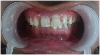
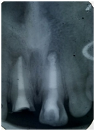
A 18 yr old patient reported to the Department of Conservative Dentistry and Endodontics, Seema dental college and hospital, Rishikesh with the complaint of an unaesthetic smile. History revealed that the patient had suffered trauma 8 years back and undergone treatment in a private clinic. The medical history was not significant. Clinical examination revealed grayish discoloration of tooth 21 and 11 attempted access preparation in both tooth. The tooth did not demonstrate any abnormal mobility or sensitivity to percussion. Both cold and electric sensibility tests failed to elicit any response. Periapical radiograph showed well-defined periapical radiolucency and wide open apex in relation to tooth 21 and 11 . The final diagnosis was pulpal necrosis with chronic apical periodontitis in relation to tooth 21 and11. After discussing different treatment options with the patient's parents, we opted to go for MTA apexification. 21 and 11 was accessed and working length was established radiographically . Root canal was chemo-mechanically debrided with circumferential filing using the International Organization for Standardization (ISO) 80 K-file in conjunction with copious amount of 0.5% sodium hypochlorite . A volume of 3 ml of 17% ethylenediaminetetraacetic acid (EDTA) solution was used for smear layer removal. Probing depths, and normal function without symptoms. Radiograph demonstrated the resorption of the apical matrix and healing periapical lesion. On second visit, an apical barrier of 3-4 mm was established using MTA. A moist cotton pellet was placed over the MTA and access cavity was sealed with IRM. Patient was recalled the next day. On third visit, the setting of MTA was confirmed using finger plugger and the root canal was obturated by sectional obturation technique.

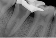
Pre Operative Radiograph
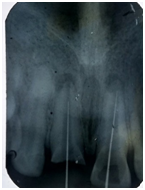
Working Length Determination
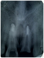
MTA Plug Formation
ObturationWith Thermoplastic Technique
CASE 2
A 23-year-old male patient presented with the chief complaint of unaesthetic smile. Patient gave a history of traumatic incident 9 years back for which he had visited a private clinic. Medical history was insignificant. Clinical examination revealed slightly grayish discolored tooth 11 and previously attempted treatment in the same tooth. The tooth was tender to percussion. Tooth also did not respond to cold and electric pulp vitality tests. Intraoral periapical radiograph showed a well-defined periapical radiolucency surrounding the wide open apex of tooth 21. Diagnosis was pulpal necrosis with chronic apical periodontitis. After detailed discussion with the patient, we decided to perform MTA apexification procedure. Root canal was chemo-mechanically debrided with circumferential filing using the International Organization for Standardization (ISO) 80 K-file in conjunction with copious amount of 0.5% sodium hypochlorite . A volume of 3 ml of 17% ethylenediaminetetraacetic acid (EDTA) solution was used for smear layer removal. Probing depths, and normal function without symptoms.Written consent was obtained from the patient's parents. After rubber dam isolation, tooth was assessed and working length determined using the radiograph. Root canal debridement was done using ISO 80 K-file and 0.5% sodium hypochlorite. 3 ml of 17% EDTA solution was used for smear layer removal. After 1 week, 4 mm of MTA plug was formed by condensing MTA.
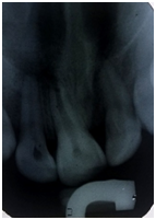
Pre Operative Radiograph
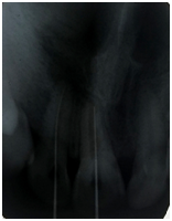
Working Length Determination
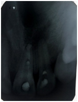
MTA Plug Formation

Obturation with Thermoplastic Technique
DISCUSSION
Apexification is defined as a method to induce a calcified barrier in a root with an open apex or the continued apical development of an incomplete root in teeth with necrotic pulp. This barrier is mandatory to allow the compaction of the root filling material. Calcium hydroxide has been considered as an efficient material for this purpose.[8] This chemical has several disadvantages such as difficulty of the patient's recall management and delay in the treatment.[9] Furthermore, there is a risk of tooth fracture after dressing with calcium hydroxide for extended periods.[2] The most promising alternative to calcium hydroxide is MTA.[10] The advantages of this material are (i) reduction in treatment time, (ii) immediate restoration of the tooth, (iii) no adverse effect on the mechanical properties of root dentin. In a prospective study, apexification treatment with MTA showed a high prevalence of healing and apical closure.[11] The main disadvantage of MTA is its difficult manipulation. Placement of the material in a wide open area is a challenging task and also there is a risk of extruding this expensive material into periapical tissues. Lemon advocated the use of a matrix when the diameter of the perforation is larger than 1 mm to prevent the extrusion of sealing material. Similarly, a matrix can be used for the predictable placement of MTA in apexification procedures. Use of matrix provides a base against which the MTA can be packed.[12] Various materials have been advocated to be used as a matrix, for example, calcium sulfate, hydroxyapatite, collagen, platelet-rich fibrin. However, the materials mentioned are either not cost-effective or technique sensitive. Calcium sulfate has a very short setting time of 2 min.[13] Hydroxyapatite is an expensive material. The common limitation shared by these materials is that once placed their position can't be adjusted as required.
CONCLUSION
Managing a tooth with open apex with a biocompatible material MTA has become a single visit procedure. This innovative procedure is predictable and less time-consuming one. Such teeth restored with this technique best serve the needs of the patients.
REFERENCES
- Goomer P, Jain RL. Comparison Of pulp revascularization & MTA apexification in adjacent central incisors with immature apex – A case report. Indian J Dent Sci 2014;6:92-1.
- Salgar AR, Chandak MG, Manwar NU. Blunder buss canal: A challenge for endodontist. Endodontology 2011;23:75-9.
- Kubasad GC, Ghivari SB. Apexification with apical plug of MTA – Report of cases. Arch Oral Sci Res 2011;1:104-7.
- Witherspoon DE, Small JC, Regan JD, Nunn M. Retrospective analysis of open apex teeth obturated with mineral trioxide aggregate. J Endod 2008;34:1171-6.
- Komabayashi T, Spångberg LS. Comparative analysis of the particle size and shape of commercially available mineral trioxide aggregates and Portland cement: A study with aflow particle image analyzer. J Endod 2008;34:94-8
- Manv MZ, Bhaskaran S, Hans M, Punga KK, Chander S, Ahluwalia A. Intraradicular rehabilitation of fractured maxillary canine with open apex. Dent Impact J 2013;5:66-70.
- Bodrumlu E. Biocompatibility of retrograde root fi lling materials: A review. AustEndod J 2008;34:30-5.
- Torabinejad M, Chivian N. Clinical applications of mineral trioxide aggregate. J Endod 1999;25:197-205.
- Takeda T, Ishigami K, Shimada A, Ohki K. A study of discoloration of the gingiva by artificial crowns. Int J Prosthodont 1996;9:197-202.
- Vats A, Loomba K, Jain A, Sharma R, Gaur TK. Intraradicular rehabilitation – A case report. Asian J Oral Health Allied Sci 2011;1:152-5.
- Kumari RA, AnithaKumari R, Vijayalakshmi L, Meena N, Mehta D. Intraradicular rehabilitation – A restorative solution to a non ideal root – A case report. Int J Clin Dent Sci 2011;2:41-5.
- Martelli R. Fourth-generation intraradicular posts for the aesthetic restoration of anterior teeth. Pract Periodontics Aesthet Dent 2000;12:579-84;586.
Disclaimer from Guident
The views and opinions expressed in articles published in Guident are solely those of the authors and do not necessarily reflect the views of the editors, the editorial board or the publisher, Ivory India. Guident does not endorse any product, technique or treatment described. Clinical content is intended for qualified dental and medical professionals and is not a substitute for individual clinical judgement. Guident, its editors and the publisher accept no liability for any consequence arising from the use of information published in the journal.
How to cite this article
Dr. Anshdeep Dr. Shalini Department of Conservative Dentistry and Endodontics. Apexification of Blunderbuss Canals with MTA : An Unusual Approach. Guident. 2018 Aug;11(9). Available from: https://guident.in/articles/endodontics/apexification-of-blunderbuss-canals-with-mta-an-unusual-approach.html