Accesories to the Dental Opearting Microscope
Director, Upskill Dental Academy Mumbai, India
Microscopes are an excellent addition to the dental armamentarium. The accessories when added to the microscopes ,improves their function multifold.
Accessories offer you the flexibility to configure your equipment specifically for your particular needs. Enlisted below are some of the accessories which play an important role in making the benefits multifold.
VARIO FOCUS/MULTI FOCAL LENS
Helps you set up faster and limits the need to reposition the microscope. This efficient tool adjusts to different heights as you work around the mouth allowing you to maintain your working position.
- Speeds up treatment
- Allows you to maintain ideal ergonomic positioning
- Provides flexibility to fine-focus at any point within its 6-inch working distance1
BINOCULAR (CARR) EXTENDER
Helps operator maintain comfortable upright position, reducing strain and fatigue.

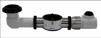
PIC COURTESY-www.seilermicro.com
The Extender and Rotation Device allows the user to change the direction of the optic pod of the microscope while keeping the binocular head in a fixed position. This allows the user to pan left and right to view different quadrants of the mouth without changing their ergonomic positioning. This allows the body of the microscope to be swung from right to left sides of the mouth ,independent of the eyepieces. This way the horizontal position of the eyepiece and subsequently the operator eyes remain unchanged and in constant position.
It gives the operator the ability to stabilize and control the patient’s head movement.2
THE ASSISTANT TEACHING TUBE
The Assistant Teaching tubes allow a second head to be attached to the scope in order for a second person to view the operating surface at the same time as the main user. The tube is attached to a beamsplitter and a binocular or monocular head is then attached.

PIC COURTESY-www.seilermicro.com
DENTAL FILTER
Prevents problem of composites curing prematurely.(orange filter)
BINOCULAR ROTATION RING
Allows for better operator positioning. Rotate the binocular +/- 25 degrees. Features a locking thumb screw
for greater stability.
DUAL IRIS DIAPHRAGM
Allows for greater depth of field - Particularly valuable for photography
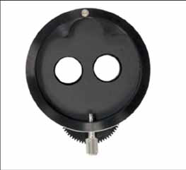
PIC COURTESY: www.seilermicro.com
BINOCULAR EYE PIECES
Additional eye pieces with different magnification configuration of 16x to 20x are also available .
OBJECTIVE LENS
Most of the microscopes come in a standard configuration of 250 to 300mm .Other objective lengths are available from 175-400mm, which can be interchanged on the scope before it is sent to the customer. (50mm=2inches) 250mm gives the user about a 10 inch working distance. 300mm= 12inches, 350mm= 14inches and so on.
PROTECTIVE LENS COVER
Protective lenses are used to keep refuse from deflecting off of the objective lens. This keeps the objective from getting cracked or scratched during surgeries.
LASER BARRIER FILTER
The laser barrier filter protects against all three different wavelengths ranging from 780-2940nm. This small compact filter has no change in visual when placed on the microscope providing superior visualization.
VIDEO AND DIGITAL CAMERA ADAPTORS
The Video and Digital Camera adapters allow Canon, Nikon, and Sony DSLR Cameras, Camcorders, and Seiler’s CCD Live Video or the new HD Live Video cameras to be mounted to any microscope. www,sieler
PIC COURTESY www.seilermicro.com
WIRELESS SHUTTER REMOTE CONTROL
This wire shutter remote control by footswitch for SLR cameras allows shutter release without touching the camera.Therefore ,problems of infection control and vibration during photography are eliminated.
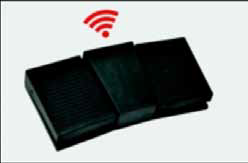
PIC COURTESY: www.zumaxmedical.com
MOBILE CAMERA ADAPTER
The mobile phone can be used to share the video and picture under the microscope at once on facebook and whatsapp on the internet. This way updating information on social media becomes easy and instant.
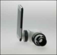
PIC COURTESY: www.zumaxmedical.com
EYE PIECE WITH INTEGRATED RETICLE
The micrometer or reticle is a round piece of glass that fits inside an eyepiece,of the operator’s dominant eye to center his or her view. Embossed or imprinted in the glass is a linear scale , used for measuring and or counting purposes.it enables the operator the possibility to take videos without the need for controlling the image section on an extern monitor. A reticle ensures the centre of field of view i.e., the object remains centred through out the procedure.
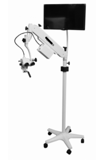
THE TV MOUNT
The TV mount allows a TV to be attached tothepole
BEAM SPLITTER (STRAIGHT & INCLINED)
The beamsplitter allows the light path to be split allowing a video camera, digital camera, or an assistant head to be attached to the microscope. The most common beamsplitter used is a 50-50 / 20-80, which means one side splits the light into 50% to the user’s eye and 50% to the digital camera. The 20-80 is 20% to the camera and light reflectence
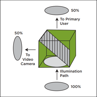
inside a 50-50 beam splitter(pic courtesy:Global surgical corporation 80% to the user’s eye.The beam splitter divides each light path in the microscope to allow digital single-lens reflex (DSLR) camera,video camera or assitant co-observation scope to be attached to the microscope.3
C MOUNT
Microscope cameras are available in several options including HD video cameras that connect to a monitor and have a high speed frame rate, tablet cameras that connect directly to the microscope, digital USB cameras that connect to a computer, and a WiFi wireless option that can stream images to up to six devices at once. The microscope digital cameras include software that allow capturing both still images and video and making measurements.4
LASER FILTER MODULE
Lasers are used in conjunction with dental microscopes to perform various surgical procedures, enabling highly precise and less invasive treatment. Dental applications range from teeth whitening to hard tissue cutting and soft tissue surgeries. The Laser Filter Module allows insertion of a Laser Filter Slide into the microscope to provide eye protection when performing laser surgeries through the microscope.
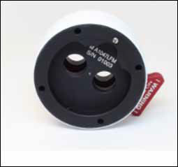
PIC COURTESY: www.sieler.com
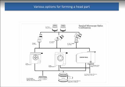
PIC COURTESY-SANMA MEDINEERS

PIC COURTESY-SANMA MEDINEERS PIC COURTESY-SANMA MEDINEERS
REFERENCES
- www.globalsurgical.com
- Enhancing Ergonomics and workflow in Micro-dentistry:OPMI pico with MORA Interface Publication for IDS 2005 by Assad F Mora ,DDS,MSD,FACP
- Best Practices in Endodontics A Desk Reference Edited by Richard S.SchwartzDDS ,Venkat Canakapalli,BDS,MDS
- www.microscopeworld.com
Disclaimer from Guident
The views and opinions expressed in articles published in Guident are solely those of the authors and do not necessarily reflect the views of the editors, the editorial board or the publisher, Ivory India. Guident does not endorse any product, technique or treatment described. Clinical content is intended for qualified dental and medical professionals and is not a substitute for individual clinical judgement. Guident, its editors and the publisher accept no liability for any consequence arising from the use of information published in the journal.
How to cite this article
Dr. Pavana Kamath. Accesories to the Dental Opearting Microscope. Guident. 2018 Oct;11(11). Available from: https://guident.in/articles/endodontics/accesories-to-the-dental-opearting-microscope.html