Occlusal Reconstruction Using Composite Resin
Victoria, Australia
Increasing numbers of people are presenting at dental offices with excessive occlusal wear and loss of vertical dimension that is both unsightly and physiologically dysfunctional. Management of these patients with indirect restorations is complicated and requires further destruction of their teeth. The use of composite resin to reconstruct these patient’s occlusions creates an ongoing stable and functional dentition and the confidence for them to smile again. Figure 1 shows a patient with excessive wear on his anterior teeth and gross loss of vertical dimension. His bite was opened on his canines and first bicuspids to generate a new vertical dimension that also alleviated his chronic TMJ pain. Following this, direct laminates were placed on his upper and lower incisors to create an attractive smile. Figure 2. Two and a half years later he presented at the office for an examination and clean. Figure 3. He then moved interstate and did not return to the office for 15 years to have a chip repaired on a central incisor. Even though there was some
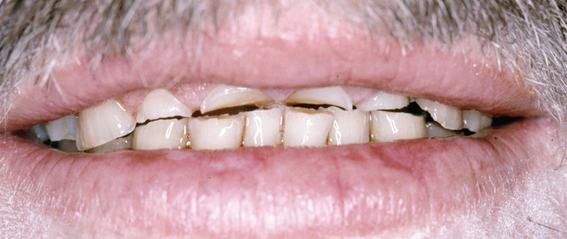
Fig 1

Fig 2
staining on the laminates (the usual culprits, cigarettes, red wine and black coffee) and some loss of his vertical dimension, his bite was still open from its original position, the lateral segments had moved into contact (apart from the lost restoration on tooth 35) and he had functioning upper and lower incisors. Figures 4 and 5

Fig 3
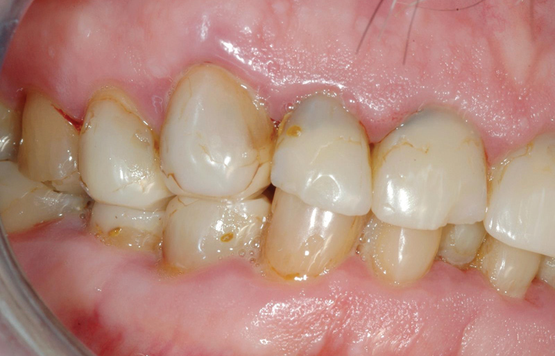
Fig 4

Fig 5
RATIONALE
The technique is founded on Okeson’s studies of the nociceptive reflex that inhibits subjects from applying excessive occlusal force to the anterior segments of their dentition. Readers can confirm this by feeling their masseter muscles contract when biting down firmly on their back teeth, compared to being unable to initiate the masseters when an object, such as a pencil, is placed between their upper and lower central incisors. In essence patients have received a fixed anterior splint. An unpublished study has shown that of 22 patients who received this treatment. Average age 54 years, average time since build up 4.4 years (range 3 to 8.5 years). Of these patients 22 % showed considerable improvements in head and neck pain (without anyone developing it) and 9 % had relief from TMJ pain (without anyone developing it). Clinical experience with this procedure for over 30 years confirms that occlusal contact occurs in the lateral segments within the next 18 months. Not by over eruption of the teeth but movements of the alveolar plates to bring the teeth back into occlusion. This is possibly due to stretching the muscles of mastication and causing them to realign the bony skeleton into equilibrium again. When the bite has been opened the mandible rotates downwards and backwards often changing an edge to edge collapsed bite into a class I open bite. For this reason it is necessary to build up the lingual aspect of the upper canines and the buccal aspects of the lower canines and first bicuspids Figure 6 (ppslide) .
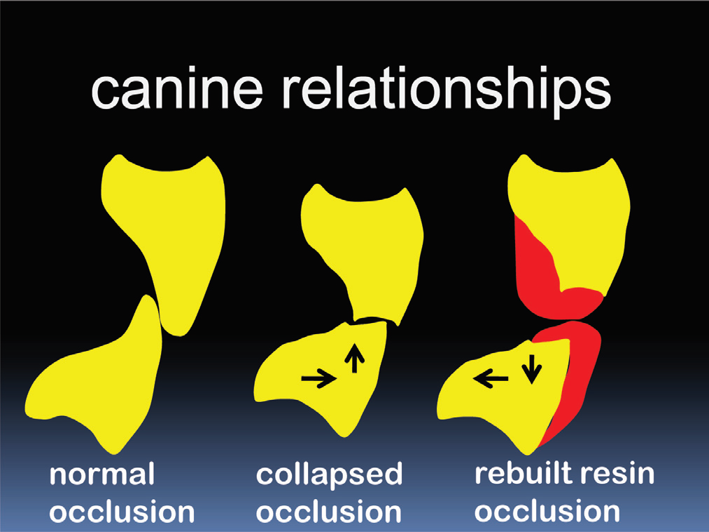
Fig 6
CLINICAL TECHNIQUE
A patient presented with a severely worn dentition and collapse of his vertical height. Figure 7. Once the occlusal problem had been diagnosed and the patient agreed to proceed with treatment, an appointment was scheduled for between 5 to 8 hours depending on the practitioner’s experience. An alternative treatment schedule (if doing this procedure for the first time) would be to give a patient 4 hours one day and 4 the following. A cheek retractor is useful to give better access to the teeth and assist patients in keeping their mouths open throughout this long procedure.
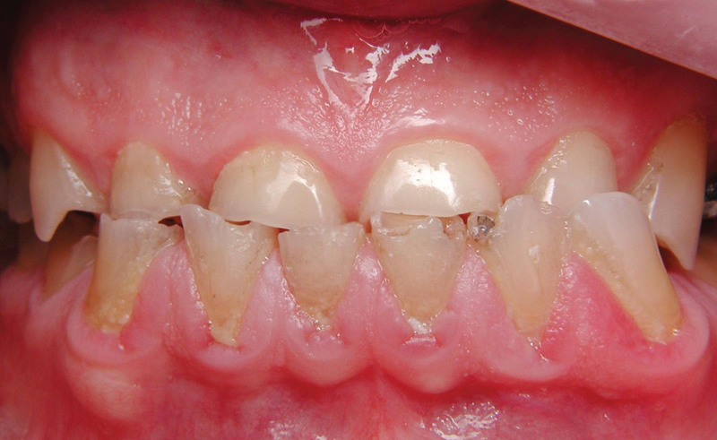
Fig 7
TOOTH PREPARATION
On the upper arch plaque and pellicle were removed with pumice and water. A slow speed diamond point was used to assure there was no remaining plaque at the gingival and interproximal margins. Caries and unsatisfactory restorations must be removed and cavities restored prior to placing the build ups. The upper teeth were then etched for 5 seconds with 35% phosphoric acid, washed with copious amounts of water and dried with oil free air.
BUILD UP PLACEMENT
A preferred bonding adhesive was applied to the upper canines and first bicuspids. Working from either the left or right hand sides; a Tofflemire matrix band was prepared so as to fit over a first bicuspid. Using a layering technique, composite resin was applied so as to slightly overfill the space between the cusps. The matrix band was then applied to the upper canine so as to extend out from the lingual aspect. Using a layering technique, composite resin was applied so as to slightly overfill this lingual area to just above the tip of the canine. Figure 6 (ppslide)This procedure was replicated again on the opposite upper arch. The canines and first bicuspids in the lower arch were next prepared after being isolated with cotton rolls. After application of a preferred bonding adhesive and prior to placement of the Tofflemire band, increments of microfill composite resin were placed freehand onto the facial margins of these teeth extending from the cervical margin to just below the contact areas of these teeth. Each increment was photo cured for 10 seconds. On a lower first bicuspid a Tofflemire matrix was placed from the lingual surface and lightly tightened. After placement of a small increment of flowable composite at the internal margin a micro hybrid resin was placed to slightly overfill the buccal space between the tooth and matrix to the cusp tip of the bicuspid. Figure 6 This process was repeated on the adjacent canine and the canine and first bicuspid in the opposite arch.
CONTOURING
Contouring commenced by reducing the composite build ups on the upper arch so as to estimate the amount of teeth that will be visible in the new smile. With the patient in a retruded occlusion, he was asked to bite together in order to estimate the location and amounts of composite removal required on the lower arch to create the new vertical dimension. Figure 8 With the aid of articulating paper, the contact surfaces were then carefully spheroid over with a fine disc so that there was an even occlusal contact on each tooth occurring at or close to the centre point of the build up. Figure 9. Spheroiding over the surfaces of the build ups prevents composite fractures at the margins and enables the lower teeth to glide smoothly over the upper teeth during protrusive and lateral movements. Figure 10.
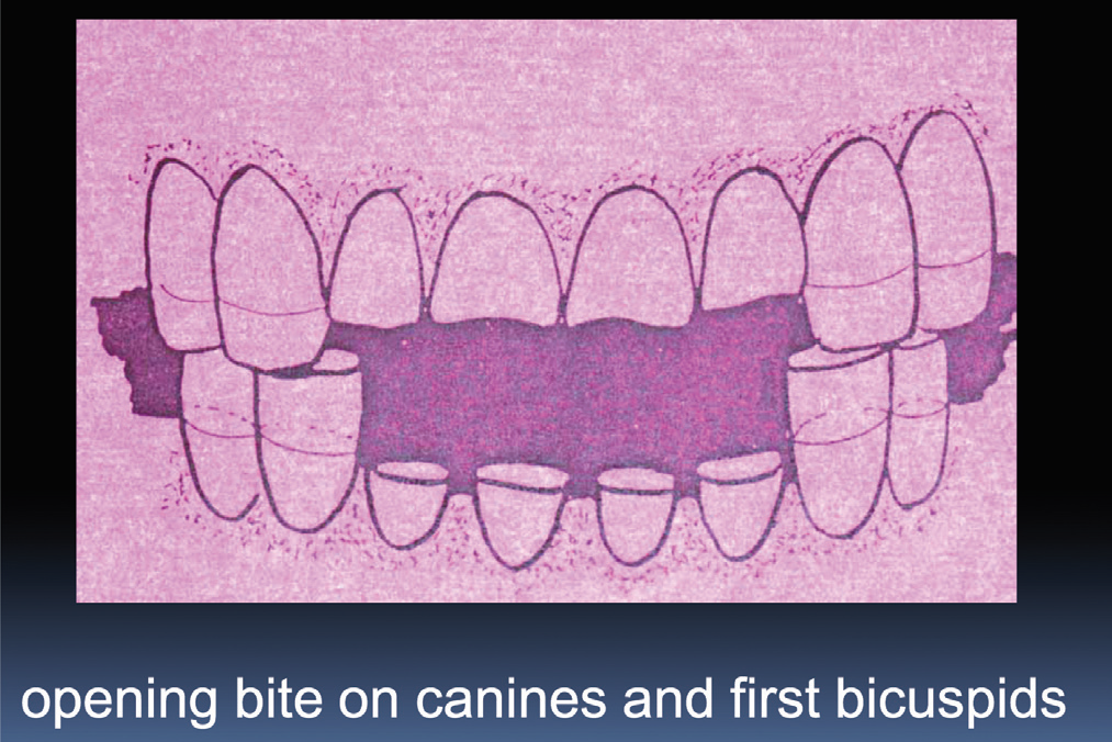
Fig 8
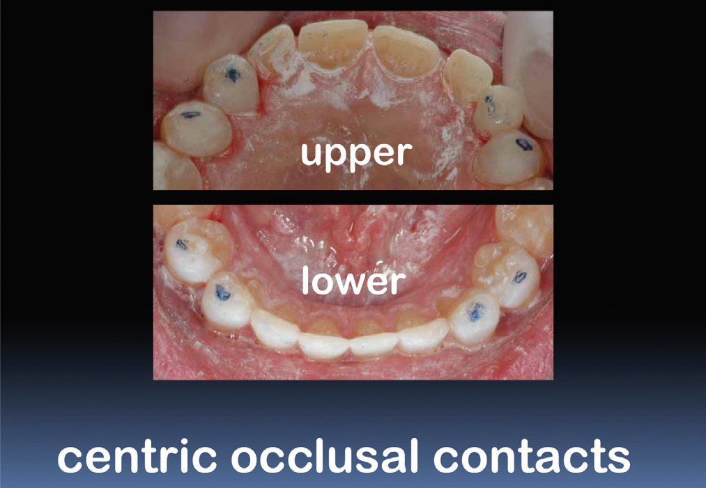
Fig 9
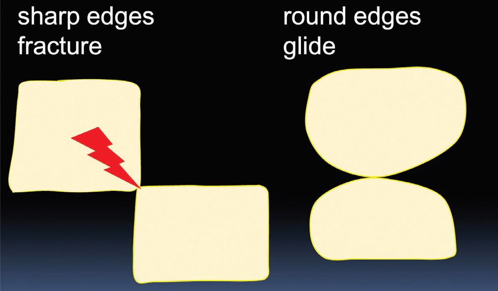
Fig 10
LAMINATE PLACEMENT
In severely worn dentitions it may be necessary to improve the bond strength of the laminates to the teeth by placing either pins or fibres as shown here. Figure 11 Composite resin laminates were then placed onto the upper arch between the canines, increasing the length of the incisal edges to the occlusion plane developed by extending the length of the canines and first bicuspids. To see the laminate technique review Aesthetic Updates May, July, November 2008 and March 09 A similar technique was used to place laminates in the lower teeth extending the plane of occlusion to fit into the new vertical dimension.
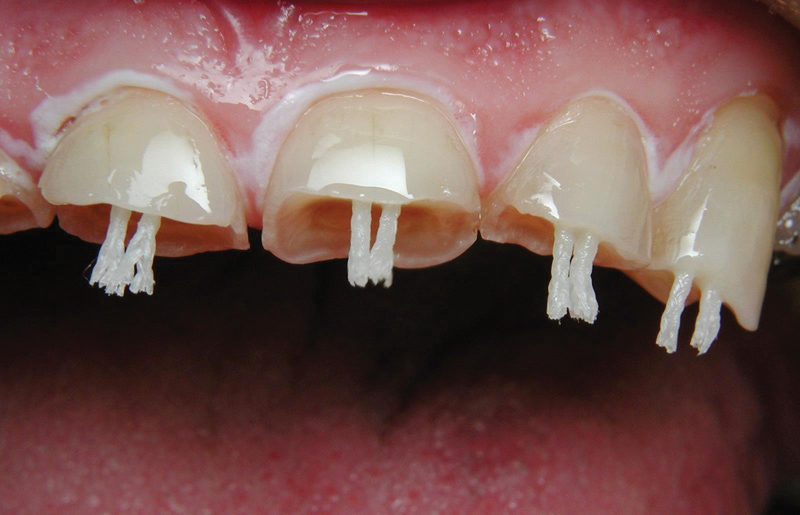
Fig 11
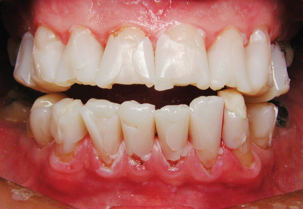
Fig 12
LAMINATE CONTOURING
Following the techniques shown in previous Aesthetic Updates the laminates were contoured and polished to fit into the occlusion. The bite must be adjusted so that the incisal edges of the lower incisors are free of the occlusion in protrusive and lateral excursions otherwise fracturing of the upper incisors may occur. This technique may result in opening a bite as much as 1 cm. As with full dentures it is essential to make sure a patient has an adequate freeway space. Figure 13.
After polishing the restorations the patient was dismissed with a 30 minute recall appointment within the next 2 weeks. Figure 14

Fig 13

Fig 14
MAINTENANCE
Over the next 2 weeks the bite has time to settle in and fine tuning of the occlusion can begin. As the composite resin continues to polymerize over this time, further polishing will improve the lustre of the finished restorations. Figure 6 Some patients may complain initially about lack of contact in their lateral segments however the author is unaware of a patient who was unable to adjust to their new occlusion. Annual maintenance visits to adjust the occlusion by spheroiding over the cusps will prevent premature contacts occurring at the edges of the build ups and reduce the chance of fracture. Furthermore it will make the bite more comfortable and provide patients with many years of improved aesthetics and function. After the lateral segments have come into the bite little ongoing maintenance will be required.
CONCLUSIONS
This relatively simple but lengthy procedure creates a fixed occlusal splint that requires minimal tooth removal and is reversible within the first month or so. Apart from improving a patient’s dental aesthetics and facial profile, this technique has been of assistance to people with chronic neck and TMJ pain. If there has been excessive wear on the upper and/or lower incisors, direct laminates may be placed on these teeth making sure that the occlusal load is maintained on the canines and first bicuspids.
DISCLAIMER
“Published in the Australian Dental Association News Bulletin November 2013 Reprinted with permission of the editor and Author.”
Disclaimer from Guident
The views and opinions expressed in articles published in Guident are solely those of the authors and do not necessarily reflect the views of the editors, the editorial board or the publisher, Ivory India. Guident does not endorse any product, technique or treatment described. Clinical content is intended for qualified dental and medical professionals and is not a substitute for individual clinical judgement. Guident, its editors and the publisher accept no liability for any consequence arising from the use of information published in the journal.
How to cite this article
Dr. Geoffrey M Knight. Occlusal Reconstruction Using Composite Resin. Guident. 2013 Dec;7(1). Available from: https://guident.in/articles/aesthetic-dentistry/occlusal-reconstruction-using-composite-resin.html