
Assessment of Skeletal Age by Using MP3 on the Same Lateral Cephalogram
Department of Orthodontics, Chandra Dental College and Hospital, Safedabad. Uttar Pradesh, India, Department of Orthodontics, BBDCODS Lucknow, Uttar Pradesh, Department of Conservative Dentistry, Chandra Dental College and Hospital, Safedabad. Uttar Pradesh, India
ABSTRACT
Knowledge of maturation status of a child plays an important role in the diagnosis, treatment planning and eventual outcome of the treatment. The main object of this clinical study is to provide a simple and practical method to assess the pubertal growth spurt stages of a subject by recording MP3 ( Middle Phalynx of Third Finger) stages on the same lateral cephalogram which is taken as a routine diagnostic aid before the initiation of any orthodontic treatment , thereby eliminating the need of taking an extra dental periapical radiograph .
KEYWORDS: MP3 Stage, Lateral cephalogram, ALARA Principal, Skeletal age.
INTRODUCTION
Radiography in orthodontics is an invaluable adjunct to the clinical examination and models of the dentition, which together form the basis for orthodontic diagnosis and treatment planning.1
Especially, a patient who reports to the orthdontist in the mixed dentition stage, understanding the skeletal maturation status of that particular individual is one of the greatest responsibilities of Functional Jaw Orthopedists and/or Orthodontists as it influences the selection and execution of treatment procedures.
Many researchers have agreed that skeletal maturity is also closely related to the craniofacial growth, and bones of hand wrist, MP3 ( Middle Phalynx of Third Finger) and cervical vertebrae are very reliable parameters in assessing it. Various methods have been used to assess the skeletal age of an individual based on the degree of maturity of different tissues of the system.2-6
The primary aim of the study was to assess the skeletal age of an individual by interpretation of the MP3 on the lateral cephalogram which is usually taken as a standard pre-treatment diagnostic record so as to eliminate the need of additional IntraOral Periapical Radiograph (IOPAR).
MATERIAL AND METHODS
The patient for the study was randomly selected from patients visiting the Department of Orthodontics and Dentofacial Orthopedics at Babu Banarsi Das College Of Dental Science, Lucknow. The patient’s criteria for the case selection was that he/she shouldnot have any history of bony deformities, bone diseases or any major illness in the past.
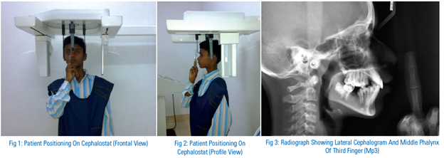
The materials used for the study was a lateral cephalogram for recording the head and neck region and Middle phalynx of the third finger (MP3) of the same patient. Planmeca proline XC machine with digital cephalostat was used to take the lateral cephalogram. The patient was made to stand in Orthoposition with the help of functional head positioners.
For recording the MP3 stage on the same radiograph, the patient’s middle finger was tied to a plastic measuring scale with the help of a cellophane tape. The patient was then asked to place the middle finger of right hand just below and approximately 5mm away from the nose with the palmar surface of the middle finger facing towards the x-ray tube. The middle finger was positioned in such a way that the x-rays are perpendicular to the film (Figure 1 and 2).
The films were exposed to 5 milliampere and 68 kilovolt power for an average of 18 seconds with a tube film distance of 6 feet ( Figure 3). All the usual protective measures for radiographic exposure were followed.
DISCUSSION
Human growth is one of the most myriad variations in nature and plays an important role in the etiology of malocclusion and also in the evaluation of diagnosis, treatment planning, retention and stability of a case.
There are wide number of growth assessment methods as suggested by many authors but the key question is reliability.6 The classical and most widely used method for skeletal-age evaluation is the highly reliable hand-wrist bone analysis performed by radiograph.
The complete use of handwrist radiograph involves 30 bones and assessment of these stages is one elaborate task which needs time and experience and also the routine use of hand-wrist radiographs has recently been questioned due to ethical issues.
Also, the present scenario in clinical orthodontics requires an accurate method with well-defined and easily identifiable stages which could be interpreted in a cross sectional study without requiring long observation periods. The method should also be non invasive, safe and economical with no elaborate armamentarium required and should be such as to be easily used in day to day orthodontic practice.
One alternatives suggested by S.M Chapman to reduce the amount of ionizing radiation is the inclusion of periapical x-ray for recording the ossification of Ulnar sesamoid bone.2 Later, Abdel –Kader used his idea successfully to record the middle finger to evaluate the middle phalanx using periapical radiograph.3 Further, many researchers MP3Rajagopal and Kansal, ,Hagg and Taranger have confirmed the use of MP3 stages as they are closely related with skeletal growth changes during puberty.5
There have been many studies to support the reliability of maturation assessment using modified MP3 stages by Kansal and Rajagopal . Their contribution replaced the need for use of a hand wrist radiographic film by a simple periapical radiograph the middle finger.
As additional radiation exposure was still the primary concern, the present method was designed with a purpose to equip the orthodontist to accurately evaluate the amount of growth remaining, using a single diagnostic tool.
In any orthodontic case a lateral cephalogram is usually taken before, during and after the commencement of treatment to assess the skeletal pattern and labial segment angulation. In the present technique, along with the routine lateral cephalgram, MP3 stage can also be recorded on the same radiograph, using single exposure. Thus, the ALARA ( as low as reasonably achievable) principle can be achieved successfully. This principal is very important in children and young adults who have proliferating cells in the crucial organs e.g. thyroid gland, bone marrow and salivary gland.3
The advantages of using the current method over the conventional are:
- It is simple and reliable.
- We can avoid taking additional IOPAR to study the skeletal maturation as it can be accomplished by using the same lateral cephalogram.
- We can avoid an additional exposure of X-rays to the patient, thus following as low as reasonably achievable (ALARA) principle.
- Ease of storage for record purpose.
- It is economical as it eliminates the additional cost of taking IOPAR.
CONCLUSION
In conclusion, we suggest that in patients considered to be treated with Functional jaw Orthopedics, MP3 maturation stages can be recorded on the same lateral cephalogram which is one of the routine diagnostic aids. Thus benefiting the patient by avoiding additional radiation exposure, reducing cost by eliminating the need of an extra IOPAR for MP3 evaluation.
REFERENCES
- Smith, N.J. : Orthodontic radiology: A Review, Int. Dent. J.37(1):16-24, 1987.
- Chapman, S.M.: Ossification of the adductor sesamoid and the adolescent growth spurt Angle Orthod. 42: 236-244,1972.
- Adbel- Kader, H.M.: The reliability of dental x-ray film in assessment of MP3 stages of the pubertal growth spurt, Am J Orthod. 114: 427-429,1998.
- Hagg U.; Taranger, J.: Maturation indicators and the pubertal growth spurt, Am. J. Orthod.82:299-309,1982.
- Rajagopal, R., Kansal, S.: A comparison of modified MP3 stages and the cervical vertebrae as growth indicators, J. Clin. Orthod.36: 398-406,2002.
- Mahajan, S. : Evaluation of skeletal maturation by comparing the hand wrist radiograph and cervical vertebrae as seen in lateral cephalogram, Indian J. Dent. Res.22:309-16,2011.
Disclaimer from Guident
The views and opinions expressed in articles published in Guident are solely those of the authors and do not necessarily reflect the views of the editors, the editorial board or the publisher, Ivory India. Guident does not endorse any product, technique or treatment described. Clinical content is intended for qualified dental and medical professionals and is not a substitute for individual clinical judgement. Guident, its editors and the publisher accept no liability for any consequence arising from the use of information published in the journal.
How to cite this article
Dr. Shally Mahajan, Dr. RP Maurya, Dr. V. K. Srivastava. Assessment of Skeletal Age by Using MP3 on the Same Lateral Cephalogram. Guident. 2013 Dec;7(1). Available from: https://guident.in/articles/endodontics/assessment-of-skeletal-age-by-using-mp3-on-the-same-lateral-cephalogram.html


