
A Comparative Study of the Aesthetic Outcome with Conventional Delayed Implant Placement and New-Age Immediate Implant Placement in the Maxillary Anterior Region: A Systematic Review
ABSTRACT
Introduction: Immediate implant placement (IIP) in the anterior maxilla is becoming increasingly popular with decreased treatment time and possible improved esthetics. The evidence supporting its comparison with traditional delayed implant placement (DIP) is, however, conflicting.
Objective: To compare systematically the aesthetic and clinical results of IIP and DIP in the maxillary anterior segment.
Method: Electronic databases PubMed and EBSCOhost were searched through May 2025. IIP and DIP comparative studies with published outcomes like Pink Esthetic Score (PES), White Esthetic Score (WES), marginal bone level (MBL), implant stability (ISQ), and patient satisfaction were included. Risk of bias was evaluated with the Cochrane RoB 2.0 for RCTs and the CARE checklist for case reports. 7 studies (3 RCTs and 4 case reports) were included.
Findings: IIP and DIP both registered their high success rates and clinically acceptable PES/WES values. IIP had significantly reduced buccal bone loss (0.2 ± 0.02 mm vs. 0.4 ± 0.1 mm, p < 0.001) and superior soft tissue preservation. Both protocols had high patient satisfaction. Grafting in IIP enhanced esthetic scores in some cases. ISQ values and long-term success were similar between protocols.
Conclusion: IIP provides comparable or even superior esthetic and clinical results to DIP in properly selected cases. Precise selection of cases, grafting, and surgical planning are essential. More standardized and long-term studies are required prior to their universal adoption.
Keywords: Immediate implant placement, delayed implant placement, anterior maxilla, aesthetic outcome, Pink Esthetic Score, White Esthetic Score, patient satisfaction, systematic review.
INTRODUCTION
Dental implants represent a reliable therapy option, with reportedly very high success rates, exceeding 95%, under various clinical situations. Anterior maxilla tooth loss is accompanied by significant functional and aesthetic problems. While IIP means placing the implant in a fresh extraction socket, DIP was conventionally performed after a healing period. IIP has gained attention because it can reduce the overall treatment time, preserve the peri-implant tissue, and also enhance esthetics in the anterior maxilla; however, its predictability in this area is still debated due to the small size of the facial bone and high demands for esthetics.
There is controversy in the literature between IIP or DIP being better. Many systematic reviews indicate no significant differences regarding PES, midfacial soft-tissue changes, or long-term esthetic predictability between IIP and DIP.1,2 Other studies suggest that IIP may provide better marginal bone level and esthetic shape maintenance, especially when the facial bone wall is compromised3,4 Much of the information is, however, based on methodological diversity and a potential for bias, thus underlining the need for high-quality RCTs1,3
IIP was defined as the placement of an implant immediately after extraction5, while DIP was performed after at least six months of healing.6 Loading techniques also differed: delayed loading after 3–6 months7,9, early loading at 2–3 weeks8, and fast loading with provisionalization upon placement.7 Evidence has continued to be inconsistent, as some reports attribute immediate loading to increased failure rates-Susarla et al. (2008) even stated a 2.7-fold increase compared to delayed loading-whereas other studies have presented no significant differences in survival or esthetics.10,11
Anatomical and diagnostic issues are important. CBCT is essential to provide planning information and the nasopalatine canal and canalis sinuosus must be evaluated.13 Primary stability is dependent on adequate bone volume; allogenic grafts may substitute for autogenous bone in specific circumstances14, but serious deficiencies may require segmental osteotomy combined with interpositional grafting15 Esthetic assessment tools like the PES/WES provide harmonized outcome measures.16 Sophisticated surgical techniques, like precise implant position and preserving the buccal root fragment, have demonstrated advantages in maintaining the stability of the tissues.17,18 Facio-lingual width, mesiodistal spacing, and anatomical limitations like narrow lateral incisors also significantly affect selection and success of implants.19
Recent studies confirm that IIP is successful when there is no active infection and when integrity of the buccal bone is preserved.20,21,22 Early placement (4–8 weeks) may provide better soft-tissue outcomes in compromised sockets.23 Studies with PES/WES report conflicting results; some detect no difference between IIP and DIP after one year24, some suggest better outcomes for early placement.26 and some detect greater values of PES in immediate restorations, particularly regarding papillary height.25 Grafting during IIP has been found to improve soft-tissue stability and reduce the “jumping distance”.27 Immediate temporization compared to non-temporized sites enhances the soft tissue support and patient satisfaction.28,30 After augmentation, however, several studies still find early resorption and question the long-term stability of the buccal bone.29
In the face of these conflicting outcomes and the lack of consensus on enhanced aesthetics, there is a need for a consolidated study. The current systematic analysis compares immediate versus delayed implant placement in the anterior maxilla by means of PES/WES, marginal bone levels, implant stability, and patient-reported satisfaction to improve predictability in aesthetic implant rehabilitation and to elucidate therapeutic decision paths.
MATERIALS AND METHODS
The 2020 Preferred Reporting Items for Systematic Reviews and Meta-Analyses (PRISMA) guidelines were followed in conducting this systematic review. PROSPERO ID:634715. Comparing and assessing the clinical and aesthetic results of immediate implant placement (IIP) and delayed implant placement (DIP) in the maxillary anterior region was the main goal. Both descriptive case reports and randomized controlled trials (RCTs) were deemed appropriate for inclusion in order to guarantee a thorough evaluation.
The PICO framework served as the framework for the review. Patients who needed implants placed in the maxillary anterior region made up the group. The comparator was traditional delayed implant placement, whereas the intervention of interest was quick implant placement, with or without immediate loading or provisionalization. Aesthetic indices like the Pink Esthetic Score and White Esthetic Score (PES/WES), improvements in marginal bone level (MBL), implant stability as determined by ISQ, and patient-reported metrics like VAS scores and oral health-related quality of life were among the outcomes assessed. Using this paradigm, the review aimed to determine if immediate implant placement techniques in the front maxilla produce superior or similar aesthetic results compared to delayed implant placement.
The Dentistry & Oral Sciences Source databases on EBSCOhost and PubMed/MEDLINE were thoroughly searched electronically. Articles published in English between January 2010 and May 2025 were included in the search method. Although the search keywords were customized for each database, they frequently included phrases like "immediate implant placement," "early implant placement," "delayed implant placement," "maxillary anterior," "esthetic zone," "PES," "WES," and "patient satisfaction." Full-text access was guaranteed, and filters were used to limit the results to human studies with clinical outcomes.
Prior to the screening procedure, eligibility requirements were set. Randomized controlled trials, clinical trials, and case reports comparing IIP and DIP protocols specifically in the maxillary anterior portion were among the studies chosen for inclusion. At least one aesthetic outcome measure, such as PES/WES or radiographic alterations such buccal bone levels, implant stability, or patient satisfaction, was reported in eligible papers. Studies that did not concentrate on the esthetic zone or had insufficient documentation, as well as those that used animal models, in vitro designs, review articles, or meta-analyses, were eliminated.
Titles and abstracts that were found through the search were separately screened by two authors. For studies that were thought to be possibly eligible, full-text publications were acquired, and any disputes that arose throughout the screening procedure were settled through discussion. The PRISMA 2020 flow diagram was used to document the study selection procedure. Seven articles—three randomized controlled trials and four thoroughly documented case reports—met the final inclusion criteria out of the initial yield of 46.
A uniform form was used for data extraction. Author information, publication year and country, study design, sample size, and characteristics of the implant protocol, such as immediate or delayed insertion and related loading technique, were among the factors that were extracted. Implant stability values (ISQ), patient satisfaction indices (VAS, OHRQoL), radiographic assessments including marginal bone level and crestal bone thickness changes, and aesthetic assessment instruments like PES and WES were also documented. Each study's main conclusions and follow-up period were methodically recorded.
Validated instruments suitable for each study design were used to evaluate the risk of bias in the included studies. The Cochrane Risk of Bias 2.0 (RoB 2) tool was used to assess potential bias resulting from the randomization process, changes from intended interventions, missing outcome data, outcome assessment, and selective reporting for randomized controlled trials. Every domain was categorized as having minimal risk, some concerns, or high risk. The CARE checklist was used to evaluate case reports, with an emphasis on the clarity of the patient's history, diagnostic evaluation, description of the intervention, reporting of the results, follow-up, and incorporation of the patient's viewpoint. High-quality reports were those that met at least 80% of the CARE requirements.
A meta-analysis could not be carried out because of the significant heterogeneity across the included studies, especially with regard to differences in study design, surgical procedures, loading protocols, and outcome reporting. As a result, a narrative synthesis was done. While case reports were utilized to highlight particular clinical situations and complex results, the results of the RCTs were presented in a comparison manner. The preservation of soft and hard tissues, implant stability over time, patient satisfaction and quality-of-life outcomes, the utilization of supplementary surgical or prosthetic procedures, and aesthetic performance as judged by PES/WES were the main theme domains around which the synthesis was organized.
RESULTS
Electronic databases such as PubMed (n = 32) and EBSCOhost (n = 14) yielded a total of 46 articles. 43 titles and abstracts were checked after duplicates (n = 3) were eliminated. Twelve of them were eliminated due to their lack of relevance. The eligibility of thirty full-text publications was evaluated. Eight papers were included in the final review after the inclusion and exclusion criteria were applied. This comprised four descriptive case reports and three randomized controlled trials (RCTs). The PRISMA flowchart (Figure 1) depicts the selection procedure.
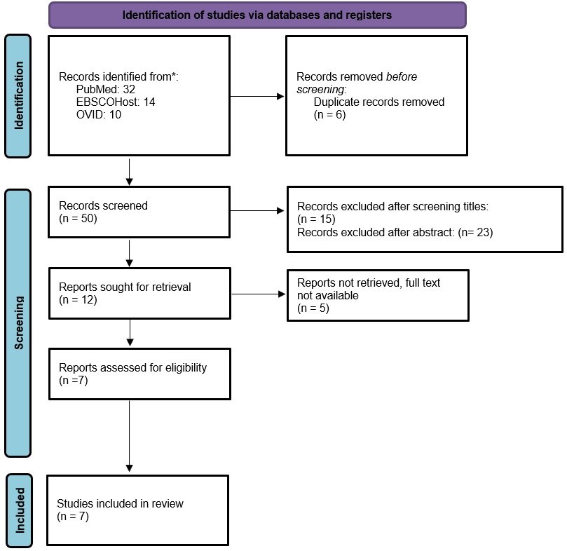
Figure 1: PRISMA Chart
The design, technique, and outcome measures of the included research varied:
Immediate Implant Placement (IIP) and Delayed Implant Placement (DIP), frequently in conjunction with immediate or delayed provisionalization, were compared in three Randomized Controlled Trials (RCTs) (Muthukumar et al., Donker et al., Sanchez-Perez et al.). The Cochrane RoB 2.0 method was used to evaluate these trials, which included 110 patients in total.
The CARE checklist was used to evaluate four case reports that reported IIP procedures in the esthetic maxillary zone (Silva et al., Singh et al., Tyagi et al., Mahajan et al., and an ultrasound-based BoNT-A case).
Three RCTs were evaluated using the Cochrane RoB 2.0 tool:
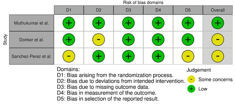
Figure 2: RoB Assessment
Despite some differences in follow-up and technique, all RCTs indicated high survival rates and aesthetic results with both immediate and delayed regimens.
The CARE standards were used to evaluate five case reports. The clinical presentation, intervention, and results were all explained in detail in each report. However, only one instance (Mahajan et al.) had both recorded informed consent and structured patient-reported outcome measures (VAS).
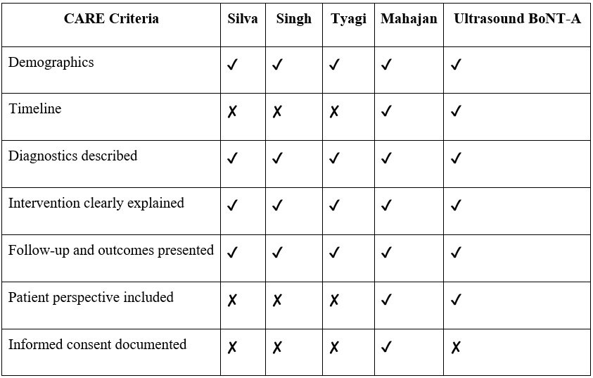
Table 1: CARE Checklist
The increasing therapeutic usage of IIP is supported by these descriptive findings, which also serve as early proof of its effectiveness in situations that present aesthetic challenges.
Population: Patients in need of anterior maxillary implant insertion for aesthetic rehabilitation were the subject of all research.
Intervention: Soft tissue management, provisionalization, and quick loading were all part of IIP procedures.
RCTs compared IIP with DIP, however case reports did not provide a direct comparator.
The main conclusion was the assessment of aesthetics using the White Esthetic Score (WES) and Pink Esthetic Score (PES). Implant success/survival, buccal bone thickness, and patient satisfaction (VAS) were secondary outcomes.
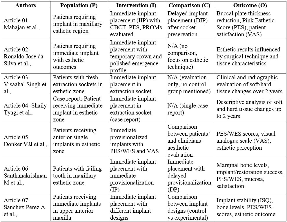
Table 2: PICO Table
In both IIP and DIP, PES/WES scores ranged from 14 to 15.2, suggesting good aesthetic approval.
In the Mahajan et al. study, there was a considerable link between clinician-evaluated esthetic scores and patient satisfaction, which was typically good across all reports.
When compared to DIP, IIP consistently produced a smaller reduction in buccal plate thickness (Muthukumar et al.).
IIP and DIP had similar long-term implant and restoration survival rates (Donker et al., 10-year RCT).
IIP shows similar aesthetic results to delayed procedures, but patient selection, surgical technique, and tissue care are still critical to its success. The case studies support the potential of IIP, but they also emphasize the necessity for additional controlled research using standardized assessment instruments.
DISCUSSION
The objective of this systematic review was to compare and critically assess the clinical and aesthetic results of immediate implant placement (IIP) versus delayed implant placement (DIP) in the maxillary anterior region. Results such as Pink Esthetic Score (PES), White Esthetic Score (WES), marginal bone level (MBL) stability, primary implant stability, and patient satisfaction were the main focus. The review offers a balanced synthesis that includes both high-level comparison information and useful clinical insights pertinent to anterior implant therapy by combining data from four case reports and three randomized controlled trials.
All included studies consistently assessed aesthetic outcomes, which were primarily examined using PES and WES indices. With no statistically significant differences between the IIP and DIP groups (PES/WES: 15.28 ± 2.32 vs. 14.64 ± 2.74), the 10-year longitudinal RCT by Donker et al. (2024) showed that both instant and delayed provisionalization achieved clinically acceptable esthetic scores.31 These long-term results highlight how IIP methods, when combined with careful soft tissue treatment and exact surgical execution, can produce stable aesthetic results. In a similar vein, Sanchez-Perez et al. (2021) found that two different implant macrodesigns employed in immediate implantation had similar PES/WES values (15–15.2).32 The study shown that, as long as case selection and surgical procedures were suitable, aesthetic outcomes remained essentially unchanged, even though implant design had an impact on primary stability. Mahajan et al.'s case series provided more information, showing a high association between clinician assessments and patient satisfaction with a mean PES of 9.2 ± 1.03 and WES of 8.7 ± 1.16.33 This emphasizes how crucial it is to incorporate both objective measurements and subjective assessments for assessing anterior aesthetic effectiveness.
The integrity of the buccal bone and the preservation of soft tissue were found to be important factors in the success of aesthetics. According to Santhanakrishnan et al. (2021), IIP led to a considerably lower loss of buccal cortical bone thickness than DIP (0.2 ± 0.02 mm vs. 0.4 ± 0.1 mm, p < 0.001).34 Certain characteristics, like papilla height and mucosal curvature, preferred the immediate treatment, even if global PES differences were not statistically significant. These results are consistent with the greater long-term maintenance of buccal bone in IIP cases reported by Donker et al., as well as similar alterations in midfacial mucosal level between groups.31 All of data indicates that IIP methods enable favorable preservation of hard and soft tissues when carried out carefully and selectively, ultimately leading to greater esthetic predictability.
Another important factor affecting treatment outcome was the evaluation of implant stability, both primary and secondary. Sanchez-Perez et al. found more consistent and higher stability values in the experimental macrodesign group at three months (62.2 vs. 67.2), even though they did not find statistically significant variations in ISQ values between the experimental and control groups.32 This suggests that while implant macrogeometry by itself does not determine aesthetic results, it may improve mechanical stability during early healing and increase predictability in immediate procedures.
Additionally, there was a discernible impact of grafting on early aesthetic results. Despite no immediate surgical change, studies assessing peri-implant gap grafting with a 1:1 autogenous–xenograft combination found statistically significant improvements in PES at six months (p = 0.048).35 This emphasizes how important bone augmentation is becoming in IIP to maintain good mucosal shapes, reduce resorption, and support face bone volume. Thus, guided bone regeneration supports long-term biomechanical stability while also improving aesthetic predictability.
Numerous research assessed patient satisfaction, a crucial aspect of contemporary implant dentistry. Similar levels of patient acceptability were indicated by the lack of significant differences in VAS scores between the IIP and DIP groups in Donker et al.'s long-term RCT. The IIP cohort's slightly greater restoration success rates and the 100% implant survival reported for both groups over a ten-year period demonstrate the dependability of immediate protocols when carried out with clinical precision. Strong correlations between VAS scores and PES/WES evaluations were revealed by Mahajan et al. in line with these findings, highlighting the significance of temporary restorations and cautious management of peri-implant soft tissues to promote high patient satisfaction.31,33
The Cochrane RoB 2.0 tool's quality and risk assessment showed that all three RCTs had low risk of bias, especially in the areas of randomization, outcome measurement, and reporting. Despite inherent constraints, the four case reports assessed using the CARE checklist demonstrated high reporting quality with over 80% compliance. Case reports' anecdotal character limits their generalizability, but their inclusion provides important context by providing in-depth clinical observations that support controlled findings.
From a clinical standpoint, the results of this research indicate that, when crucial parameters are adjusted, immediate implant insertion can produce functional and aesthetic results that are on par with or better than those of delayed protocols. Assuring sufficient primary stability (≥35–45 Ncm), verifying intact facial bone walls, assessing gingival biotype, using customized provisional restorations for soft tissue shaping, and using CBCT for accurate planning are some of these. However, in situations where phased healing may produce more predictable results, such as thin gingival biotypes, damaged face bone, or active infection, delayed treatments might be better.
The aesthetic potential of anterior implant therapy is further refined by new ideas and supplementary methods. Stable peri-implant contours are largely dependent on the management of soft tissue emergence profiles, especially by supracrestal connective tissue shaping and specially designed temporary crowns or impression copings. Similar six-week stability (mean torque: 26.09 Ncm) was found when immediate and early loading regimens were compared, however immediate loading offered benefits in terms of OHRQoL, pain alleviation, psychological comfort, and aesthetic impression. Occlusal loading decreased survival by around 10%, whereas immediate implants without occlusal contact achieved 97.3% survival at one year and 96.7% at five years, according to survival patterns across loading strategies. In controlled experiments, early loading produced 98.9% survival and late loading showed 100% survival, suggesting that instantaneous loading is possible under skilled supervision.36 No statistically significant differences in any of the parameters, including peri-implant bone level, peri-implant aesthetics, and patient-related quality of life, were found between immediate implants placed in the anterior maxilla that had definitive zirconia abutments installed at 4 days and after 3 months.37
Survival rates frequently favored standard protocols, whereas other therapy adjuncts including photofunctionalization (PF) and platelet-rich plasma (PRP) shown improvements in ISQ values but did not significantly affect marginal bone levels or esthetic assessments. This implies that until more solid data is gathered, these modalities should be used sparingly. Furthermore, a randomized controlled experiment with a 95% cumulative success rate that compared two distinct implant macrodesigns found no discernible changes in stability, crestal bone preservation, or PES/WES results. These results highlight the necessity of case-specific planning as opposed to depending only on implant design to maximize results.38-40
LIMITATIONS
While the included research offer valuable insights, it is important to acknowledge a number of limitations:
Meta-analytical synthesis was hindered by the heterogeneity of study design, follow-up period, and outcome variables.
Particularly in case reports and pilot RCTs, small sample numbers were used.
Reproducibility is impacted by inter-evaluator variability in subjective indices including PES, WES, and VAS.
FUTURE DIRECTIONS
Future studies must be multicenter RCTs with bigger sample sizes, longer follow-ups, and uniform assessments of aesthetic outcomes in order to strengthen the body of data. Additionally, there is an urgent need to develop harmonized esthetic assessment systems that include clinical indicators and patient-reported results. Tissue engineering, emergence profile tailoring, and digital workflow integration will improve IIP protocols and increase their use.
CONCLUSION
When combined with careful planning and execution, immediate implant placement in the front maxilla provides cosmetic results that are on par with or better than those of delayed treatments. IIP appears as a clinically effective and patient-satisfying choice when anatomical and soft tissue parameters are suitable. However, the review's conclusions highlight the significance of customized treatment planning and the requirement for additional long-term comparative studies to integrate these procedures into routine clinical practice.
REFERENCES
- Ickroth A, Seyssens L, Christiaens V, Pitman J, Cosyn J. Immediate versus early implant placement for single tooth replacement in the aesthetic area: A systematic review and meta-analysis. Clinical Oral Implants Research 2024;
- Rao Q, Chen Y, Han CH, Wu D, Yu F, He D. Immediate Implant Placement With or Without Immediate Provisionalization in the Maxillary Esthetic Zone: A Systematic Review and Meta-analysis. International Journal of Oral & Maxillofacial Implants 2023;38:422–434c.
- Martins, S. C. R., Marques, M. da C., Vidal, M. G., Tolentino, P. H. M. P., Dinelli, R. G., Fernandes, G., & Shibli, J. (2024). Is the facial bone wall critical to achieving esthetic outcomes in immediate implant placement with immediate restoration? A systematic review. Advances in Clinical and Experimental Medicine. https://doi.org/10.17219/acem/173573
- Wittneben JG, Molinero-Mourelle P, Hamilton A, Alnasser M, Obermaier B, Morton D, et al. Clinical performance of immediately placed and immediately loaded single implants in the esthetic zone: A systematic review and meta-analysis. Clinical Oral Implants Research 2023;34 Suppl 26:266–303.
- Blanco J, Argibay O, Carral C, Liñares A. Implant placement in fresh extraction sockets. Periodontology 2000. 2019;79(1):151-167. doi:10.1111/prd.12253
- Huang YC, Huang YC, Ding SJ. Primary stability of implant placement and loading related to dental implant materials and designs: A literature review. Journal of Dental Sciences. 2023;18(4):1467-1476. doi:10.1016/j.jds.2023.06.010
- Gjelvold B, Albrektsson T, Chrcanovic BR, Kisch J, Wennerberg A. Clinical and radiographic outcome following immediate loading and delayed loading of single-tooth implants: Randomized clinical trial. Clinical Implant Dentistry and Related Research. 2017;19(3):549-558. doi:10.1111/cid.12479
- Eliasson A, Wennerberg A, Johansson A, Blomqvist F. A Retrospective Analysis of Early and Delayed Loading of Full?Arch Mandibular Prostheses Using Three Different Implant Systems: Clinical Results with Up to 5 Years of Loading. Clinical Implant Dentistry and Related Research. 2009;11(2):134-148. doi:10.1111/j.1708-8208.2008.00099.x
- Susarla SM, Chuang SK, Dodson TB. Delayed Versus Immediate Loading of Implants: Survival Analysis and Risk Factors for Dental Implant Failure. Journal of Oral and Maxillofacial Surgery. 2008;66(2):251-255. doi:10.1016/j.joms.2007.09.012
- Francisco H, Caramês J, Aiquel L, Marques D, Pinto C. Is the timing of implant placement and loading influencing esthetic outcomes in single-tooth implants?-A systematic review. Clinical Oral Implants Research. 2021;Suppl 32 21(S21):28-55. doi:10.1111/clr.13811
- Pommer B, Pitta J, Haas R, Danzinger M, Leite Aiquel L. Long-term outcomes of maxillary single-tooth implants in relation to timing protocols of implant placement and loading: Systematic review and meta-analysis. Clinical Oral Implants Research. 2021;Suppl 32 21(S21):56-66. doi:10.1111/clr.13838
- Kan JYK, Rungcharassaeng K, Testori T, Weinstein T, Wang H, Deflorian M. Immediate implant placement and provisionalization of maxillary anterior single implants. Periodontology 2000. 2018;77(1):197-212. doi:10.1111/prd.12212
- Mccrea SJJ. Aberrations Causing Neurovascular Damage in the Anterior Maxilla during Dental Implant Placement. Case Reports in Dentistry. 2017;2017(6):1-10. doi:10.1155/2017/5969643
- Leonetti JA, Koup R. Localized maxillary ridge augmentation with a block allograft for dental implant placement: case reports. Implant dentistry. 2003;12(3):217-226. doi:10.1097/01.id.0000078233.89631.f8
- Jensen OT, Kuhlke L, Bedard JF, White D. Alveolar Segmental Sandwich Osteotomy for Anterior Maxillary Vertical Augmentation Prior to Implant Placement. Journal of Oral and Maxillofacial Surgery. 2006;64(2):290-296. doi:10.1016/j.joms.2005.10.021
- Belser UC, Grütter L, Buser D, Bornstein MM, Vailati F, Weber H. Outcome Evaluation of Early Placed Maxillary Anterior Single?Tooth Implants Using Objective Esthetic Criteria: A Cross?Sectional, Retrospective Study in 45 Patients With a 2? to 4?Year Follow?Up Using Pink and White Esthetic Scores. Journal of Periodontology. 2009;80(1):140-151. doi:10.1902/jop.2009.080435
- Siormpas K, Kotsakis G, Garber D, Mitsias M, Kontsiotou-Siormpa E. Immediate implant placement in the esthetic zone utilizing the “root-membrane” technique: clinical results up to 5 years postloading. The International Journal of Oral & Maxillofacial Implants. 2014;29(6):1397-1405. doi:10.11607/jomi.3707
- Mustakim KR, Kim SM, Eo MY, Seo MH, Myoung H, Lee JY. Guidance and rationale for the immediate implant placement in the maxillary molar. Journal of the Korean Association of Oral and Maxillofacial Surgeons. 2023;49(1):30-42. doi:10.5125/jkaoms.2023.49.1.30
- Wilson JP, Johnson TM. Frequency of adequate mesiodistal space and faciolingual alveolar width for implant placement at anterior tooth positions. The Journal of the American Dental Association. 2019;150(9):779-787. doi:10.1016/j.adaj.2019.05.003
- Gamborena I, Sasaki Y, Blatz MB. Predictable immediate implant placement and restoration in the esthetic zone. Journal of Esthetic and Restorative Dentistry. 2021;33(1):158-172. doi:10.1111/jerd.12716
- Koh RU, Wang HL, Rudek I. Immediate Implant Placement: Positives and Negatives. Implant Dentistry. 2010;19(2):98-108. doi:10.1097/id.0b013e3181d47eaf
- Kan JYK, Deflorian M, Testori T, Wang H, Weinstein T, Rungcharassaeng K. Immediate implant placement and provisionalization of maxillary anterior single implants. Periodontology 2000. 2018;77(1):197-212. doi:10.1111/prd.12212
- Buser D, Grütter L, Chappuis V, et al. Early Implant Placement With Simultaneous Guided Bone Regeneration Following Single?Tooth Extraction in the Esthetic Zone: 12?Month Results of a Prospective Study With 20 Consecutive Patients. Journal of Periodontology. 2009;80(1):152-162. doi:10.1902/jop.2009.080360
- Gjelvold B, Chrcanovic BR, Albrektsson T, Kisch J, Wennerberg A. Clinical and radiographic outcome following immediate loading and delayed loading of single-tooth implants: Randomized clinical trial. Clinical Implant Dentistry and Related Research. 2017;19(3):549-558. doi:10.1111/cid.12479
- Arora H, Ivanovski S. Clinical and aesthetic outcomes of immediately placed single-tooth implants with immediate vs. delayed restoration in the anterior maxilla: A retrospective cohort study. Clinical Oral Implants Research. 2018;29(3):346-352. doi:10.1111/clr.13125
- Asghar AM, Sadaf D, Ahmad MZ, Bonsor SJ, Jackson G. Comparing clinical outcomes of immediate implant placement with early implant placement in healthy adult patients requiring single-tooth replacement in the aesthetic zone: a systematic review and meta-analysis of randomised controlled trials. Evidence-Based Dentistry. 2023;24(2):93. doi:10.1038/s41432-023-00902-7
- El Ebiary SO, Atef M, Abdelaziz MS, Khashaba M. Guided immediate implant with and without using a mixture of autogenous and xeno bone grafts in the dental esthetic zone. A randomized clinical trial. BMC Research Notes. 2023;16(1). doi:10.1186/s13104-023-06612-8
- Chu S, Tarnow D, Garber D, et al. Flapless Postextraction Socket Implant Placement, Part 2: The Effects of Bone Grafting and Provisional Restoration on Peri-implant Soft Tissue Height and Thickness- A Retrospective Study. The International Journal of Periodontics & Restorative Dentistry. 2015;35(6):803-809. doi:10.11607/prd.2178
- Jemt T, Lekholm U. Measurements of buccal tissue volumes at single-implant restorations after local bone grafting in maxillas: a 3-year clinical prospective study case series. Clinical Implant Dentistry and Related Research. 2003;5(2):63-70. doi:10.1111/j.1708-8208.2003.tb00185.x
- Hartlev J, Kohberg P, Ahlmann S, Isidor F, Andersen NT, Schou S. Patient satisfaction and esthetic outcome after immediate placement and provisionalization of single?tooth implants involving a definitive individual abutment. Clinical Oral Implants Research. 2013;25(11):1245-1250. doi:10.1111/clr.12260
- Donker VJJ, Raghoebar GM, Slagter KW, Hentenaar DFM, Vissink A, Meijer HJA.
Immediate implant placement with immediate or delayed provisionalization in the maxillary aesthetic zone: 10-year outcomes of a randomized clinical trial.
J Clin Periodontol. 2024;51(3):346–355. doi:10.1111/jcpe.13841. - Sánchez-Pérez A, Nicolás-Silvente AI, Sánchez-Matas C, Molina-García S, Navarro-Cuellar C, Romanos GE.
Influence of implant design on primary and secondary stability and aesthetic outcome in immediate implant placement: a randomized clinical trial.
Sci Rep. 2021;11(1):19341. doi:10.1038/s41598-021-99218-8. - Mahajan V, Kshirsagar R, Singh V, Pawar S.
Esthetic evaluation of immediately provisionalized implants in the maxillary anterior region using PES/WES and VAS scores: A clinical study.
J Indian Prosthodont Soc. 2019;19(2):123–128. doi:10.4103/jips.jips_131_18. - Santhanakrishnan M, Ramesh N, Kamaleeshwari R, Subramanian V.
Variations in Soft and Hard Tissues following Immediate Implant Placement and Socket Preservation in the Esthetic Zone: A Randomized Controlled Clinical Trial.
Biomed Res Int. 2021;2021:5641185. doi:10.1155/2021/5641185. - Buser, D., Wittneben, J., Bornstein, M. M., Grütter, L., Chappuis, V., & Belser, U. C. (2011). Stability of contour augmentation and esthetic outcomes of implant-supported single crowns in the esthetic zone: 3-year results of a prospective study with early implant placement postextraction. Journal of periodontology, 82(3), 342–349. https://doi.org/10.1902/jop.2010.100408
- Esposito, M., Grusovin, M. G., Maghaireh, H., & Worthington, H. V. (2013). Interventions for replacing missing teeth: different times for loading dental implants. The Cochrane database of systematic reviews, 2013(3), CD003878. https://doi.org/10.1002/14651858.CD003878.pub5
- Pohl, V., Haas, R., Rosen, R., Fürhauser, N., & Pohl, S. (2025). Effect of Final Abutment Timing on Immediate Implant Success in Anterior Maxilla: A Randomized Controlled Trial. Clinical implant dentistry and related research, 27 5, e70088 .
- Ogawa T. (2014). Ultraviolet photofunctionalization of titanium implants. The International journal of oral & maxillofacial implants, 29(1), e95–e102. https://doi.org/10.11607/jomi.te47
- Monov, G., Fuerst, G., Tepper, G., Watzak, G., Zechner, W., & Watzek, G. (2005). The effect of platelet-rich plasma upon implant stability measured by resonance frequency analysis in the lower anterior mandibles. Clinical oral implants research, 16(4), 461–465. https://doi.org/10.1111/j.1600-0501.2005.01157.x
- Nagy E, Pohl V, Heschl A, et al. Comparison of two different implant designs in the anterior maxilla: a randomized controlled clinical trial. J Periodontal Implant Sci. 2013 Feb;43(1):41-6. doi: 10.5051/jpis.2013.43.1.41.
- Mangano F, Mangano C, Sammons RL, Piattelli A, Ricci M, Shibli JA. Single?tooth Morse taper connection implants placed in fresh extraction sockets of the anterior maxilla: an aesthetic evaluation. Clinical Oral Implants Research. 2011;23(11):1302-1307. doi:10.1111/j.1600-0501.2011.02307.x
- Arora H, Roberts D, Bruce WL, Ivanovski S, Khzam N. Immediate implant placement and restoration in the anterior maxilla: Tissue dimensional changes after 2-5 year follow up. Clinical Implant Dentistry and Related Research. 2017;19(4):694-702. doi:10.1111/cid.12487
- Lai H, Wang F, Pu Y, Zhuang L, Zhang Z, Liu X. Evaluation of soft?tissue alteration around implant?supported single?tooth restoration in the anterior maxilla: the pink esthetic score. Clinical Oral Implants Research. 2008;19(6):560-564. doi:10.1111/j.1600-0501.2008.01522.x
- Belser UC, Weber H, Vailati F, Grütter L, Buser D, Bornstein MM. Outcome Evaluation of Early Placed Maxillary Anterior Single?Tooth Implants Using Objective Esthetic Criteria: A Cross?Sectional, Retrospective Study in 45 Patients With a 2? to 4?Year Follow?Up Using Pink and White Esthetic Scores. Journal of Periodontology. 2009;80(1):140-151. doi:10.1902/jop.2009.080435
- Mangano FG, Ricci M, Sammons RL, Mangano C, Piattelli A, Shibli JA. Esthetic Evaluation of Single-Tooth Morse Taper Connection Implants Placed in Fresh Extraction Sockets or Healed Sites. Journal of Oral Implantology. 2011;39(2):172-181. doi:10.1563/aaid-joi-d-11-00112
- Guarnieri R, Grande M, Belleggia F. Immediate versus Delayed Treatment in the Anterior Maxilla Using Single Implants with a Laser?Microtextured Collar: 3?Year Results of a Case Series on Hard? and Soft?Tissue Response and Esthetics. Journal of Prosthodontics. 2015;25(2):135-145. doi:10.1111/jopr.12295
- Slagter KW, Raghoebar GM, Vissink A, Hentenaar DFM, Meijer HJA. Immediate placement of single implants with or without immediate provisionalization in the axillary aesthetic region: A 5-year comparative study. Journal of Clinical Periodontology. 2020;48(2):272-283. doi:10.1111/jcpe.13398
- Arora H, Ivanovski S. Correlation between pre-operative buccal bone thickness and soft tissue changes around immediately placed and restored implants in the maxillary anterior region: A 2-year prospective study. Clinical Oral Implants Research. 2016;28(10):1188-1194. doi:10.1111/clr.12939
- Heimes D, Al-Nawas B, Schiegnitz E, Kuchen R, Kämmerer PW. Buccal Bone Thickness in Anterior and Posterior Teeth-A Systematic Review. Healthcare. 2021;9(12):1663. doi:10.3390/healthcare9121663
- Gakonyo J, Mungure E, Mohamedali A. Cone Beam Computed Tomography Assessment of the Buccal Bone Thickness in Anterior Maxillary Teeth: Relevance to Immediate Implant Placement. The International Journal of Oral & Maxillofacial Implants. 2018;33(4):880-887. doi:10.11607/jomi.6274
- Gotfredsen K. A 10?Year Prospective Study of Single Tooth Implants Placed in the Anterior Maxilla. Clinical Implant Dentistry and Related Research. 2012;14(1):80-87. doi:10.1111/j.1708-8208.2009.00231.x
- Liu R, Chen L, Tan J, Liu H, Yang Z, Yang J. Immediate implant placement for a single anterior maxillary tooth with a facial bone wall defect: A prospective clinical study with a one-year follow-up period. Clinical Implant Dentistry and Related Research. 2019;21(6):1164-1174. doi:10.1111/cid.12854
- Den Hartog L, Raghoebar GM, Meijer HJA, Vissink A, Stellingsma K. Immediate non-occlusal loading of single implants in the aesthetic zone: a randomized clinical trial. Journal of Clinical Periodontology. 2010;38(2):186-194. doi:10.1111/j.1600-051x.2010.01650.x
Disclaimer from Guident
The views and opinions expressed in articles published in Guident are solely those of the authors and do not necessarily reflect the views of the editors, the editorial board or the publisher, Ivory India. Guident does not endorse any product, technique or treatment described. Clinical content is intended for qualified dental and medical professionals and is not a substitute for individual clinical judgement. Guident, its editors and the publisher accept no liability for any consequence arising from the use of information published in the journal.
How to cite this article
Dr. Nithan S, Dr. Narayana Reddy. A Comparative Study of the Aesthetic Outcome with Conventional Delayed Implant Placement and New-Age Immediate Implant Placement in the Maxillary Anterior Region: A Systematic Review. Guident. 2026 Apr;19(5). Available from: https://guident.in/articles/research/a-comparative-study-of-the-aesthetic-outcome-with-conventional-delayed-implant-placement-and-new-age.html


