Oral Manifestations of Neurofibromatosis
Department of Prosthodontics A.B. Shetty Memorial Institute of Dental Sciences, Nitte University, Deralakatte, Mangalore, India
ABSTRACT
Neurofibromatosis 1 (NF1) is a common nerve associated tumor with a frequency of 1 in 3000 patients. Although the systemic features of this disorder have been widely documented, the literature on the oral manifestations is vague. Several studies have shown the hard tissue findings while soft tissue findings haven’t been acknowledged. This case report discuses the neurofibromatosis patient rehabilitated with complete dentures. The hard and soft tissue findings in this particular case are discussed with reference to previous studies, in order to compare the frequency of their clinical presence.
KEYWORDS: Neurofibroma, complete denture, oral manifestations.
INTRODUCTION
The neurofibromatoses are a group of genetically distinct disorders that cause tumors to grow in the nervous system. Tumors begin in the supporting cells that make up the nerve and the myelin sheath (the thin membrane that envelops and protects the nerves). Scientists have classified the disorders as neurofibromatosis type 1 (NF1, also called von Recklinghausen disease), neurofibromatosis type 2 (NF2), and a type that was once considered to be a variation of NF2 but is now called schwannomatosis.
The most common nerve-associated tumors in NF1 are neurofibromas (tumors of the peripheral nerves), whereas schwannomas (tumors that begin in Schwann cells that helps to form the myelin sheath) are most common in NF2 and schwannomatosis. Most tumors are benign, although occasionally they may become cancerous.
The cause behind these tumors isn’t completely known, but it appears to be related mainly to mutations in genes that play key roles in suppressing cell growth in the nervous system. These mutations keep the genes—identified as NF1—from making normal proteins that control cell production. Without the normal function of these proteins, cells multiply out of control and form tumors.
Neurofibromatosis (NF1) or Von Recklinghausen’s disease is a hereditary autosomal dominant neurocutaneous syndrome with a frequency of about 1 in 3000 persons. Only one half of those affected have a positive family history, the remainder having the disease as a result of spontaneous mutation. Therefore, if only one parent has neurofibromatosis, his or her children have a 50% chance of developing the condition as well.1 Thus, the disease has the highest mutation rates among genetic disease in human beings.
SIGNS AND SYMPTOMS OF NF1
The National Institutes of Health Consensus Development Conference formulated the diagnostic criteria for neurofibromatosis 1 (NF1), underlining the involvement of the skin, bone and the nervous system in the condition.1

OTHER SIGNS AND SYMPTOMS ASSOCIATED WITH NF1
Patients have multiple neurofibromas that can occur anywhere in the body but are most common on the skin. There is high variability in the expression of the disease. Some patients have only few fibromas; others have literally hundreds and thousands of tumors.
The clinical appearance may vary from small papules to large soft nodules to massive baggy pendulous masses on the skin
Another highly characteristic feature is the presence of café au lait pigmentation on the skin. These spots are smooth edged yellow tan to dark brown macules that vary in diameter from 1 to 2 mm to several centimeters.
ORAL LESIONS
There have been conflicting reports with respect to the extent of oral involvement in this disease.2
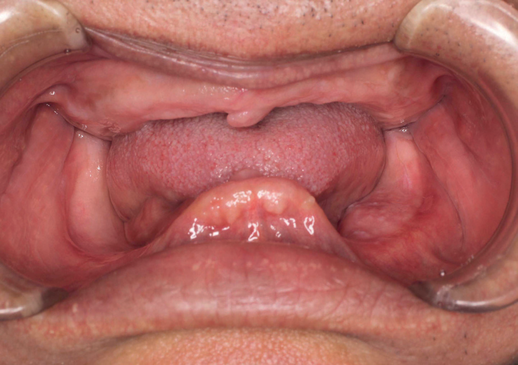
Hypertrophy of the fungiform papillae of the tongue has been observed.2 Impacted, displaced, supernumerary or missing teeth particularly in the mandible, widening of the alveolar canal and overgrowth of the alveolar ridge are recognized oral manifestations of NF1.3
The most common radiological finding was enlargement of the mandibular canal. It is logical to assume that the enlargement can be caused by the overgrowth of the mandibular nerve.3
APPEARANCE OF SYMPTOMS
Symptoms, particularly the most common skin abnormalities like café-au-lait spots, neurofibromas, Lisch nodules, and freckling in the arm pit and groin are often evident at birth or shortly afterwards and almost always by the time a child is 10 years old.
CASE REPORT
The patient is a 56 yrs. old lady with NF1 diagnosed at the age of 18 yrs. Patient is married and had 5 children of which 2 expired. Only the second child has similar kind of lesion. Patient claims no history of similar condition in the family other than the second child.
GENERAL EXAMINATION
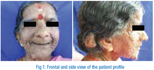
General examination of the patient revealed well defined swellings all over the body including hands, feet, trunk, and face suggestive of neurofibromas. (Fig.1)
INTRAORAL EXAMINATION
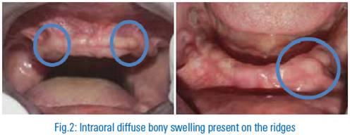
Intraoral examination revealed completely edentulous maxillary and mandibular arches. Patient had undergone extraction of all teeth 6 yrs. ago due to caries involvement. Bony enlargement is evident in some areas. (Fig.2)
SOFT TISSUE EXAMINATION
Soft tissue examination revealed a diffuse swelling in the lower lip measuring 1x1 cm on the left side, soft in consistency and non tender on palpation. Patient also had enlargement of the fungiform papilla. (Fig. 3)

RADIOLOGICAL EXAMINATION
The most common radiologic finding was enlargement of the mandibular canal.3 This is supported by the fact that the literature contains reports in which neurofibromatous tissue is found in association with osseous deformities and others when it is not. It is logical to assume that enlargement of the mandibular canal can be caused by the overgrowth of the mandibular nerve, although no direct evidence on the nerve structure is available.3
The change of the shape of the coronoid notch may be a pathognomonic feature.
In a study by Linda lee, Yat hang et al, the most common findings included increase in bone density, enlarged mandibular foramen, lateral bowing of the mandibular ramus, increase in dimensions of the coronoid notch and a decrease in the mandibular angle.4
Baden in 1955 classified the bone changes as
(1) Deformity such as asymmetry or hypertrophy,
(2) Atrophy from pressure of a soft tissue tumor, and
(3) Radiolucent effects from central bone tumors or secondary bone destruction.
Jaffe stated that the skeletal abnormalities seen in NF represent
(1) Direct destruction from neurofibromatous tissue proliferation
(2) Aberrations of skeletal development and growth either localized or systemic.4
In this patient, in the OPG (Fig. 4), we can see diffuse radio-opacity corresponding to the regions of intra oral diffuse bony swellings. The mandibular canal shows slight enlargement and there is deepening of the coronoid notch.
The side view photograph of the patient is compared with the lateral cephalogram to see the overlapping of the neurofibromas and the internal osseous defects. (Fig 5)
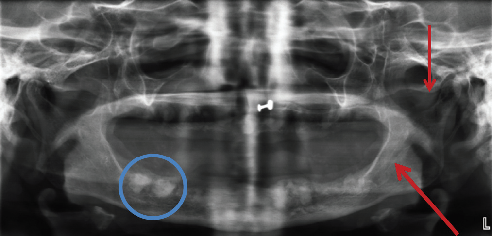
Fig 4: OPG findings

TREATMENT
The patient was given explanation regarding the advantages and the limitations of the prosthesis to avoid unrealistic expectations by the patient. Informed consent was obtained as a part of treatment protocol.
TRAY SELECTION AND PRIMARY IMPRESSION:
After preliminary examination of the patient’s oral cavity, primary stock trays were selected for maxillary and mandibular arches. The trays were beaded with wax to avoid any sharp edges that could harm the intraoral tissues. Primary impressions of the maxillary and mandibular arch were made with Alginate as impression material and were sent to the laboratory area for pouring the primary casts and preparation of special trays.
SECONDARY IMPRESSION:
The special trays were checked in the patient’s mouth and overextensions if any were trimmed and smoothened. Border moulding was done using low fusing green stick compound. The tissues (checks and lips) were displaced within physiological limits in order to allow the maximum extension to the denture bearing area without impinging on the oral tissues while in function.
After the wax spacers were removed, the secondary impressions were made with zinc oxide eugenol impression paste / light body addition silicone. The negative likenesses of the bony protrusions were noticed in the impressions. (Fig.6)
The master cast (Fig.7) obtained from the secondary impression was used to fabricate a temporary denture base of self-cure acrylic resin. An occlusal rim was made out of modelling wax and it followed the shape of the residual ridge of the patient.
JAW RELATION
The jaw relation procedure for the maxillary arch were recorded by the adjustment of occlusal rims for facial fullness, anterior and posterior planes, amount of visibility, the facial midline, canine lines and smile line, which were corrected and recorded.
The vertical dimension was determined and adequate freeway space was given by using Niswonger’s technique or phonetic technique5. Finally, the patient was guided into centric relation and the occlusal rims are sealed in this position. Teeth were selected according to various criteria such as shape of the face, gender and complexion of the patient.
The master casts along with the occlusal rims were mounted on a mean value articulator. Characterized teeth arrangements were done.
TRY IN
Once the teeth arrangement was completed and waxing and carving was done and try in of waxed up denture was carried out in the patients mouth. Once again, facial fullness, midline and occlusal plane were inspected and necessary corrections were carried out. Phonetics and esthetics were assessed and patient’s satisfaction and approval were considered before processing the complete dentures.FLASKING AND CURING:
The waxed-up denture was flasked. This was followed by dewaxing, packing of the heat cure acrylic material and then curing. Once the curing cycle was completed, the flasks are allowed to cool to room temperature. The complete denture prosthesis was retrieved from the flasks and trimming, finishing and polishing were carried out.
COMPLETE DENTURE PROSTHESIS FIT AND INSERTION:
The prostheses were inserted into the patient’s mouth and necessary corrections were made to suit the comfort of the patient. Occlusal adjustments and selective grinding were also done.POST INSERTION INSTRUCTIONS:
The patient was instructed to wear the complete denture prosthesis only during daytime and to place it in a container with water while not in use. She was told that the food cannot be incised using the anterior teeth and it had to be masticated using the posterior teeth of the prosthesis. The patient was recalled after 48 hours for initial follow up and then after 7 days for evaluation and then subsequently for long term follow up.

DISCUSSION
NF1 is a progressive disorder, which means most symptoms will worsen over time, although a small number of people may have symptoms that remain constant. It isn’t possible to predict the course of an individual’s disorder. Most people with NF1 have a normal life expectancy.
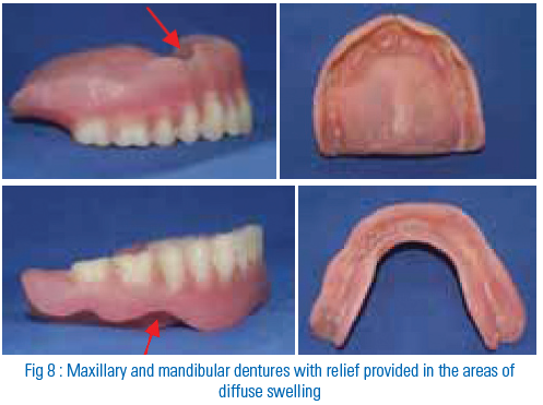
Neurofibromas on or under the skin can increase with age and cause cosmetic and psychological issues. The complete dentures gave the patient a new lease of life but due to the presence of diffuse osseous swelling along the ridges, the borders had to be trimmed to provide relief in these areas (Fig.8, 9). This in turn, slightly compromised the retention of the complete dentures. It was a limitation in this case. However, the patient is content with complete dentures.
SUMMARY AND CONCLUSION
Neurofibromatosis has its changes on the appearance of the patient and due to fibromatous growth within the oral cavity can lead to compromised retention of complete or partial dentures in the patient .However within the boundries of treatment options available and with interdisciplinary help we can proceed with providing the necessary rehabilitation to improve the appearance of the patient and also providing the patient with comfort of replaced teeth .Thus we will help the patient to perform the masticatory and phonetic functions which call for the socialization of the patient and increase his/her self confidence.
REFERENCES
- Neville, Damm, Allen, Bouquot. Oral and Maxillofacial Pathology. 3rd ED, Elsevier.
- Joseph, Robert Langlais et al. Jaw and Skull changes in neurofibromatosis. Oral Surg Oral Med Oral Pathol 1988;66:391-6
- Vivian, Sirkku et al. Radiographic findings in the jaws of patients with Neurofibromatosis 1. J oral Maxillofac Surg 70:1351-1357,2012
- Linda lee, Yat hang et al. Radiographic features of the mandible in Neurofibromatosis. Oral Surg Oral Med Oral Pathol oral radiol Endod 1996;81:361-7
- Zarb-Bolender. Prosthodontic treatment for edentulous patients, complete dentures and implant supported prostheses. 12th Ed., Elsevier, page 278-279.
Disclaimer from Guident
The views and opinions expressed in articles published in Guident are solely those of the authors and do not necessarily reflect the views of the editors, the editorial board or the publisher, Ivory India. Guident does not endorse any product, technique or treatment described. Clinical content is intended for qualified dental and medical professionals and is not a substitute for individual clinical judgement. Guident, its editors and the publisher accept no liability for any consequence arising from the use of information published in the journal.
How to cite this article
Dr. Krishna Prasad, Dr. Anupama Prasad, Dr. Archana Ashok S.. Oral Manifestations of Neurofibromatosis. Guident. 2013 Dec;7(1). Available from: https://guident.in/articles/prosthodontics/oral-manifestations-of-neurofibromatosis.html