Maxillary Antroplasty Case Selection Protocol
Department of Prosthodotics K.D. Dental College & Hospital, Mathura, (U.P), India
Abstract
For many years, open-field aluminum–nickle–cobalt magnets have been used in prosthodontics, but limited success has been achieved because of their susceptibility to corrosion by the saliva. Also their retentive force is weaker relative to the initial retention offered by mechanical attachments. Magnets made from alloys of the rare earth elements samarium and neodymium provide stronger magnetic force per unit size. In addition laser-welded containers have improved protection from salivary corrosion. The magnetic attachments have immense application because, unlike mechanical attachments, they have potential for unlimited durability and might therefore be superior to mechanical ball or bar attachments for
the retention of removable prostheses on implants such as overdentures.
Key Words: Magnets, overdentures, retentive force
INTRODUCTION
Maxillary Antroplasty is a procedure which allows surgical placement of bone grafts at the sinus floor enabling placement of implants and optimal restoration in patients missing posterior maxillary teeth.
Restoration of the posterior edentulous maxilla using dental implants can be difficult because of increased pneumatisation of the maxillary sinus or inadequate ridge width, often the bone partition between the alveolar mucosa and the maxillary sinus is as thin as 1mm.However, with the advent of sinus lift and grafting procedures implant treatment is as feasible as in any other site.
Maxillary Antroplasty with bone grafting as a procedure has evolved with new and more refined surgical techniques, instrumentation and also the availability of new grafting materials.
BENEFITS
Maxillary Antroplasty with bone graft augmentation enables the surgeon to:
- Reconstruct the highly atrophic posterior maxilla
- Replace the patients removable prosthesis
- Stabilize the anterior residual dentition by reconstructing the entire arch
- Reduce the continuous progressive atrophy of the posterior alveolar ridge.
LIMITATIONS
Certain factors may limit the desirability of performing an antroplasty with augmentation bone grafting.
- 1. Any acute and chronic sinusitis other pathologic state must be eliminated before performing the surgical procedure.
- The procedure is relatively difficult. The surgeon must have proper training and knowledge of the anatomy of the area, along with capabilities of managing and treating potential interoperative and postoperative complications. A lack of adequate instrumentation can also make this procedure extremely difficult.
- Perforation of the Schneiderian membrane may occur. The membrane must either be repaired or the procedure must be aborted if the graft material cannot be contained.
CONTRAINDICATIONS
- Patients who have undergone irradiation in the zone to maxilla
- Sepsis.
- Uncontrolled systemic diseases.
- Excessive consumption of tobacco, alcohol or drugs.
- Psychophobias.
- Scar by ablation
CASE SELECTION
Preoperative Patient Evaluation
a) Clinical Assessment
a.1 Patient should be evaluated for:
-
- Acute sinusitis which includes unilateral headaches/ throbbing and constant pain in the maxillary sinus area, often radiating to the teeth and /ears. The teeth are vital, but may be sore and painful to percussion. The skin overlying the infraorbital foramen may be swollen or red. This is a result of inflammation to the neuromuscular structures as they proceed through the superior wall of the maxillary sinus.
- Chronic sinusitis is an infection with a duration usually longer than three months and symptoms usually similar to acute sinusitis but milder, such as chronic cough and bronchitis, caused by a polymicrobial origin such as peptostreptococcus, corneybacterium, bacteroides, and veillonella spp. Vague headaches, fullness of the face, and mucopurulent hypersecretion are also associated with chronic sinusitis. The excretions from the nose are thick and often have a yellow or green hue.
- Sinusitis of odontogenic origin occurs in approximately 10% of the cases and presents a different clinical and microbiologic picture, facilitating the differential diagnosis. Polymicrobial population; contain anaerobic streptococci, bacteroides , and coliform bacilli. Clinical symptoms may be minimal despite extensive radiographic findings.
- Allergic rhinitis may be the most common form encountered, with 15% to 56% of patients undergoing endoscopy showing evidence of allergy. In cases of allergic sinusitis, sneezing, and watering eyes may be observed. Secretions from the nose are clear and thin.
- Sinus congestion upon waking, all of which may indicate potential sinus pathosis. A patient with sinusitis, sinus disease, or invasive lesions should be referred to an appropriate medical therapist for treatment before surgery proceeds.
- Patient should be asked about tobacco use and the ability to refrain from use before and after surgery, as this can severely impact the success of bone grafting. Nicotine impairs bone healing, diminishes osteoblast function, causes autogenous bone graft morbidity, and decreases graft biomechanical properties.
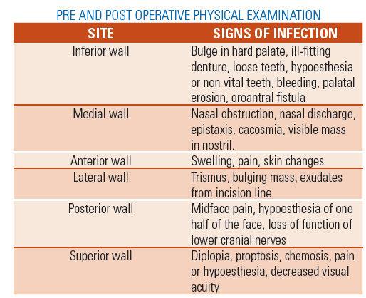
a.2 Physical examination of the maxillary sinus:-
- A physical examination of the maxillary sinus evaluates the middle one third of the face for the presence of asymmetry, deformity, swelling, erythema, ecchymosis, hematoma, or facial tenderness. The examination is conducted to assess each wall separately.
- Nasal congestion or obstruction, prevalent nasal discharge, epistaxis, anosmia, and/ or halitosis are noted.
- The infraorbital foramen is palpated through the soft tissue of the cheeks or intraorally to determine if tenderness or discomfort is present.
- The intraoral examination assesses alveolar ulceration, expansion, tenderness, paresthesia, and oroantral fistulae.
- The eyes are examined for proptosis, papillary level, lack of movement, and diplopia.
- The nasal fluids may be evaluated by asking the patient to blow the nose in a waxed paper. The mucous should be clear and thin in nature. A yellow or greenish yellow tint or thickened discharge indicates infection.
- Other methods of examination include transillumination, rhinoscopy, nasoendoscopy, bacteriology, cytology, fiberoptic antroscopy, radiography or magnetic resonance imaging. Infected maxillary sinuses typically exhibit pus in the middle meatus and can be inspected with a nasal speculum through the nares.
b) Pathologic Assessment
- The pathologic conditions that involve the mucosa of the maxillary sinus include inflammatory conditions, neoplasms and cysts.
- Because of its proximity to the oral cavity, oral disease may extend into the antral cavity.
- Fibrous dysplasia, ossifying fibromas or giant cell lesions may also be diagnosed in this area.
c) Radiographic Assessment
Maxillary sinus is traditionally studied by a
- Waters projection (face down or modified),
Advantages
- Represents a better view to illustrate cloudiness and sclerotic changes of the maxillary sinus.
Disadvantages
-
- Contour and detection of cyst like densities of the sinus are illustrated better on a panoramic x-ray film or CT scan.
- The floor of the antrum and the amount of available bone between the alveolar crest and sinus floor cannot be determined with waters view.
- Panoramic Radiograph
- It can demonstrate the maxillary sinus as multicompartmented, with radiopaque bony septa. These septa rarely completely divide the antrum. They are composed of cortical bone and can be used for augmentation. They act as additional walls of bone for blood vessels to grow into the graft and generate bone within the graft site. If future implants can engage these bony septa, a denser, stronger immediate fixation and bone implant interface likely will occur. However, they are the most likely area of membrane tear during its elevation.
- The innominate line is a thin, vertical, radiopaque line on a panoramic x ray film in the posterior third of the antrum. The line is an artefact corresponding in its lower half to the posterior surface of the zygomatic process of the maxilla and in its upper half to the posterior surface of the frontal process of the zygoma. It should be distinguished from the vertical septa, reinforcement webs, or the posterior wall of the antrum.
- The hard palate and floor of the nose are two radiographic lines observed in the lower third of the maxillary antrum. The lower line corresponds to the actual image whereas the upper line represents the ghost image of the contra lateral side. The distance between the palatal lines and the floor of the antrum gives a good indication of the facial height of the bone below the zygoma for direct surgical access for the sinus graft. In long term edentulous maxilla, the crest of the ridge may be superimposed on the palatal lines. In this case, the lateral access window may require preparation within the zygomatic arch.
- Healthy maxillary sinus radiographic image should appear radiolucent. Any pathologic condition in the antrum will present radiopaque.
- To obtain a proper diagnostic image of the antrum the patients tongue should be kept in contact with the palate. Improper obliteration of the airway results in a radiolucent image superimposed on the sinus leading to misdiagnosis.
- Occasional radiopaque projections of the inferior and middle turbinates or of the inferior, superior, or middle meatus also may be present within the maxillary sinus image.
- Maxillary tori create a bilateral, dense radiopaque image within the antrum.
- Occasionally one may detect the presence of oroantral fistula.
CT Scans
Advantages
Clear diagnostic images of the maxillary sinus in all dimensions, view of all the walls and potential associated pathologic conditions can be visualized.
- The images show details of the regional anatomy, illustrate any mucosal thickening and their extent, and one can clearly identify the presence of septa.
- Use of interactive CT (2D 3D CT -SIM/PLANT) for treatment planning in the maxillary posterior region enables the surgeon and restorative doctor to visualize all surgical and prosthetic parameters during the treatment planning phase. The information acquired about the volume of graft, ridge relationship before and after grafting and resulting implant angulations are valuable assets.
- Because of individual patient variations, the crest of the ridge may be parallel to the floor of the antrum. CT scans allow for three dimensional treatment planning.
- MRI
a.It is sensitive for diagnosis of maxillary sinus mucosal pathologic conditions and may be an adjunct for definitive diagnosis.
TREATMENT SELECTION
The treatment plan approach to providing additional prosthodontic abutments in the maxillary posterior edentulous region has been organized by Misch into four alternative treatment options dependent on the available bone height between the floor of the antrum and the crest of the residual ridge in the region of the ideal implant location. Once the height is determined, the width of the bone at the crest provides the key for implant selection and the surgical approach.

*Subantral augmentation option^ Evaluate at implant insertion.
SUBANTRAL OPTION 1: CONVENTIONAL IMPLANT PLACEMENT
- Available bone height should be12mm or more.
- Standard protocol is followed for implant placement.
- Surgical approach is dictated by bone density. Patients with narrower volume bone may be treated with osteoplasty or augmentation to increase the width
- Augmentation for width may be accomplished with bone spreading and intrapositional grafts, leaving the lateral periosteum intact. Autogenous grafts also may be onlay or appositional.
- Augmentation in width is less often in posterior maxilla because of the available residual width and the area not being an aesthetic zone.
- No contraindications exist to the preparation or placement of implants at the level of or even through the sinus floor cortical plate.
- Endosteal implants in this category are left to heal in a non-functional environment for 4-8 months before the abutment posts are added for prosthodontic reconstruction.
- One must take care that the implants are not traumatized in any way during the initial healing period, which is followed by the progressive loading phases of treatment.
SUBANTRAL OPTION 2: SINUS LIFT (INDIRECT)/SUMMERS TECHNIQUE
- This technique is used when 0-2mm less than sufficient available bone is present.
- An implant osteotomy is made below the sinus floor.
- With the help of flat end osteotomes; by tapping them through the prepared implant osteotomy, the sinus floor is elevated.
- Bone grafting material can be carried to the site by placing it on the flat end of the osteotome.
- The implant is then placed in the osteotomy provided there is no tear in the membrane.
- The success of the sinus lift through the osteotomy cannot be confirmed at the time of implant placement. Six to eight months later, radiographs are taken to indicate the success of the 0-2 mm of increased vertical height.
- If perforation occurs during manipulation of the osteotome or implant, 2 months are added to the usual healing time to allow integration of bone with the implant and bone formation.
- Prosthodontic treatment is similar to the SA-1 category.
REFERENCES
- TISSUE ENGINEERED BONEAuthored by Zizelmann C, Schoen R, Metzger MC, Schmelzeisen R, Schramm A, Dott B, Bormann KH, Gellrich NC. Department of Oral and Maxillofacial Surgery, University Hospital Freiburg,Germany.
- A new technique for the transcrestal sinus floor elevation and alveolar ridge augmentation with press-fit bone cylinders: A technical noteMINIMALLY INVASIVE TECH USING CYLINDRICAL BONE GRAFTSPublished in Clin Oral Implants Res. 2007 Feb;18(1):69-73.
- Draenert GF, Eisenmenger W. Clinic for Maxillofacial Surgery (Head: Prof. Dr. Dr. M. Ehrenfeld), University of Munich, Lindwurmstr. 2a, 80337 Munich,Germany.Published in J Craniomaxillofac Surg. 2007 Jun 18
- A histologic and histomorphometric evaluation of anorganic bovine bone retrieved 9 years after a sinus augmentation procedureAuthored by Traini T, Valentini P, Iezzi G, Piattelli A. Dental School, University of Chieti-Pescara,Chieti,Italy.Published in J Periodontol. 2007 May;78(5):955-61
- Flapless sinus floor augmentation using endoscopy combined with CT scan-designed surgical templates: method and report of
- consecutive casesAuthored by Engelke W, Capobianco M. Department of Oral Surgery, Georg-August-Universitat, Gottingen, Germany.wengelke@med.uni-goettingen.de Published in Int J Oral Maxillofac Implants. 2005 Nov-Dec;20(6):891-7.
Disclaimer from Guident
The views and opinions expressed in articles published in Guident are solely those of the authors and do not necessarily reflect the views of the editors, the editorial board or the publisher, Ivory India. Guident does not endorse any product, technique or treatment described. Clinical content is intended for qualified dental and medical professionals and is not a substitute for individual clinical judgement. Guident, its editors and the publisher accept no liability for any consequence arising from the use of information published in the journal.
How to cite this article
Dr. Manesh Lahori, Dr. Prerna Kaushik. Maxillary Antroplasty Case Selection Protocol. Guident. 2013 Dec;7(1). Available from: https://guident.in/articles/implantology/maxillary-antroplasty-case-selection-protocol.html